
- school Campus Bookshelves
- menu_book Bookshelves
- perm_media Learning Objects
- login Login
- how_to_reg Request Instructor Account
- hub Instructor Commons
- Download Page (PDF)
- Download Full Book (PDF)
- Periodic Table
- Physics Constants
- Scientific Calculator
- Reference & Cite
- Tools expand_more
- Readability
selected template will load here
This action is not available.

Chapter 19: The Cardiovascular System - The Heart
- Last updated
- Save as PDF
- Page ID 68896

Chapter Objectives
- Identify and describe the interior and exterior parts of the human heart
- Describe the path of blood through the cardiac circuits
- Describe the size, shape, and location of the heart
- Compare cardiac muscle to skeletal and smooth muscle
- Explain the cardiac conduction system
- Describe the process and purpose of an electrocardiogram
- Explain the cardiac cycle
- Calculate cardiac output
- Describe the effects of exercise on cardiac output and heart rate
- Name the centers of the brain that control heart rate and describe their function
- Identify other factors affecting heart rate
- Describe fetal heart development
In this chapter, you will explore the remarkable pump that propels the blood into the vessels. There is no single better word to describe the function of the heart other than “pump,” since its contraction develops the pressure that ejects blood into the major vessels: the aorta and pulmonary trunk. From these vessels, the blood is distributed to the remainder of the body. Although the connotation of the term “pump” suggests a mechanical device made of steel and plastic, the anatomical structure is a living, sophisticated muscle. As you read this chapter, try to keep these twin concepts in mind: pump and muscle.
- 19.1: Introduction Although the term “heart” is an English word, cardiac (heart-related) terminology can be traced back to the Latin term, “kardia.” Cardiology is the study of the heart, and cardiologists are the physicians who deal primarily with the heart.
- 19.2: Heart Anatomy The vital importance of the heart is obvious. If one assumes an average rate of contraction of 75 contractions per minute, a human heart would contract approximately 108,000 times in one day, more than 39 million times in one year, and nearly 3 billion times during a 75-year lifespan. Each of the major pumping chambers of the heart ejects approximately 70 mL blood per contraction in a resting adult. This would be equal to 5.25 liters of fluid per minute and approximately 14,000 liters per day.
- 19.3: Cardiac Muscle and Electrical Activity Recall that cardiac muscle shares a few characteristics with both skeletal muscle and smooth muscle, but it has some unique properties of its own. Not the least of these exceptional properties is its ability to initiate an electrical potential at a fixed rate that spreads rapidly from cell to cell to trigger the contractile mechanism. This property is known as autorhythmicity. Neither smooth nor skeletal muscle can do this. Heart rate is modulated by the endocrine and nervous systems.
- 19.4: Cardiac Cycle The period of time that begins with contraction of the atria and ends with ventricular relaxation is known as the cardiac cycle. The period of contraction that the heart undergoes while it pumps blood into circulation is called systole. The period of relaxation that occurs as the chambers fill with blood is called diastole. Both the atria and ventricles undergo systole and diastole, and it is essential that these components be carefully regulated and coordinated.
- 19.5: Cardiac Physiology The autorhythmicity inherent in cardiac cells keeps the heart beating at a regular pace; however, the heart is regulated by and responds to outside influences as well. Neural and endocrine controls are vital to the regulation of cardiac function. In addition, the heart is sensitive to several environmental factors, including electrolytes.
- 19.6: Development of the Heart The human heart is the first functional organ to develop. It begins beating and pumping blood around day 21 or 22, a mere three weeks after fertilization. This emphasizes the critical nature of the heart in distributing blood through the vessels and the vital exchange of nutrients, oxygen, and wastes both to and from the developing baby. The critical early development of the heart is reflected by the prominent heart bulge that appears on the anterior surface of the embryo.
- 19.7: Key Terms
- 19.8: Chapter Review
- 19.9: Interactive Link Questions
- 19.10: Review Questions
- 19.11: Critical Thinking Questions

Want to create or adapt books like this? Learn more about how Pressbooks supports open publishing practices.
19 Chapter 19 The Cardiovascular System: The Heart
By Aylin Marz
Motivation.
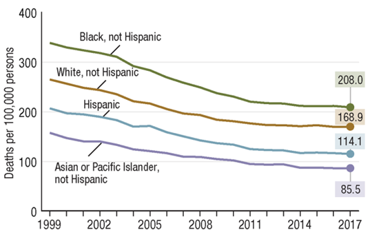
Heart disease is the number one cause of death in the U.S.racial and ethnic disparities observed in heart disease deaths also shows a disproportionately higher number of deaths due to heart disease are in Black, non-Hispanic persons. Some risk factors such as obesity, hypertension, diabetes, and high cholesterol that contribute to heart disease deaths are disproportionately higher in African American communities. Both biological factors and non-biological societal issues contribute to these disparities. As health practitioners in your communities, it is very important to be aware of the risks facing your patients and advise them for heart-healthy lifestyles. Caring and good choices can lower risk.
Learning Objectives
Upon completion of the work in this chapter students should be able to:
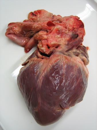
- Dissect a pig’s or sheep’s heart and label the main chambers, valves, vessels, and other structures.
- Identify and trace the path of blood flow through the heart using the dissected heart
- Relate electrocardiogram (ECG) peaks to the electrical activity, systole/diastole of the heart chambers, and the “lub” and “dub” sounds of the heart beat
- Compare the ECG of a normal heart to a diseased heart’s ECG
Background.
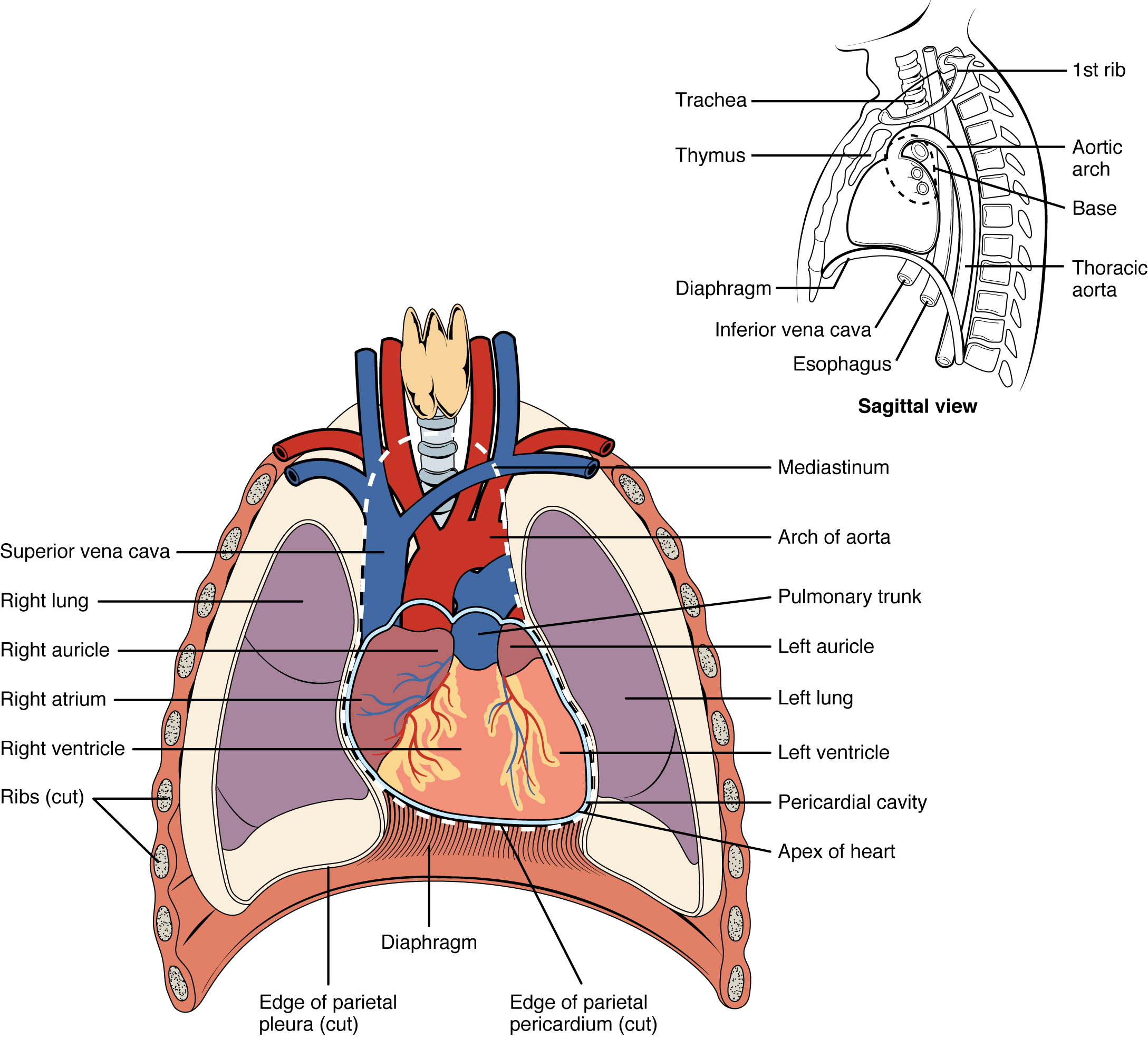
Heart Anatomy . The heart resides within the pericardial sac and is located in the mediastinal space within the thoracic cavity (Figure 19.2).
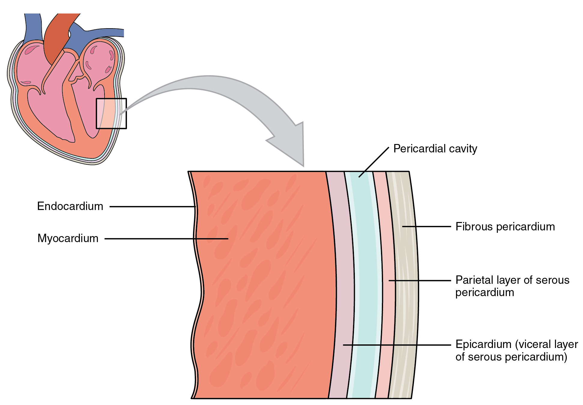
The pericardial sac consists of two fused layers: an outer fibrous capsule and an inner parietal pericardium lined with a serous membrane. Between the pericardial sac and the heart is the pericardial cavity, which is filled with lubricating serous fluid. The walls of the heart are composed of an outer epicardium, a thick myocardium (cardiac muscle layer), and an inner lining layer of endocardium (Figure 19.3).
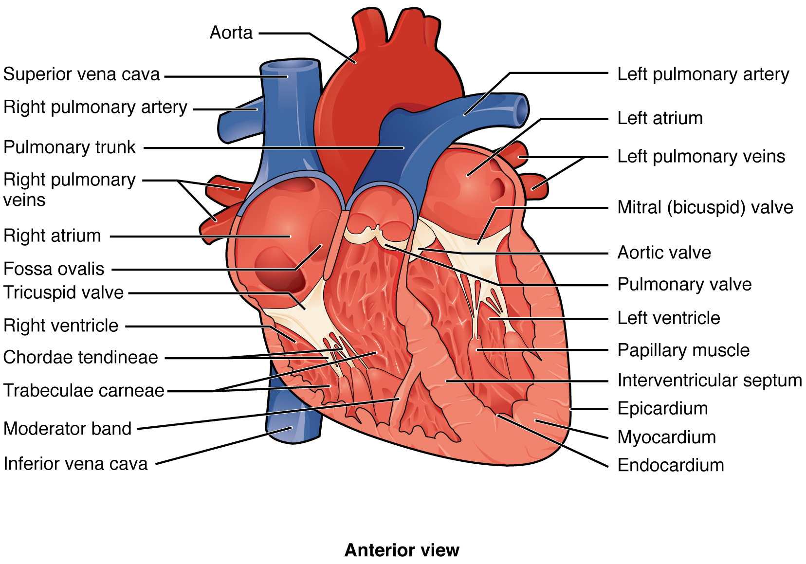
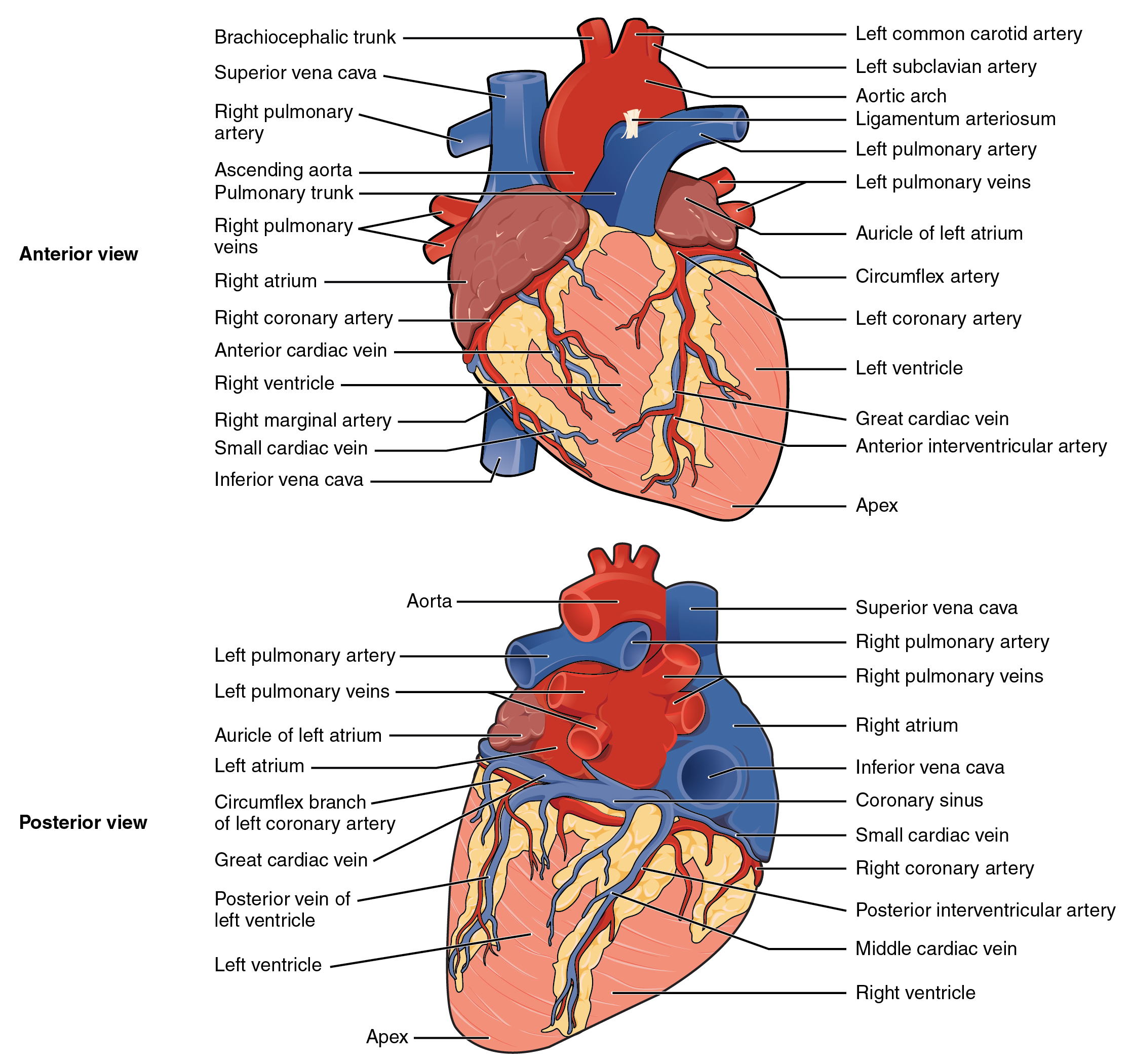
The human heart consists of a pair of atria, which receive blood and pump it into a pair of ventricles, which pump blood into the vessels. The right atrium receives systemic blood relatively low in oxygen and pumps it into the right ventricle, which pumps it into the pulmonary circuit. Exchange of oxygen and carbon dioxide occurs in the lungs, and blood high in oxygen returns to the left atrium, which pumps blood into the left ventricle, which in turn pumps blood into the aorta and the remainder of the systemic circuit. The septa are the partitions that separate the chambers of the heart. They include the interatrial septum, the interventricular septum, and the atrioventricular septum. Two of these openings are guarded by the atrioventricular valves, the right tricuspid valve and the left mitral valve, which prevent the backflow of blood. Each is attached to chordae tendineae that extend to the papillary muscles, which are extensions of the myocardium, to prevent the valves from being blown back into the atria. The pulmonary valve is located at the base of the pulmonary trunk, and the left semilunar valve is located at the base of the aorta (Figure 19.4).
The right and left coronary arteries are the first to branch off the aorta and arise from two of the three sinuses located near the base of the aorta and are generally located in the sulci. Cardiac veins parallel the small cardiac arteries and generally drain into the coronary sinus (Figure 19.5).
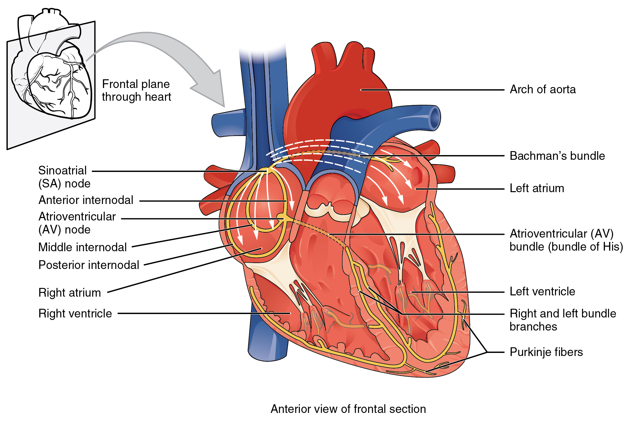
Cardiac Muscle and Electrical Activity. The heart is regulated by both neural and endocrine control, yet it is capable of initiating its own action potential followed by muscular contraction. The conductive cells within the heart establish the heart rate and transmit it through the myocardium (cardiac muscle). The contractile cells (cardiac muscle cells) contract and propel the blood. The normal path of transmission for the conductive cells is the sinoatrial (SA) node, internodal pathways, atrioventricular (AV) node, atrioventricular (AV) bundle of His, bundle branches, and Purkinje fibers (Figure 19.6).
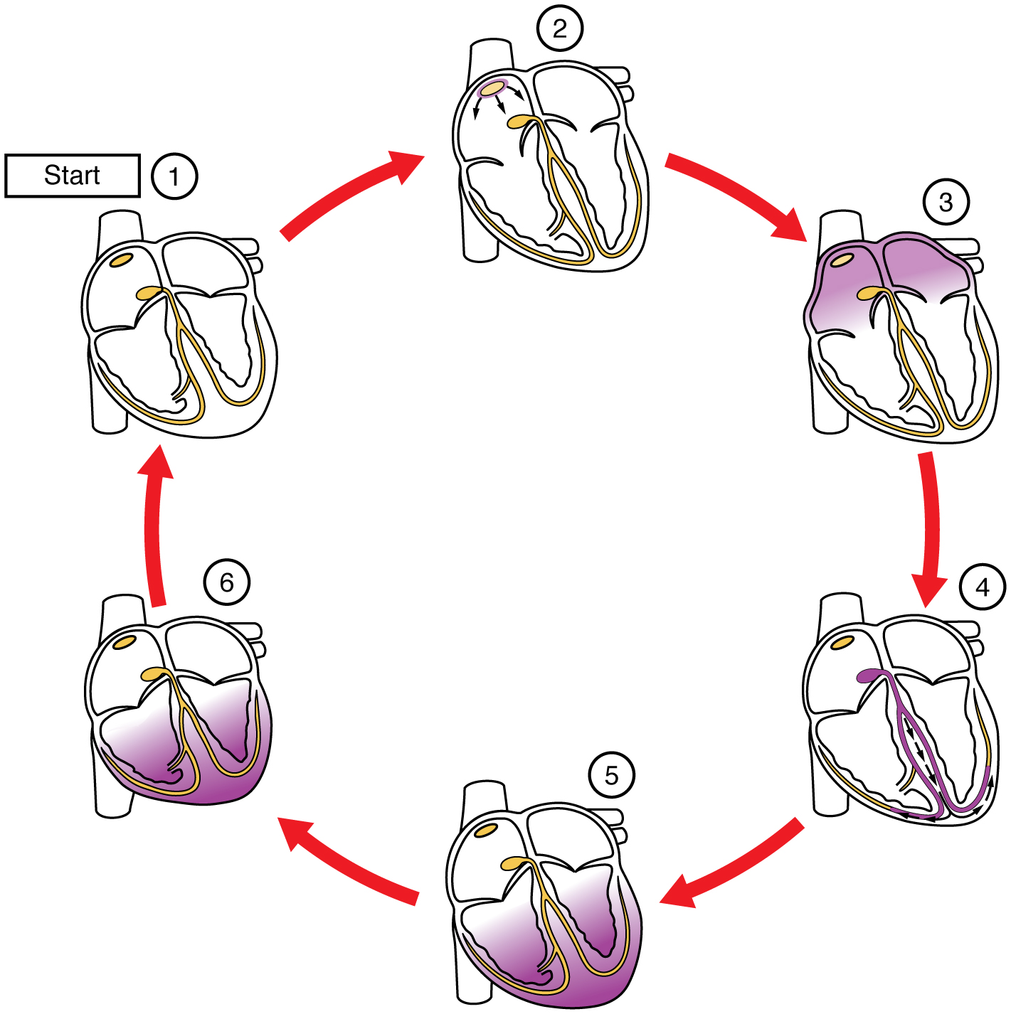
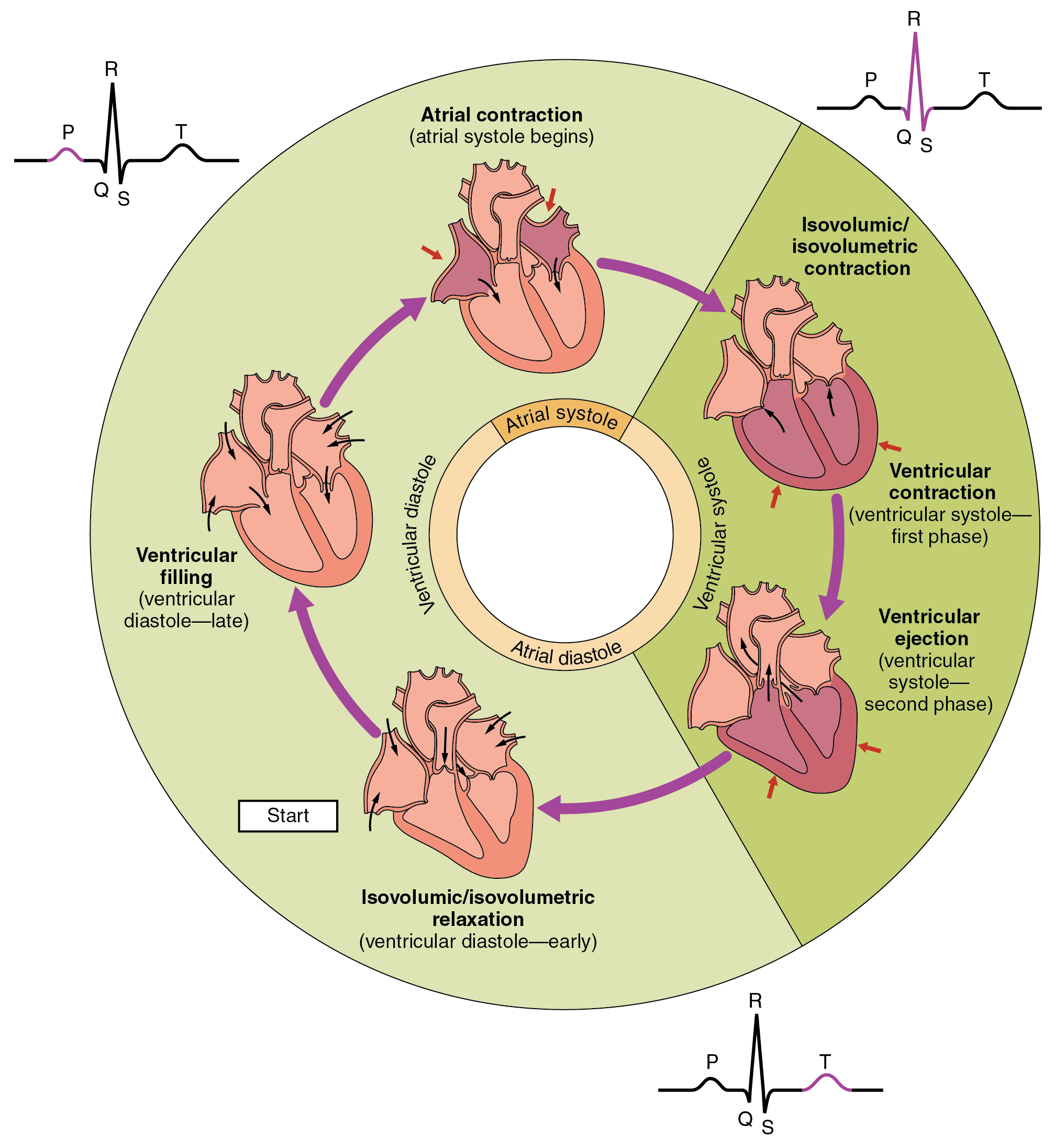
Cardiac conduction occurs in a cycle starting with the SA node that initiates atrial contraction. The electrical signal then is passed to the AV node and AV bundle and Purkinje fibers to initiate ventricular contraction. As the ventricular contraction is initiated, the atria relax (Figure 19.7).
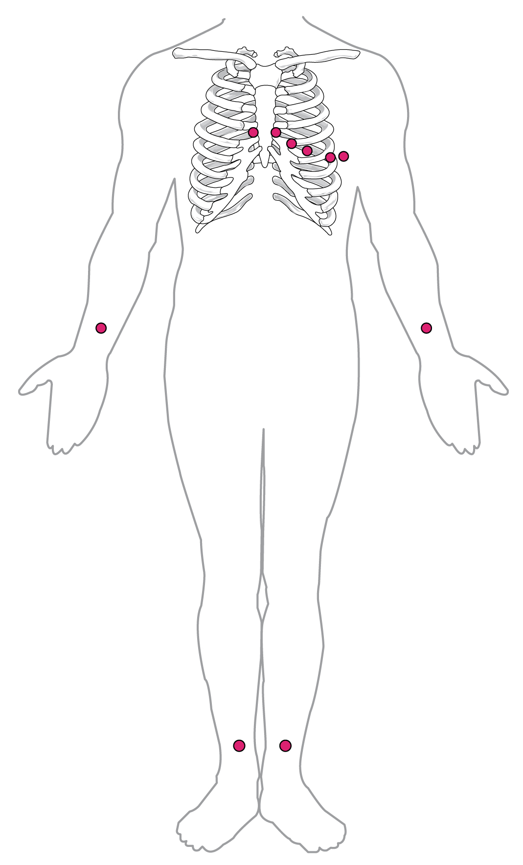
The Electrocardiogram (ECG) is used to record the electrical signals generated by the heart’s conducting cells (SA node, AV node, AV bundle, Purkinje cells) and contracting cells (myocardium or cardiac muscle cells). Electrodes are placed as shown in Figure 19.8 and the recorded traces are used to distinguish normal and abnormal heart function.
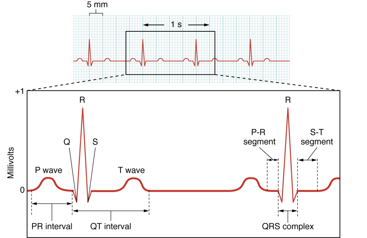
Recognizable points on the ECG include the P wave that corresponds to atrial depolarization, the QRS complex that corresponds to ventricular depolarization, and the T wave that corresponds to ventricular repolarization (Figures 19.9 and 19.10).
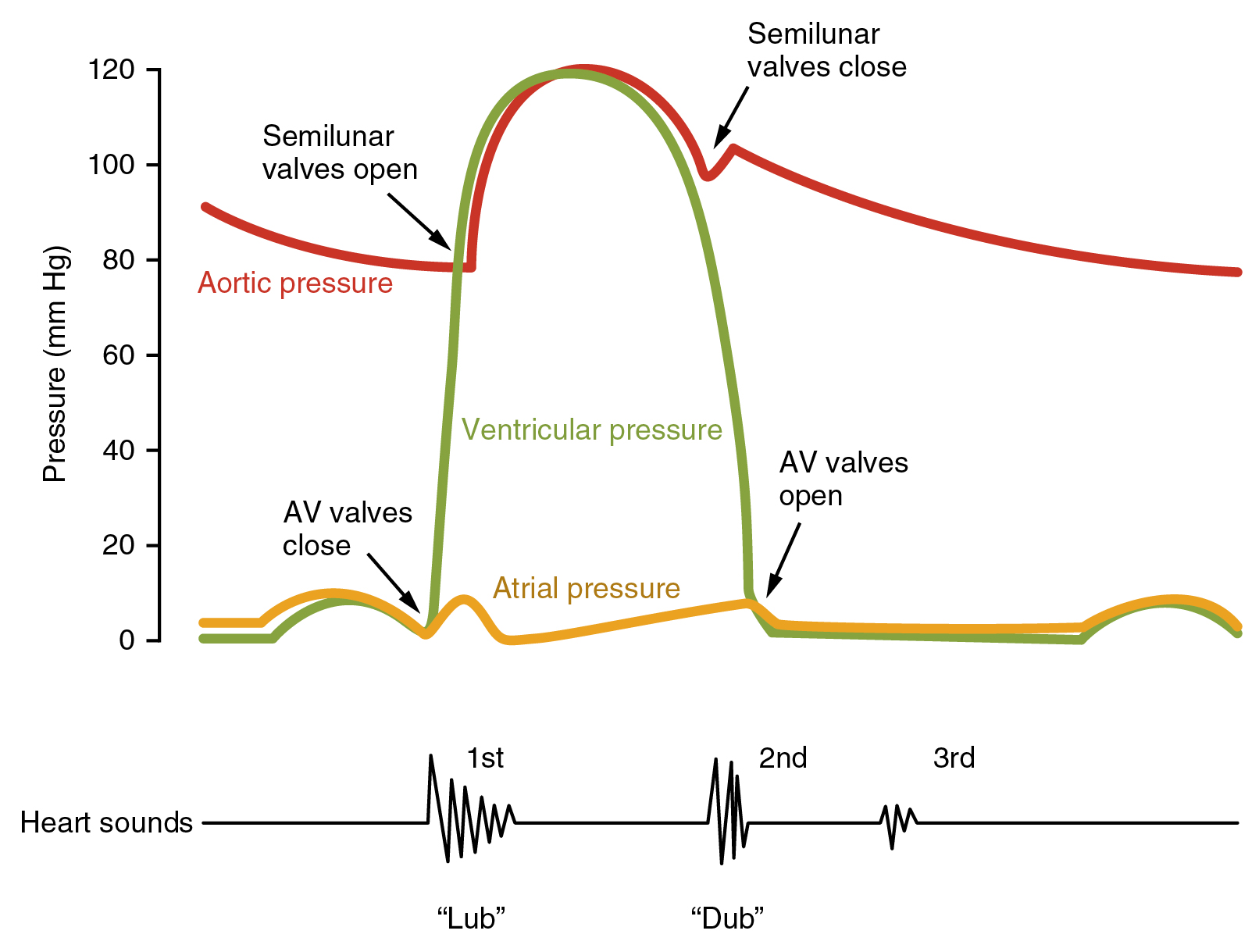
Cardiac Cycle. The cardiac cycle comprises a complete relaxation and contraction of both the atria and ventricles, and lasts approximately 0.8 seconds. Beginning with all chambers in diastole (relaxation), blood flows passively from the veins into the atria and past the atrioventricular valves into the ventricles. The atria begin to contract (atrial systole), following depolarization of the atria, and pump blood into the ventricles. The ventricles begin to contract (ventricular systole), raising pressure within the ventricles. When ventricular pressure rises above the pressure in the atria, blood flows toward the atria, producing the first heart sound, S1 or lub . As pressure in the ventricles rises above two major arteries, blood pushes open the two semilunar valves and moves into the pulmonary trunk and aorta in the ventricular ejection phase. Following ventricular repolarization, the ventricles begin to relax (ventricular diastole), and pressure within the ventricles drops. As ventricular pressure drops, there is a tendency for blood to flow back into the atria from the major arteries, producing the dicrotic notch in the ECG and closing the two semilunar valves. The second heart sound, S2 or dub , occurs when the semilunar valves close. When the ventricular pressure falls below that of the atria, blood moves from the atria into the ventricles, opening the atrioventricular valves and marking one complete heart cycle (Figure 19.11).
The valves prevent backflow of blood. Failure of the valves to operate properly produces turbulent blood flow within the heart; the resulting heart murmur can often be heard with a stethoscope. Normal and abnormal heart sounds can be heard using a stethoscope and a method called auscultation (Figure 19.12 and 19.13).
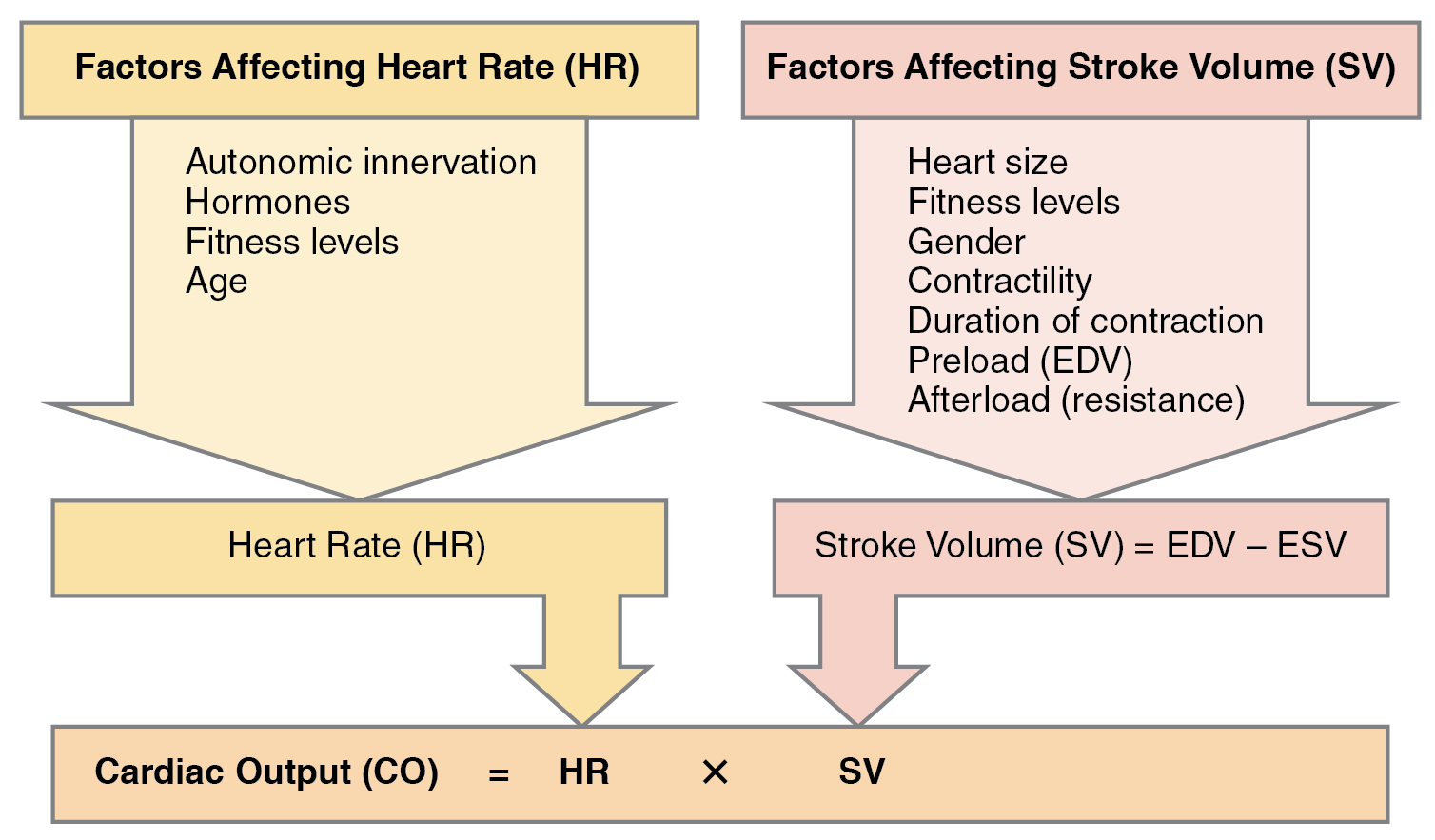
Cardiac Physiology. Cardiac output (CO) is determined by multiplying the Heart Rate (HR) by Stroke Volume (SV). HR is beats per minute and can be determined by counting the number of “lub” and “dub” sounds per minute by auscultation or by taking the pulse from the wrist (brachial artery) or neck (carotid artery). Heart rate can also be determined by using the ECG and counting the number of QRS peaks per minute. SV is the volume of blood pumped by the ventricles. SV is the difference between End Diastolic Volume (EDV) and End Systolic Volume (ESV).
Many factors affect HR and SV and together, they contribute to cardiac function. HR is largely determined and regulated by autonomic stimulation and hormones. There are several feedback loops that contribute to maintaining homeostasis dependent upon activity levels. SV is regulated by autonomic innervation and hormones, but also by venous return. Venous return is the volume of blood that returns to the atria of the heart and is determined by activity of the skeletal muscles, blood volume, and changes in peripheral circulation. Figure 19.14 summarized the main influencers of cardiac output.
Pre-Laboratory Questions
After you review the Background information above, answer the following questions before attempting the Exercises in the laboratory.
- What are the main chambers of the heart? Sketch and label each.
- What are the main blood vessels that bring blood into and pump it out of the heart? Sketch and label each. Indicate which one brings blood “into” and which one takes blood “out of” the heart.
- What is the function of the coronary arteries and veins? Where are these located?
- List the nodes and fibers involved in cardiac conduction starting with the pacemaker and listing these structures in the order of activation.
- List the steps in the cardiac cycle starting with a heart that has all chambers relaxed.
- What do the P, QRS and T designation on an ECG correlate to in atrial and ventricular depolarization/repolarization and contraction/relaxation?
- Which event produces the “lub” and “dub” sounds of the heart beat?
- What is auscultation and the name of the instrument used for it?
- What is the formula for cardiac output?
- List one way in which you can determine heart rate.
Exercise 1 Pig or Sheep Heart Dissection – Heart Anatomy
Exercise 2 blood flow through the heart (optional), exercise 3 electrocardiogram (ecg) analysis in normal and diseased hearts.
Dissection guide: https://www.biologycorner.com/anatomy/circulatory/heart/heart_dissection.html
Required Materials
- dissection tray,
- dissection tools (knife or scalpel; forceps; scissors; dissection pins for labeling)
- preserved pig or sheep heart
- labeling tape
- Heart Model on a Stand
- Giant Heart Model
- Heart Bismount
- dissected pig or sheep heart
Use the dissected heart to label the path of the heart from the entry of blood into the heart chambers to the exit of blood from these chambers and out of the heart. Use the dissecting pins and tape to label the following.
1.Find and label the veins that bring blood to the heart. Take a picture and paste it below.

2. Label the heart chambers into which blood flows. Take a picture and paste it below .
3. Label the two heart valves through which blood flows from the entry chambers to the exit chambers. Take a picture and insert it below.
4. Label the two chambers from which blood gets pumped out. Take a picture and insert it below .
5. Identify the arteries that carry blood out of the heart from the two chambers you labeled above. Label each artery. Take a picture and insert it below .
6. Using the pictures you took as a guide, create a drawing to trace the path of blood flow from entry into to exit from the heart. Label the veins, the heart chambers, the heart valves, and the arteries. Use red color to show oxygenated blood and blue color to show deoxygenated blood.
Required Materials
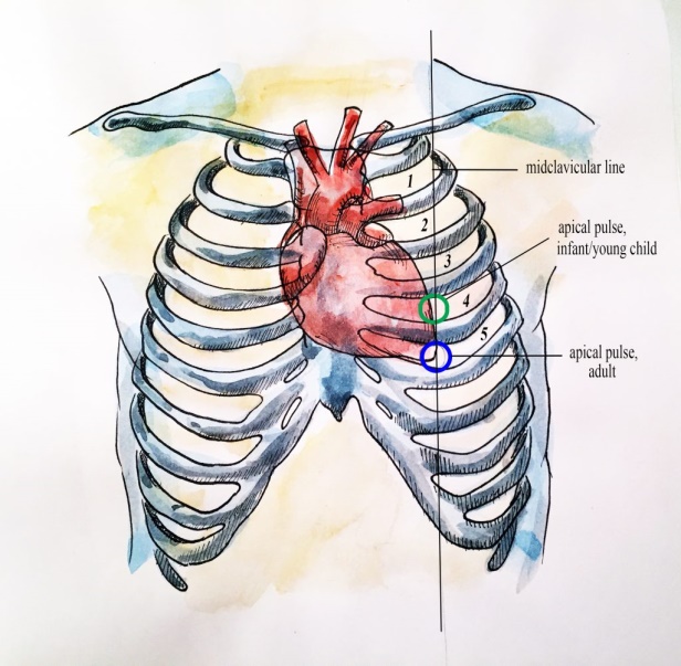
- stethoscope
1.Use the stethoscope as shown in Figure 19.17 to listen to the heart sounds of a classmate. Count the number of beats per 10 seconds. Record this value here: __________
2. Calculate how many seconds it takes from the start of one heart beat to the next. Record: __________
3. Calculate how many beats the heart beats per minute (60 seconds) in BPM (beats per minute). Record: ______
4. Examine the following EKG (also known as ECG):
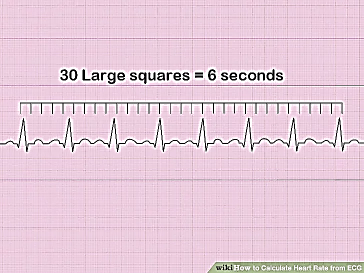
5. How many QRS complexes do you count within the 6 seconds indicated by the 30 large squares of the EKG paper in Figure 19.18? Record:__________
6. What is the heart rate corresponding to this EKG trace? Calculate it in BPM . Record: __________
7. If this EKG were taken from the friend whose heart rate you determined above, how many QRS complexes would you expect to see within the 6 second window of the EKG ? Record:_________
8. How many squares from the left edge would you expect to hear the first “lub” sound? Last “lub” sound (in Figure 19.18)? Record:__________
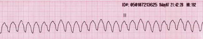
9. In Figure 19.19, you have the ECG recording of a patient with a condition called tachycardia. Count the number of peaks per 30 squares to determine the 6 second and then the 1 minute heart rate in BPM. Record: ______, ________
10. How does the heart rate in tachycardia compare to the heart rate in the normal ECG shown in Figure 19.18?
11. If you were to guess, what do you think the word Tachycardia refers to based on your findings above?
Post-laboratory Questions
- What would be the effect of this hole on the oxygenation level of the blood pumped out of the heart into the systemic circulation?
- Explain the reasoning behind your answer. Use a sketch of the heart to help.
- What effect will this have on the ability of the heart to pump blood out of the heart?
- Will this affect pulmonary circulation or systemic circulation?
- Explain the reasoning behind your answers to (a) and (b). Use a sketch of the heart to help.
- If you hear 20 “lub” and “dub” sounds in 15 seconds, what is the heart rate of your friend in BPM?
- In the cardiac cycle, what does the “lub” sound you hear correspond to? How about the “dub” sound?
- If you obtain an EKG of the same classmate, how many QRS complexes do you expect to see in the 30 large squares of the EKG paper that correspond to 6 seconds?
Anatomy and Physiology Laboratory Manual for Nursing and Allied Health Copyright © by Aylin Marz; Ganesan Kamatchi; Joseph D'Silva; Krishnan Prabhakaran; Rajeev Chandra; and Solomon Isekeije is licensed under a Creative Commons Attribution 4.0 International License , except where otherwise noted.
Share This Book

- school Campus Bookshelves
- menu_book Bookshelves
- perm_media Learning Objects
- login Login
- how_to_reg Request Instructor Account
- hub Instructor Commons
- Download Page (PDF)
- Download Full Book (PDF)
- Periodic Table
- Physics Constants
- Scientific Calculator
- Reference & Cite
- Tools expand_more
- Readability
selected template will load here
This action is not available.
5: Module 3- The Cardiovascular System- The Heart
- Last updated
- Save as PDF
- Page ID 34595
- 5.1: Introduction to the Cardiovascular System- The Heart
- 5.2: Heart Anatomy
- 5.3: Cardiac Muscle and Electrical Activity
- 5.4: Cardiac Cycle
- 5.5: Cardiac Physiology
- 5.6: Development of the Heart

An official website of the United States government
The .gov means it's official. Federal government websites often end in .gov or .mil. Before sharing sensitive information, make sure you're on a federal government site.
The site is secure. The https:// ensures that you are connecting to the official website and that any information you provide is encrypted and transmitted securely.
- Publications
- Account settings
- Browse Titles
NCBI Bookshelf. A service of the National Library of Medicine, National Institutes of Health.
StatPearls [Internet]. Treasure Island (FL): StatPearls Publishing; 2024 Jan-.

StatPearls [Internet].
Physiology, cardiovascular.
Raheel Chaudhry ; Julia H. Miao ; Afzal Rehman .
Affiliations
Last Update: October 16, 2022 .
- Introduction
The cardiovascular system provides blood supply throughout the body. By responding to various stimuli, it can control the velocity and amount of blood carried through the vessels. The cardiovascular system consists of the heart, arteries, veins, and capillaries. The heart and vessels work together intricately to provide adequate blood flow to all parts of the body. The regulation of the cardiovascular system occurs via a myriad of stimuli, including changing blood volume, hormones, electrolytes, osmolarity, medications, adrenal glands, kidneys, and much more. The parasympathetic and sympathetic nervous systems also play a key role in the regulation of the cardiovascular system. [1] [2] [3]
- Organ Systems Involved
The heart is the organ that pumps blood through the vessels. It pumps blood directly into arteries, more specifically, the aorta or the pulmonary artery. Blood vessels are critical because they control the amount of blood flow to specific parts of the body. Blood vessels include arteries, capillaries, and veins. Arteries carry blood away from the heart and can divide into large and small arteries. Large arteries receive the highest pressure of blood flow and are thicker and more elastic to accommodate the high pressures. Smaller arteries, such as arterioles, have more smooth muscle, which contracts or relaxes to regulate blood flow to specific portions of the body. Arterioles face a smaller blood pressure, meaning they don't need to be as elastic. Arterioles account for most of the resistance in pulmonary circulation because they are more rigid than larger arteries. Furthermore, the capillaries branch off of arterioles and are a single-cell layer. This thin layer allows for the exchange of nutrients, gases, and waste with tissues and organs. Also, the veins transport blood back to the heart. They contain valves to prevent the backflow of blood.
The cardiovascular system consists of two main loops, systemic circulation, and pulmonary circulation. The purpose of the cardiovascular system is to provide adequate circulation of blood through the body. Pulmonary circulation allows for the oxygenation of the blood, and systemic circulation provides for oxygenated blood and nutrients to reach the rest of the body.
It is important to understand the concept of cardiac output, stroke volume, preload, Frank-Starling law, afterload, and ejection fraction to understand the physiology of the heart. The cardiac output (CO) is the amount of blood ejected from the left ventricle, and normally it is equal to the venous return. The calculation is CO = stroke volume (SV) x heart rate (HR). CO also equals the rate of oxygen consumption divided by the difference in arterial and venous oxygen content. The stroke volume is the amount of blood pumped out of the heart after one contraction. It is the difference in end-diastolic (EDV) and end-systolic volume (ESV). It increases with increased contractility, increased preload, and decreased afterload. Also, contractility of the left ventricle increases with catecholamines by increasing intracellular calcium ions and lowering extracellular sodium. The preload is the pressure on the ventricular muscle by the ventricular EDV. Frank-Starling law describes the relationship between EDV and SV. This law states that the heart attempts to equalize CO with venous return. As venous return increases, there is a larger EDV in the left ventricle, which leads to further stretching of the ventricle. Further stretching of the ventricle leads to a larger contraction force and a larger SV. A larger stroke volume leads to a larger CO, thus equalizing CO with venous return. Next, the afterload is the pressure that the left ventricular pressure must exceed to push blood forward. Mean arterial pressure best estimates this. Also, afterload can be estimated by the minimum amount of pressure needed to open the aortic valve, which is equivalent to the diastolic pressure. Thus, diastolic blood pressure is one of the better ways to index afterload. Finally, the ejection fraction (EF) is equal to SV/EDV. EF of the left ventricle is an index for contractility. A normal EF is greater than 55%. A low EF indicates heart failure. [4] [5] [6] [7]
The cardiac cycle describes the path of the blood through the heart. It runs in the following order:
- Atrial contraction closure of the mitral valve
- Isovolumetric phase
- Opening of the aortic valve
- Ejection phase (rapid and reduced ejection), emptying of the left ventricle
- Closure of the aortic valve
- Isovolumetric relaxation
- The opening of the mitral valve
- Filling phase (rapid and reduced filling) of the left ventricle
Vasculature plays a significant role in the regulation of blood flow throughout the body. In general, blood pressure decreases from arteries to veins, and this is because of the pressure overcoming the resistance of the vessels. The greater the change in resistance at any point in the vasculature, the greater the loss of pressure at that point. Arterioles have the most increase in resistance and cause the largest decrease in blood pressure. The constriction of arterioles increases resistance, which causes a decrease in blood flow to downstream capillaries and a larger decrease in blood pressure. Dilation of arterioles causes a decrease in resistance, increasing blood flow to downstream capillaries and a smaller decrease in blood pressure. Diastolic blood pressure (DP) is the lowest pressure in an artery at the beginning of the cardiac cycle while the ventricles are relaxing and filling. DP is directly proportional to total peripheral resistance (TPR). Also, the energy stored in the compliant aorta during systole is now released by the recoil of the aortic wall during diastole, thus increasing diastolic pressure. Systolic blood pressure (SP) is the peak pressure in an artery at the end of the cardiac cycle while the ventricles are contracting. Directly related to stroke volume, as stroke volume increases, SP also increases. SP is also affected by aortic compliance. Because the aorta is elastic, it stretches and stores the energy caused by ventricular contraction and decreases the systolic pressure. Pulse pressure is the difference between SP and DP. Pulse pressure is proportional to SV and inversely proportional to arterial compliance. Thus the stiffer the artery, the larger the pulse pressure. Mean arterial pressure (MAP) is the average pressure in the arteries throughout the cardiac cycle. The MAP is always closer to DP. MAP is calculated by MAP= DP + 1/3 (pulse pressure). Also, MAP = CO x TPR, where CO is cardiac output. This value is significant because whenever there is a decrease in CO, to maintain the MAP, the TPR will increase, which is relevant in many pathophysiology problems.
Systemic veins have a lower decrease in pressure because it has low resistance. The venous system is very compliant and contains up to 70% of the circulating blood at once. A small change in venous pressure can mobilize the blood stored in the venous system. Velocity of blood in the vasculature has an inverse relationship with cross-sectional area (volumetric flow rate (Q) = flow velocity (v) x cross-sectional area (A)). As the cross-sectional area increases, velocity decreases. Arteries and veins have smaller cross-sectional areas and the highest velocities, whereas capillaries have the most cross-sectional area and the lowest velocities. The vasculature also gives resistance. Resistance is R= (8*viscosity*length)/(πr^4). Viscosity depends on hematocrit and increases in multiple myeloma or polycythemia. As tube length increases, the resistance increases. As the tube radius increases, the resistance decreases. The fact that the radius is to the power of 4 means that slight changes in the radius have a profound effect on resistance. The total resistance of vessels in a series is R1 + R2 + R3, and so on, and the total resistance of arteries in parallel is 1/TR = 1/R1+1/R2+1/R and so on, where TR is the total resistance.
The Poiseuille equation measures the flow of blood through a vessel. It is measured by the change in pressure divided by resistance: Flow = (P1 - P2)/R, where P is pressure, and R is resistance. Increasing resistance in a vessel, such as the constriction of an arteriole, causes a decrease in blood flow across the arteriole. At the same time, there is a larger decrease in pressure across this point because the pressure is lost by overcoming the resistance. Increasing the resistance at any point increases upstream pressure but decreases downstream pressure. The Poiseuille equation applies to the systemic circulation such that F is the cardiac output (CO), P1 is the mean arterial pressure (MAP), P2 is the right atrial pressure (RAP), and R is the total peripheral resistance (TPR). Because RAP is close to 0 and very small in comparison to MAP, the equation approximates as F=P1/R or CO=MAP/TPR where MAP=CO*TPR - this means that cardiac output and total peripheral resistance control MAP. Its application is important because in trauma situations with hemorrhage, there is also a decrease in cardiac output, but at times the blood pressure is near normal. This is because the TPR at the level of the arterioles has increased. This equation, as applied to the pulmonary vasculature, is used to determine the cause of pulmonary hypertension. As related to the pulmonary vasculature, F represents CO, P1 represents pulmonary artery pressure (PAP), and P2 represents left atrial pressure (LAP), and R is pulmonary vascular resistance (PR); CO=(PAP-LAP)/PR. A Swan-Ganz catheter helps to measure both PAP and LAP, allowing for the measurement of PR and, thus, the etiology of pulmonary hypertension.
The nervous system regulates the cardiovascular system with the help of baroreceptors and chemoreceptors. Both receptors are located in the carotids and aortic arch. Also, both have afferent signals through the vagus nerve from the aortic arch and afferent signals through the glossopharyngeal nerve from the carotids.
- Baroreceptors are more specifically located in the carotid sinus and aortic arch. They respond quickly to changes in blood pressure.
- A decrease in blood pressure or blood volume causes hypotension, which leads to a decrease in arterial pressure, which creates a decrease in the stretch of the baroreceptors and decreases afferent baroreceptor signaling. This decrease in afferent signaling from the baroreceptor causes an increase in efferent sympathetic activity and a reduction in parasympathetic activity, which leads to vasoconstriction, increased heart rate, increased contractility, and an increase in BP. The vasoconstriction increases TPR in the equation MAP=CO*TPR to bring pressure (MAP) back up.
- An increase in blood pressure or blood volume causes hypertension which increases the stretch of the baroreceptors
- Chemoreceptors come in 2 types: peripheral and central. Peripheral chemoreceptors are specifically located in the carotid body and aortic arch. They respond to oxygen levels, carbon dioxide levels, and the pH of the blood. They become stimulated when oxygen decreases, carbon dioxide increases, and the pH decreases. Central chemoreceptors are located in the medulla oblongata and measure the pH and carbon dioxide changes in the cerebral spinal fluid.
Autoregulation
Autoregulation is the method by which an organ or tissue maintains blood flow despite a change in perfusion pressure. When blood flow becomes decreased to an organ, arterioles dilate to reduce resistance.
- Myogenic theory: Myogenic regulation is intrinsic to the vascular smooth muscle. When there is an increase in perfusion, the vascular smooth muscle is stretched. This causes it to constrict the artery. If there is a decrease in perfusion to the arteriole, then there is decreased stretching of the smooth muscle. This leads to the relaxation of the smooth muscles and dilation of the arteriole.
- Metabolic theory: Blood flow is closely related to metabolic activity. When there is an increase in metabolism to muscle or any tissue, there is an increase in blood flow to that location. Metabolic activity creates substances that are vasoactive and stimulate vasodilation. The increase or decrease in metabolism leads to an increase or decrease in metabolic byproducts that cause vasodilation. Increased adenosine, carbon dioxide, potassium, hydrogen ion, lactic acid levels, and decreased oxygen levels, and increased oxygen demand all lead to vasodilation. Adenosine is from AMP, which derives from the hydrolysis of ATP and increases during hypoxia or increased oxygen consumption. Potassium is increased extracellularly during metabolic activity (muscle contraction) and has a direct effect on relaxing smooth muscles. Carbon dioxide is produced as a byproduct of the oxidative pathway and increases with metabolic activity. Carbon dioxide diffuses to vascular smooth muscle and triggers an intracellular pathway to relax the vascular smooth muscle.
- Heart: Metabolites that cause coronary vasodilation include adenosine, NO, carbon dioxide, and low oxygen.
- Brain: The primary metabolite controlling cerebral blood flow is carbon dioxide. An increase in arterial carbon dioxide causes vasodilation of cerebral vasculature. A decrease in arterial carbon dioxide causes vasoconstriction of the cerebral vasculature. Hydrogen ions do not cross the blood-brain barrier and thus are not a factor in regulating cerebral blood flow. A decrease in oxygen pressure in arteries causes vasodilation of the cerebral arteries; however, an increase in oxygen pressure in arteries does not cause vasoconstriction.
- Kidneys: Autoregulation of the kidneys is myogenic and with tubuloglomerular feedback. In severe cases of hypotension, kidney arterioles constrict, and renal function is lost.
- Lungs: Hypoxia of the lungs causes vasoconstriction, creating a shunt away from poorly ventilated areas of the lung and redirecting perfusion to ventilated portions of the lung.
- Skeletal muscle: Adenosine, potassium, hydrogen ion, lactate, and carbon dioxide all increase during exercise and cause vasodilation. When resting, the skeletal muscle is controlled extrinsically by sympathetic activity and not by metabolites.
- Skin: Regulation of the skin occurs through sympathetic stimulation. The purpose of regulating blood flow in the skin is to regulate body temperature. In a warm environment, skin vasculature dilates due to a decrease in sympathetic stimulation. In cold environments, skin vasculature constricts due to an increase in sympathetic activity. During fever, regulation of the body temperature is at a higher setpoint.
The starling equation can explain the capillary fluid exchange. This equation describes the forces of oncotic and hydrostatic pressure on the movement of fluid across the capillary membrane. Edema can result from an increase in capillary pressure (heart failure), a decrease in plasma proteins (liver failure), an increase in the interstitial fluid due to lymphatic blockage, or an increase in capillary permeability due to infections or burns.
- Related Testing
Swan-Ganz catheter is a thin tube that is inserted peripherally and passed to the right side of the heart and into the pulmonary artery. This catheterization is to measure the pressures in the pulmonary vasculature and the left atrium. Pulmonary capillary wedge pressure (PCWP) is an estimate of the pressure in the left atrium given by the Swanz-Ganz catheter. It is significant because it helps to differentiate pathologies. In cardiac shock, there is an increase in PCWP, whereas in hemorrhagic shock, there is a decrease in PCWP.
- Pathophysiology
Chronic hypertension is a common pathological process related to the cardiovascular system. This condition is significant because, with hypertension, there is an increase in afterload. A long-term increase in afterload leads to concentric hypertrophy of the heart and eventual left-sided diastolic heart failure. Also, an S4 heart sound will be audible at the apex of the heart. Another type of heart disease is alcoholic cardiomyopathy, which occurs in alcoholics and causes dilated cardiomyopathy, which means the ventricles become dilated, leading to systolic failure. It can be reversible if the patient stops drinking alcohol.
Heart failure or cardiac tamponade can cause cardiogenic shock. In cardiogenic shock, there is an increase in PCWP because there is a backup of blood; the heart is not able to pump blood forward because it is not able to overcome the afterload. Subsequently, there is a decrease in CO. In response to low CO, the SVR increases.
In hemorrhagic shock, there is a loss of blood, thus a loss in total volume. Because there is a loss of volume, there is a decrease in pressure and, therefore, a decrease in PCWP. Also, there is an increase in cardiac output because there is a need for more blood in the periphery. While there is an increase in CO, there is also an increase in SVR to maintain MAP.
- Clinical Significance
Blood pressure (BP) is an essential clinical value because it describes the status of the vasculature in acute and chronic states. If a patient has elevated blood pressure in the clinic on more than two occasions, the clinician can diagnose the patient with essential hypertension. BP can also be significant in acute settings, such as in the emergency room, after a patient is brought in by an ambulance due to a motor vehicle accident. At this point, it is important to assess the patient's BP because if it is low, it might indicate the patient is bleeding somewhere, and the clinician must determine the location of the bleeding as soon as possible. [8]
S1 and S2 heart sounds are normal heart sounds heard on auscultation of the heart. S1 is the sound made due to the closure of the mitral and tricuspid valves. This is followed by systole. Then the S2 sounds are heard, which are the closure of the aortic and pulmonary valves. Diastole follows this. It is important to recognize these normal heart sounds on auscultation because abnormal heart sounds such as S3, S4, and murmurs can be signs of a pathological condition.
- Review Questions
- Access free multiple choice questions on this topic.
- Comment on this article.
Disclosure: Raheel Chaudhry declares no relevant financial relationships with ineligible companies.
Disclosure: Julia Miao declares no relevant financial relationships with ineligible companies.
Disclosure: Afzal Rehman declares no relevant financial relationships with ineligible companies.
This book is distributed under the terms of the Creative Commons Attribution-NonCommercial-NoDerivatives 4.0 International (CC BY-NC-ND 4.0) ( http://creativecommons.org/licenses/by-nc-nd/4.0/ ), which permits others to distribute the work, provided that the article is not altered or used commercially. You are not required to obtain permission to distribute this article, provided that you credit the author and journal.
- Cite this Page Chaudhry R, Miao JH, Rehman A. Physiology, Cardiovascular. [Updated 2022 Oct 16]. In: StatPearls [Internet]. Treasure Island (FL): StatPearls Publishing; 2024 Jan-.
In this Page
Bulk download.
- Bulk download StatPearls data from FTP
Related information
- PMC PubMed Central citations
- PubMed Links to PubMed
Similar articles in PubMed
- Lower Genitourinary Trauma. [StatPearls. 2024] Lower Genitourinary Trauma. Tullington JE, Blecker N. StatPearls. 2024 Jan
- Deciphering the neural signature of human cardiovascular regulation. [Elife. 2020] Deciphering the neural signature of human cardiovascular regulation. Manuel J, Färber N, Gerlach DA, Heusser K, Jordan J, Tank J, Beissner F. Elife. 2020 Jul 28; 9. Epub 2020 Jul 28.
- Review Regulation of coronary blood flow during exercise. [Physiol Rev. 2008] Review Regulation of coronary blood flow during exercise. Duncker DJ, Bache RJ. Physiol Rev. 2008 Jul; 88(3):1009-86.
- Review Sympathetic nervous system overactivity and its role in the development of cardiovascular disease. [Physiol Rev. 2010] Review Sympathetic nervous system overactivity and its role in the development of cardiovascular disease. Malpas SC. Physiol Rev. 2010 Apr; 90(2):513-57.
- Electrolytes. [StatPearls. 2024] Electrolytes. Shrimanker I, Bhattarai S. StatPearls. 2024 Jan
Recent Activity
- Physiology, Cardiovascular - StatPearls Physiology, Cardiovascular - StatPearls
Your browsing activity is empty.
Activity recording is turned off.
Turn recording back on
Connect with NLM
National Library of Medicine 8600 Rockville Pike Bethesda, MD 20894
Web Policies FOIA HHS Vulnerability Disclosure
Help Accessibility Careers
COMMENTS
Usha is giving a presentation on the cardiovascular system. One of her slides shows a diagram of the blood moving from the heart to the arms and legs. Which type of circulation is the slide illustrating? systemic circulation pulmonary circulation systolic circulation diastolic circulation The best answer is : Systolic circulation
b. "Only around one quarter of your blood is in your heart at any given time." c. "Blood pressure and blood volume roughly mimic one another at any given location in the circulatory system." d. "Left-sided and right-sided pumping action on each beat of the heart must equal each other to ensure adequate blood distribution."
The human circulatory system consists of several circuits: The pulmonary circuit provides blood flow between the heart and lungs. The systemic circuit allows blood to flow to and from the rest of the body. The coronary circuit strictly provides blood to the heart (not pictured in the figure below). Image credit: Blood flow from the heart by ...
Summary. The cardiovascular system consists of the heart, blood vessels, and blood. Its primary function is to transport nutrients and oxygen-rich blood to all parts of the body and to carry ...
Circulatory system. The circulatory system, also called cardiovascular system, is a vital organ system that delivers essential substances to all cells for basic functions to occur. Also commonly known as the cardiovascular system, is a network composed of the heart as a centralised pump, blood vessels that distribute blood throughout the body, and the blood itself, for transportation of ...
The circulatory system is also called the cardiovascular system, where "cardi" refers to the heart, and "vascular" refers to the blood vessels. So, these are the two key parts: the heart, which pumps blood, and the blood vessels, which carry blood to the body and return it back to the heart again. Ultimately, this is how nutrients like ...
What is the Cardiovascular System? The cardiovascular system, also called the circulatory system, is the organ system that transports materials to and from all the cells of the body.The materials carried by the cardiovascular system include oxygen from the lungs, nutrients from the digestive system, hormones from glands of the endocrine system, and waste materials from cells throughout the body.
This condition—a patent ductus arteriosus— creates a "right-to-left shunt.". Because the circulating blood is not adequately oxygenated, it develops a deep red color. The skin then develops the blue tones typical of cyanosis and the infant is known as a "blue baby.". Tetralogy of Fallot.
Figure 1. This artist's conception of the human heart suggests a powerful engine—not inappropriate for a muscular pump that keeps the body continually supplied with blood. (credit: Patrick J. Lynch) In this chapter, you will explore the remarkable pump that propels the blood into the vessels. There is no single better word to describe the ...
human cardiovascular system, organ system that conveys blood through vessels to and from all parts of the body, carrying nutrients and oxygen to tissues and removing carbon dioxide and other wastes. It is a closed tubular system in which the blood is propelled by a muscular heart. Two circuits, the pulmonary and the systemic, consist of ...
The circulatory system (cardiovascular system) pumps blood from the heart to the lungs to get oxygen. The heart then sends oxygenated blood through arteries to the rest of the body. The veins carry oxygen-poor blood back to the heart to start the circulation process over. Your circulatory system is critical to healthy organs, muscles and tissues.
Usha is giving a presentation on the cardiovascular system. One of her slides shows a diagram of the blood moving from the heart to the arms and legs. Which type of circulation is the slide illustrating? systemic circulation pulmonary circulation systolic circulation diastolic circulation
The cardiovascular system is one of the eleven organ systems of the human body. Its main function is to transport nutrients to cells and wastes from cells (Figure 3.5.1 3.5. 1 ). Figure 3.5.1 3.5. 1: The Cardiovascular system. "Simplified diagram of the human Circulatory system in anterior view" by Mariana Ruiz / Public Domain.
Chapter Objectives. Identify and describe the interior and exterior parts of the human heart. Describe the path of blood through the cardiac circuits. Describe the size, shape, and location of the heart. Compare cardiac muscle to skeletal and smooth muscle. Explain the cardiac conduction system. Describe the process and purpose of an ...
The heart is composed of cardiac muscle, an involuntary muscle tissue that is found only within this organ. The term "cardiac" (as in cardiology) means "related to the heart" and comes from the Greek word kardia, for "heart." It has a four-chambered, double pump and is located in the thoracic cavity between the lungs.
Select one: A. Our normal heart rate at rest is more than 100 beats per minute (bmp). B. The pulmonary circulation is longer with a higher blood pressure. C. The average cardiac output at rest is about 5 L/min. D. Capillaries carry blood back to the heart. The average cardiac output at rest is about 5 L/min.
2. The Cardiovascular System Cardiovascular system: organ system that distributes blood to all parts of the body Major function - transportation, using blood as the transport vehicle. 3. The Cardiovascular System This system carries oxygen, nutrients, cell wastes, hormones and other substances vital for body homeostasis to and form cells The ...
Figure 19.6 The Conduction System of the Heart. The SA node (pacemaker) to the AV node, to the bundle of His (AV bundle) and Purkinje fibers, the heart generates and conducts electrical signals to control the order in which cardiac muscle areas contract. (Credit: OpenStax Anatomy and Physiology, CC-BY 4.0 license).
5.1: Introduction to the Cardiovascular System- The Heart; 5.2: Heart Anatomy; 5.3: Cardiac Muscle and Electrical Activity; 5.4: Cardiac Cycle; 5.5: Cardiac Physiology; 5.6: Development of the Heart; 5: Module 3- The Cardiovascular System- The Heart is shared under a not declared license and was authored, remixed, and/or curated by LibreTexts.
The cardiovascular system provides blood supply throughout the body. By responding to various stimuli, it can control the velocity and amount of blood carried through the vessels. The cardiovascular system consists of the heart, arteries, veins, and capillaries. The heart and vessels work together intricately to provide adequate blood flow to all parts of the body. The regulation of the ...
Study with Quizlet and memorize flashcards containing terms like Chapter 21 Cardiovascular System Function, Assessment, and Therapeutic Measures Questions 1-24, 1. Which part of the heart pumps deoxygenated blood toward the lungs? a. Right atrium b. Right ventricle c. Left atrium d. Left ventricle, 2. The LPN/LVN is recording the resting heart rate of a marathon runner.
Study with Quizlet and memorize flashcards containing terms like Pulmonary circulation is the key component that supports both respiratory and cardiovascular systems. Examine this diagram of a human heart as seen while looking at the front of the body. Which correctly identifies where the blood is flowing to and from?, Which of these are common symptoms during a myocardial infarction?, A ...
American Heart Association