
- Types of Bones
- Axial Skeleton
- Appendicular Skeleton
- Skeletal Pathologies
- Muscle Tissue Types
- Muscle Attachments & Actions
- Muscle Contractions
- Muscular Pathologies
- Functions of Blood
- Blood Vessels
- Circulation
- Circulatory Pathologies
- Respiratory Functions
- Upper Respiratory System
- Lower Respiratory System
- Respiratory Pathologies
- Oral Cavity
- Alimentary Canal
- Accessory Organs
- Absorption & Elimination
- Digestive Pathologies
- Lymphatic Structures
- Immune System
- Urinary System Structures
- Urine Formation
- Urine Storage & Elimination
- Urinary Pathologies
- Female Reproductive System
- Male Reproductive System
- Reproductive Process
- Endocrine Glands

Welcome to the Visible Body Learn Site
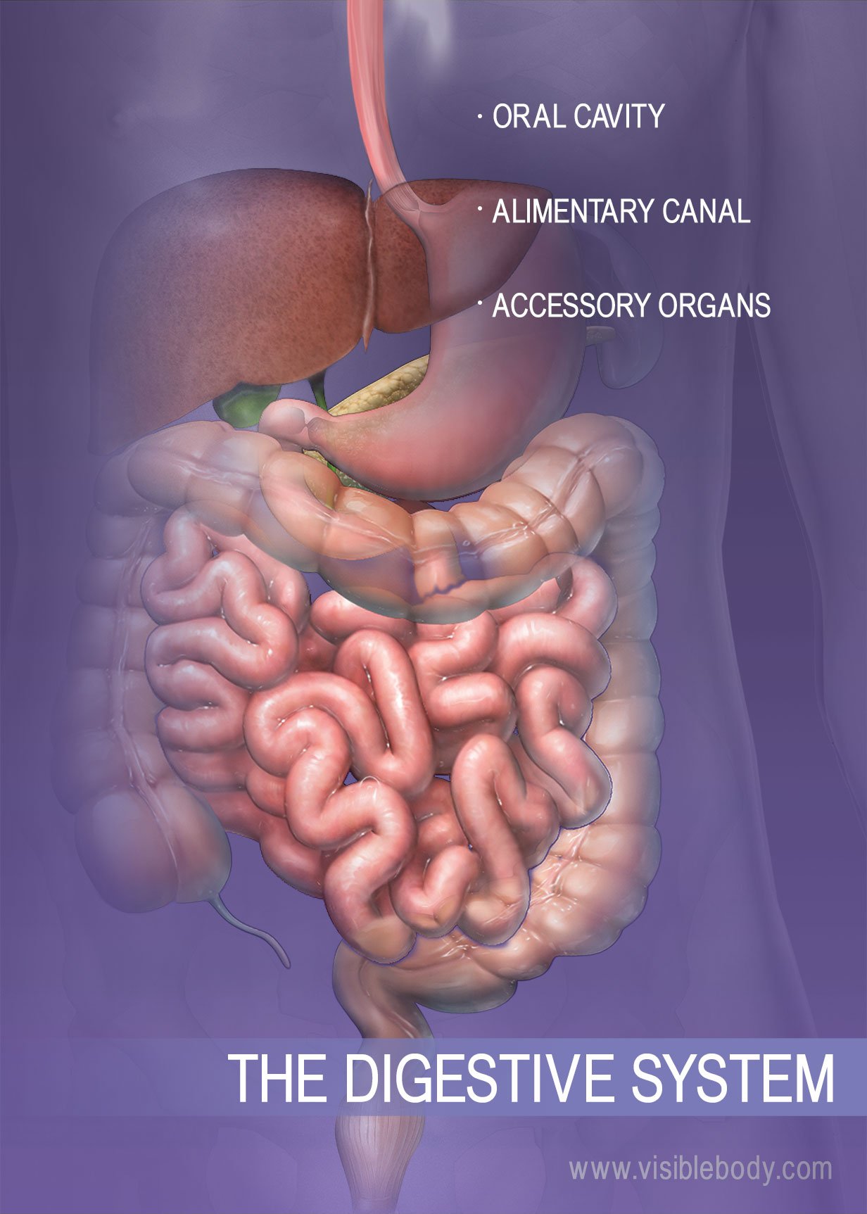
Anatomy & Physiology of Digestion: 10 Facts That Explain How the Body Absorbs Nutrients
What does the digestive system do? The digestive system is a kind of processing plant inside the body. It pulls in food and pushes it through organs and structures where the processing happens. The fuels we need are extracted, and the digestive system discards the rest. What are the 6 processes of the digestive system? What is the alimentary canal? How does the stomach break down food? What is peristalsis?
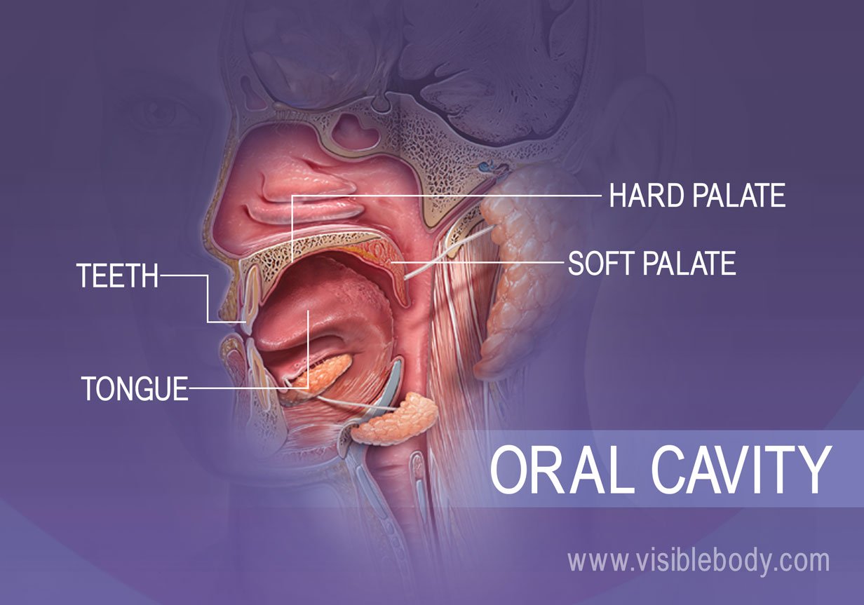
Structures of the Oral Cavity Are Responsible for the First Step of Digestion: Ingestion
- Teeth and the process of mastication in the oral cavity
- The salivary glands and the role of saliva in chemical digestion
- Churning ingested food into a bolus
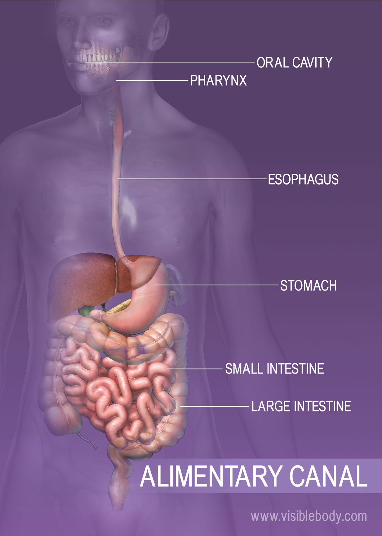
Peristalsis Creates Propulsion: How Food Moves Through the Alimentary Canal
- The trachea, epiglottis, esophagus and the process of swallowing
- What is peristalsis?
- How peristalsis moves food through the digestive tract
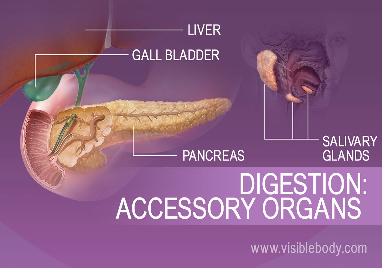
Accessory Organs: Glands and Organs That Facilitate the Process of Digestion
- Where are the salivary glands located?
- Is saliva all water?
- How does the liver facilitate digestion?
- What does bile do in the body?
- What is the connection between bile and the gall bladder?
- What is the role of the pancreas in the process of digestion?
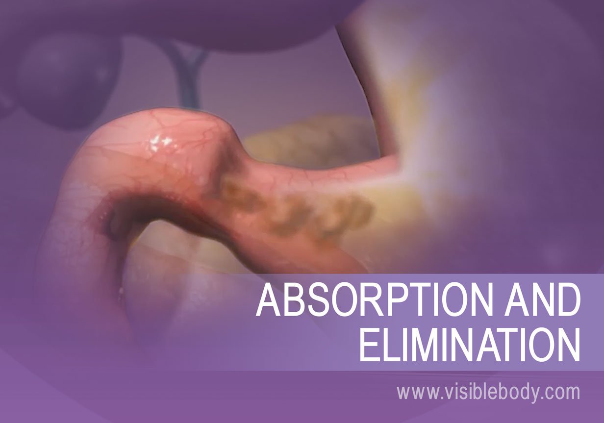
Nutrients In, Waste Out: How the Human Body Absorbs Nutrients and Eliminates Waste
- How do villi absorb nutrients?
- The role of the large intestine
- Defecation: When the body expels waste products from digestion
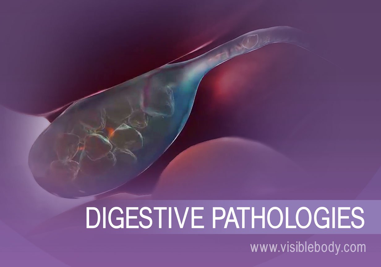
Digestive System Pathologies: A Few Common Diseases and Disorders
- Appendicitis
- Hemorrhoids
- Ulcerative Colitis
- Diverticulitis
For students
For instructors
Get our awesome anatomy emails!
When you select "Subscribe" you will start receiving our email newsletter. Use the links at the bottom of any email to manage the type of emails you receive or to unsubscribe. See our privacy policy for additional details.
- User Agreement
- Permissions
- Type 2 Diabetes
- Heart Disease
- Digestive Health
- Multiple Sclerosis
- COVID-19 Vaccines
- Occupational Therapy
- Healthy Aging
- Health Insurance
- Public Health
- Patient Rights
- Caregivers & Loved Ones
- End of Life Concerns
- Health News
- Thyroid Test Analyzer
- Doctor Discussion Guides
- Hemoglobin A1c Test Analyzer
- Lipid Test Analyzer
- Complete Blood Count (CBC) Analyzer
- What to Buy
- Editorial Process
- Meet Our Medical Expert Board
Your Digestive System in Pictures
It can be scary to experience unusual stomach and digestive system problems. While you are waiting to see your healthcare provider, or as you work with your healthcare provider on a treatment plan, it can be helpful to educate yourself about how your digestive system actually works.
Learn About Your Insides
You will find that you may be able to ease some of the anxiety that goes along with not feeling well by having a good understanding of what your digestive system looks like inside of you. Looking at pictures of your GI tract can help you to pinpoint where symptoms such as abdominal pain may be coming from. This understanding can also help you to better describe your symptoms to your healthcare provider. Here you will find pictures of the primary organs of your digestive system. They may bring back memories of high school biology class, and they will certainly help to make you a more educated patient.
If you experience unusual and ongoing digestive system symptoms, see your healthcare provider to get an accurate diagnosis and develop an optimal treatment plan.
Your Upper Digestive System
The process of digestion begins in your mouth as you chew food. Saliva not only adds moisture to food but also adds enzymes that begin the process of breaking down the components of food.
As you swallow, food moves into your esophagus , where it travels downward to your stomach .
In your stomach , the act of digestion begins in earnest. Your stomach stores and churns the food you have consumed and releases pepsin and hydrochloric acid, both of which break down the components of food, resulting in a substance called chyme. After approximately two to three hours, the chyme is moved out of your stomach as it makes its way along your GI tract.
Your Small Intestine
The digestive process continues as chyme from the stomach enters the small intestine. The main job of the small intestine is to absorb essential nutrients into the bloodstream. The small intestine is made up of three parts:
The small intestine is aided in its work by the liver, gallbladder, and pancreas. In the duodenum , bile from the gallbladder and pancreatic secretions are added to the chyme. The jejunum and ileum are responsible for the breakdown and absorption of most nutrients, including fats, starches, proteins, vitamins, and minerals.
Your Liver, Gallbladder, and Pancreas
The liver , gallbladder, and pancreas all play an important role in the digestion of food. The liver produces bile, which is then stored in the gallbladder . Bile is then released into the small intestine as needed, where it dissolves fat so that it can be absorbed into the body.
The pancreas secretes bicarbonate, which neutralizes the hydrochloric acid from the stomach, as well as enzymes that break down proteins, carbohydrates, and fats.
Your Large Intestine
The contents of your small intestine empty into your large intestine , which also goes by the terms "bowel" or "colon." As you can see in the picture, intestinal contents move through the ascending colon , across the transverse colon and down through the descending colon and sigmoid colon . As material moves through the various parts of the large intestine, water and salt are absorbed by the lining and the material is compacted into the stool.
Typically, the stool is moved into the rectum once or twice a day; pressure from this process stimulates the urge for a bowel movement. This process is not quite so simple in digestive disorders such as irritable bowel syndrome (IBS), in which problems with motility (movements of the muscles in the large intestine) results in symptoms such as diarrhea and constipation . Pelvic floor dyssynergia, which involves problems with coordination between rectum and pelvic floor muscles, can also result in constipation or incomplete evacuation.
Putting It All Together
As you look at the above picture of your entire digestive system, you now have a better understanding of how food gets digested and where your digestive organs are located. This knowledge can empower you to work with your medical professionals to come up with an optimal treatment plan for addressing your digestive symptoms, whatever they may be.
Frequently Asked Questions
The gastrointestinal (GI) tract is a collection of organs that allow for food to be swallowed, digested, absorbed, and removed from the body. The organs that make up the GI tract are the mouth, throat, esophagus, stomach, small intestine, large intestine, rectum, and anus. The GI tract is one part of the digestive system.
The small intestine is responsible for absorbing nutrients. As food is broken down by the stomach and small intestine, nutrients are absorbed into the bloodstream.
The exact size of the stomach will differ from one person to another. Generally, the average stomach can comfortably hold 1 or 2 cups of food. If we overeat, it's possible for the stomach to stretch and expand, making extra room for more food.
NIH National Institute of Diabetes and Digestive and Kidney Diseases. Irritable bowel syndrome (IBS) .
National Cancer Institute (NIH). Gastrointestinal tract .
Michigan Health. 4 ways to stop digestive discomfort after a supersized meal .
" Your Digestive System and How It Works " National Digestive Diseases Information Clearinghouse
Minocha, A. & Adamec, C. The Encyclopedia of the Digestive System and Digestive Disorders (2nd Ed.) New York:Facts on File.
By Barbara Bolen, PhD Barbara Bolen, PhD, is a licensed clinical psychologist and health coach. She has written multiple books focused on living with irritable bowel syndrome.
Diagram of the Human Digestive System (Infographic)
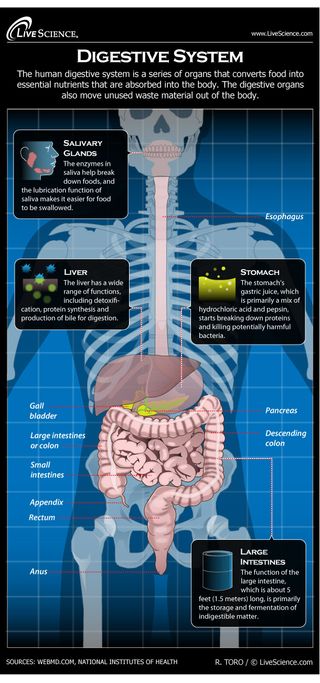
The human digestive system is a series of organs that converts food into essential nutrients that are absorbed into the body. The digestive organs also move waste material out of the body.
The enzymes in saliva help break down foods, and the lubrication function of saliva makes it easier for food to be swallowed.
The stomach's gastric juice, which is primarily a mix of hydrochloric acid and pepsin, starts breaking down proteins and killing potentially harmful bacteria.
The liver has a wide range of functions, including detoxification, protein synthesis and production of bile for digestion.
The function of the large intestine, which is about five feet long (1.5 meters), is primarily the storage and fermentation of indigestible matter.
- Digestive System: Facts, Function & Diseases
Sign up for the Live Science daily newsletter now
Get the world’s most fascinating discoveries delivered straight to your inbox.
New UTI vaccine wards off infection for years, early studies suggest
Blood test powered by AI could catch osteoarthritis 8 years earlier than X-ray, early data show
PTSD tied to 95 'risk hotspots' in the genome
- 2 Giant, 82-foot lizard fish discovered on UK beach could be largest marine reptile ever found
- 3 Global 'time signals' subtly shifted as the total solar eclipse reshaped Earth's upper atmosphere, new data shows
- 4 'I nearly fell out of my chair': 1,800-year-old mini portrait of Alexander the Great found in a field in Denmark
- 5 NASA reveals 'glass-smooth lake of cooling lava' on surface of Jupiter's moon Io
- 2 China green-lights mass production of autonomous flying taxis — with commercial flights set for 2025
- 3 George Washington's stash of centuries-old cherries found hidden under Mount Vernon floor
- 4 5 catastrophic megathrust earthquakes led to the demise of the pre-Aztec city of Teotihuacan, new study suggests
- 5 Scientists find one of the oldest stars in the universe in a galaxy right next to ours
Medical Information Illustrated
Medical and health topics fully illustrated with pictures and videos for easy understanding..

The Digestive System, with Animation.

Related posts:
- Swallowing and Dysphagia (with Animation)
- Bariatric surgery
- Le Réflexe de Déglutition, Phases et Contrôle Neuronal, avec Animation.
- Gallstones and Cholecystectomy
- Vertical Sleeve Gastrectomy and Gastric Lap Band Surgeries
- GERD and Heartburn (with video)


- Visual Dictionary

- Virtual Human Body

- Qu’est-ce que Le Visuel ?
- L'histoire d’un succès
- Le Visuel dans le monde
- Livres grand public
- Livres jeunesse
- Jeu de la semaine

digestive system

sphincter muscle of anus
Sigmoid colon, vermiform appendix, ascending colon, descending colon, transverse colon, gallbladder, salivary glands.
oral cavity
Small intestine, large intestine.
- Press releases

- Le Dictionnaire Visuel (Fr)
- The Visual Dictionary
- Diccionario Visual (es)
- External links
- Image purchase
- Advertising and partnerships
- Institutional sales
- Terms and conditions of use
- Privacy policy

- school Campus Bookshelves
- menu_book Bookshelves
- perm_media Learning Objects
- login Login
- how_to_reg Request Instructor Account
- hub Instructor Commons
- Download Page (PDF)
- Download Full Book (PDF)
- Periodic Table
- Physics Constants
- Scientific Calculator
- Reference & Cite
- Tools expand_more
- Readability
selected template will load here
This action is not available.

21.8: Anatomical Atlas - Digestive System
- Last updated
- Save as PDF
- Page ID 65216
A compendium of anatomical images: slides, models, and/or cadavers.
Images of models by John Polos and shared under CC 4.0
Histology photos John Polos and shared under CC 4.0
All cadaver images from the University of Michigan and are made available by the regents under CC 4.0
For larger images, right click(PC)/command click (Mac) and click open image in new window.
- DAILY PRACTICE
- IMAIOS DICOM Viewer
- vet-Anatomy
- Breast imaging learning tool
- Blog News, opinions & thoughts of anatomy, medical imaging
Articles talking about IMAIOS and its products
What our users say about us
Our commitments
Get help with your subscription, account and more
- Contact us Other questions?

e-Anatomy is better on the mobile app!
Faster, more ergonomic, more features.
Abdomen and digestive system anatomy
- Antoine MICHEAU, MD , Denis HOA, MD
- Antoine MICHEAU, MD : 2 Allée Charles Darwin, 34170 Castelnau-le-lez
- Denis HOA, MD : 2 Allée Charles Darwin, 34170 Castelnau-le-lez
- Publication date: Mar 20, 2012 | Last update: Oct 3, 2022
- https://doi.org/10.37019/e-anatomy/166969 ISSN 2534-5079
This e-Anatomy illustrates the gross anatomy of the digestive system. We focused especially on the diagrams of the abdominal digestive system (oesophagus is described on the modules about the thorax and oral cavity/pharynx on the ENT modules).
84 anatomical diagrams and histological images with over 300 labeled anatomical parts.
This digestive anatomy tool is especially designed for students of medicine and paramedical studies.
Anatomical illustrations of the digestive system
All the anatomical illustrations are original and created by Antoine Micheau - MD. They were drawn on Photoshop or Illustrator using 3D models derived from medical imaging (principally abdominal scan). The anatomical drawings were organized in a fairly classical way to be easily used as a standard anatomical atlas. The user can browse between different groups of images using the "Series" tab:
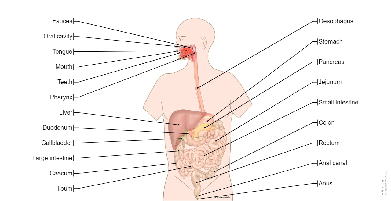
- Digestive system: general diagram showing all the major structures included in this system: mouth, tongue, oral cavity, teeth, buccal glands, throat, pharynx, oesophagus, stomach, small intestine (duodenum, jejunum and ileum), large intestine (colon, rectum, anal canal and anus), liver, gall bladder and pancreas. Please note that the spleen is not part of the digestive system but belongs to the lymphoid system, however it still appears on this module because of its location close to the various digestive organs.
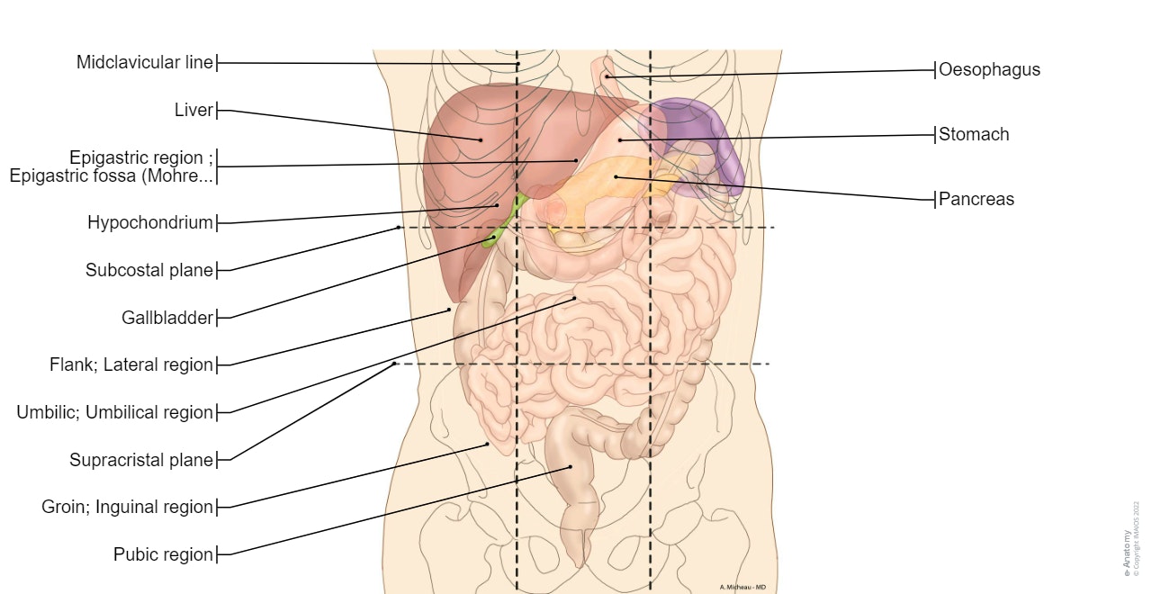
- Abdominal cavity: illustration showing the main organs of the abdomen with their projections on the different quadrants of the abdominal wall (upper quadrant, flank, groin, epigastrium, umbilical region and hypogastrium).
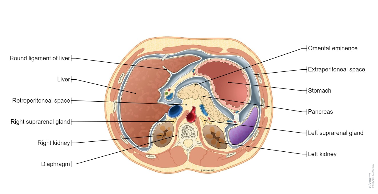
- Peritoneal cavity: series of anatomical charts to view the architecture and organisation of the abdominal cavity from the superficial to deeper planes, especially to view the greater and lesser omenta, the omental bursa, the mesocolons, the mesentery and its root. An axial and a sagittal section allows the study of the recess of the peritoneal cavity.
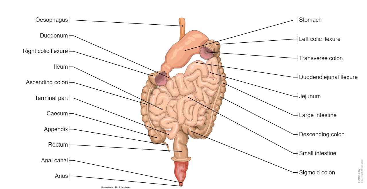
- Intestinal tract: 2 illustrations of gross anatomy to introduce the different parts of the digestive tract.
- Stomach: anatomical images of the gastric anatomy, from the serous membrane to the gastric mucosa, with a diagram of the histology of the stomach lining.
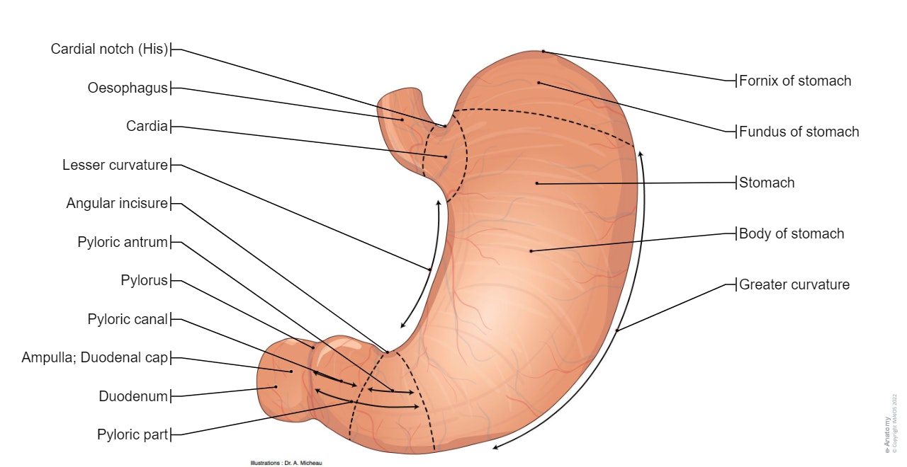
- Duodenum: an overall diagram of duodenal anatomy and an illustration of the duodenojejunal flexure with its duodenal recesses.
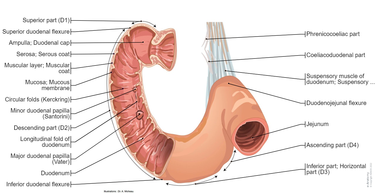
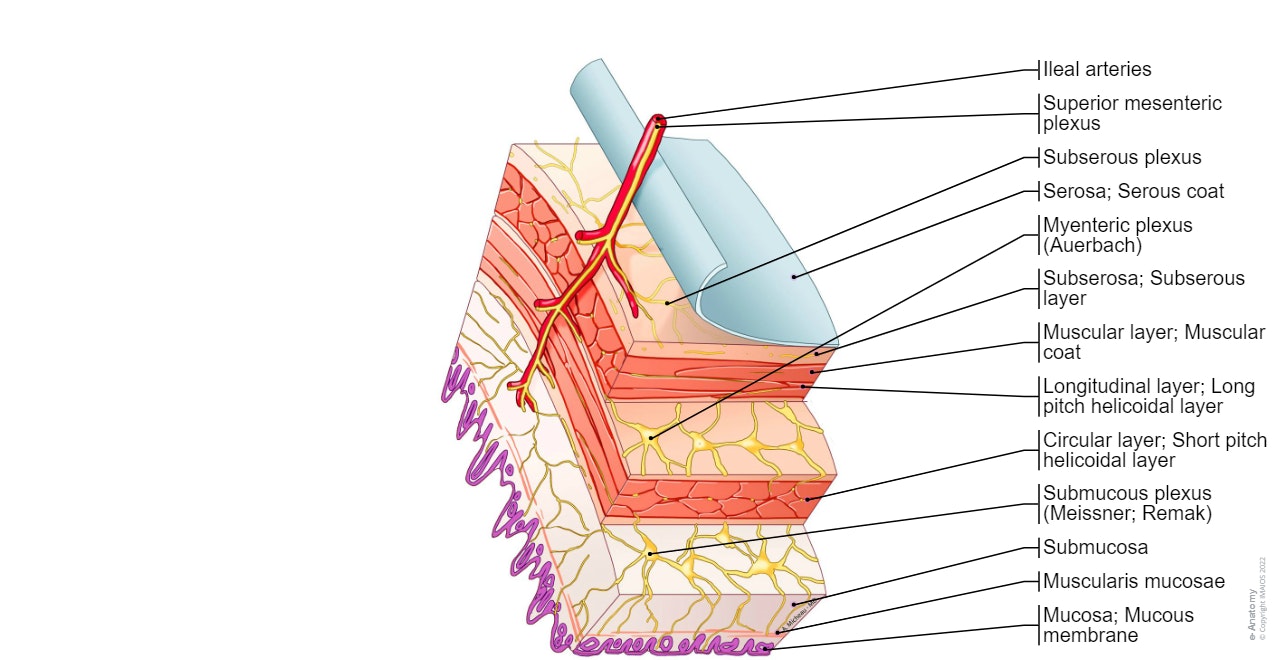
- Jejunum: diagram of a jejunal loop with its different parietal layers
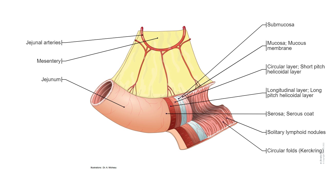
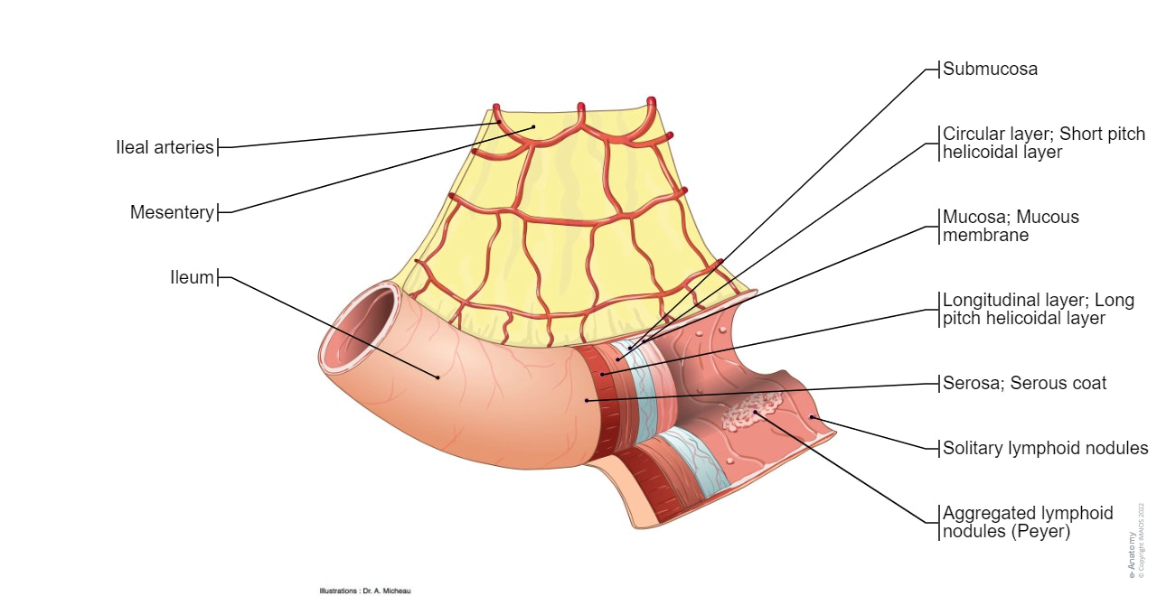
- Ileum: diagram of an ileal loop similar to the previous diagram of the jejunum, allowing to view the differences between these two parts of the small intestine (arterial vascularisation with the arteria rectae and anastomotic arches, circular folds, etc). A histological pattern individualises different histological layers of the jejunum (serosa, subserosa, muscularis, submucosa, mucosa).
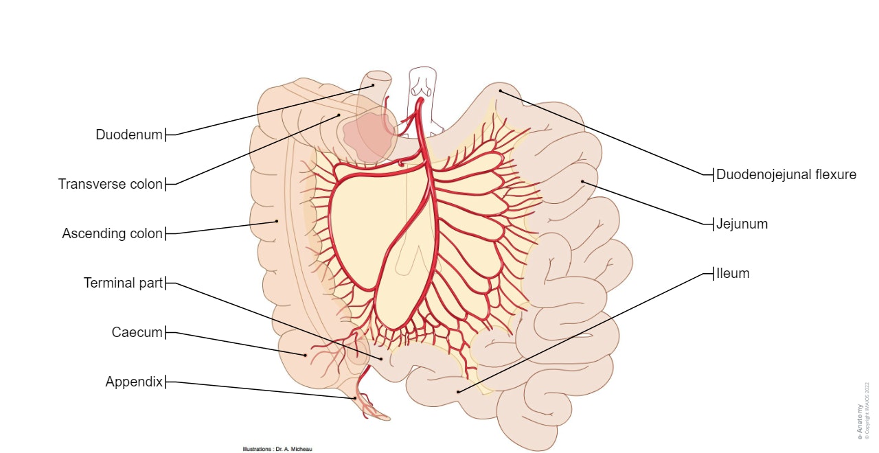
- Mesentery: general diagram of the appearance of the mesentery and its root.
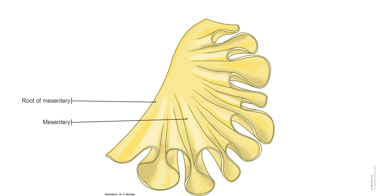
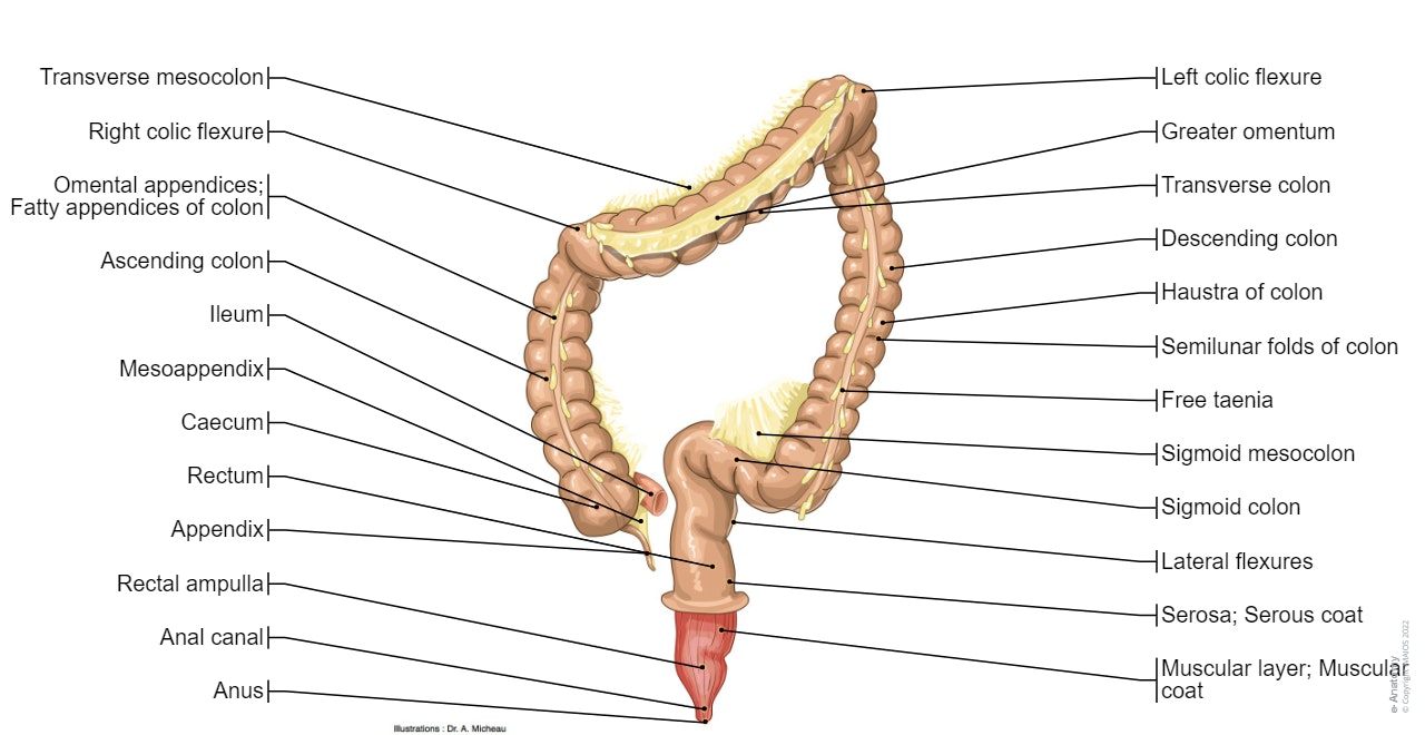
- Colon: three diagrams of the face of the colon and rectum, with or without the mesentery of the colon and taenia coli as well as a detailed illustration of the right colonic flexure.
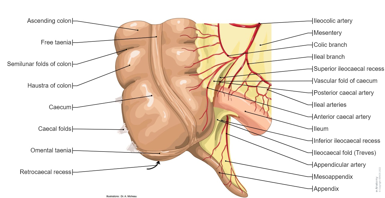
- Cecum: illustration of the cecum and its appendix, front view with its mesenteries and its recesses, and then a diagram of an open cecum (on the body because the ileal papilla is flat and elongated).
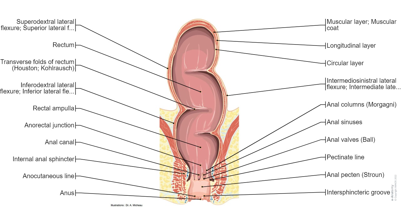
- Rectum: coronal section of the rectum with the main curvatures and stages of the rectum. The rectum will be illustrated in a more detailed way on the modules about the pelvic region.
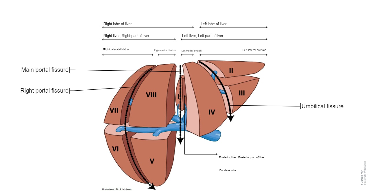
Liver: a first diagram presents the nomenclature of parts, divisions and liver segments. The following diagram summarizes the venous, arterial and biliary systems of the liver. Illustrations of the hepatic anatomy showing the different faces of the liver (visceral, diaphragmatic...) and the hepatic pedicle. Hepatic segmentation is then detailed on two summarized recapitulative diagrams. Finally, photos and diagrams resume liver histology.
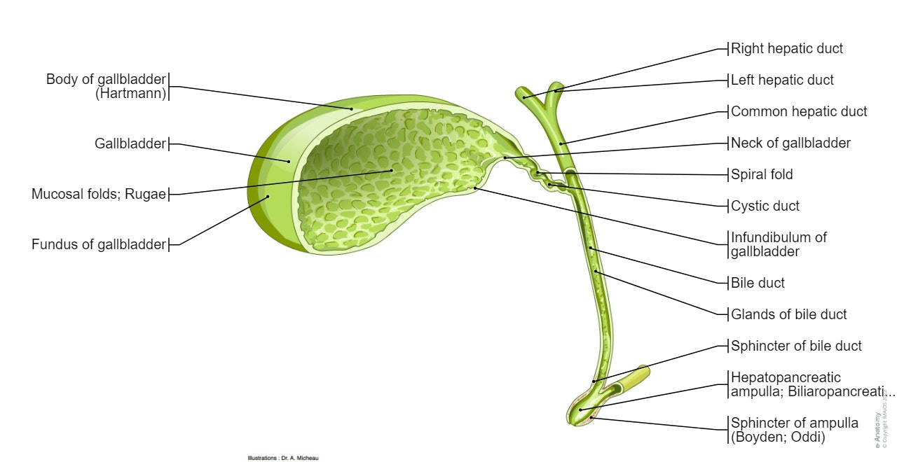
- Gallbladder: the different parts of the bile ducts inside and outside the liver are detailed in these three diagrams.
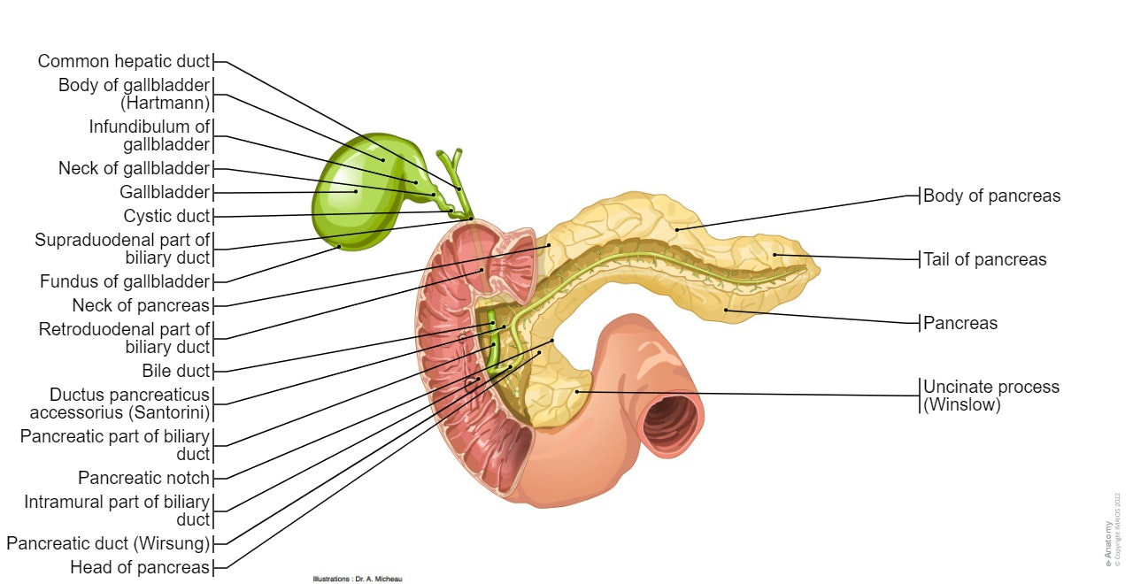
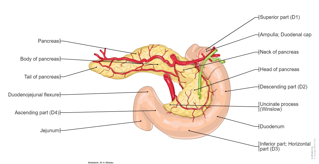
- Pancreas: the first diagram shows the different parts of the pancreatic anatomy in anterior view, following up by: the links of the duodenum with the pancreas, the ampulla of Vater (hepatopancreatic ampulla), the pancreas in posterior view with and without retropancreatic vessels.
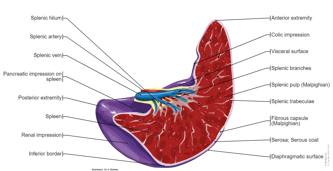
- Spleen: the splenic anatomy is detailed in anterior and posterior views, as well as in an anatomical section of the spleen followed up by a diagram showing splenic histology.
- Arteries: the initial anatomical drawings show the aorta, the venous quadrilateral and the aorto-mesenteric clamp, then the different arteries of the abdomen are detailed (celiac trunk, hepatic artery, splenic artery, superior and inferior mesenteric arteries).
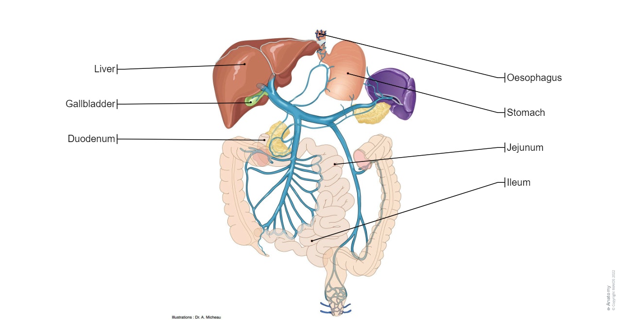
- Veins: the diagrams are focused on the anatomy of the portal system, first with an overall pattern of the portal vein and its branches, then with illustrations of the venous vascularisation of the digestive organs.
- Lymph nodes: we reviewed the diagrams of the arteries to which the different lymph nodes in the abdomen were added as their nomenclature is based on the underlying arteries.
- Autonomic nervous system: three diagrams on the main autonomic nerve structures (sympathetic and parasympathetic) of the digestive system, including the celiac plexus, the mesenteric nerve plexus and enteric plexus.
Labels of the anatomical structures of the digestive system
125 anatomical structures of the digestive system were labeled. The labels are grouped into subcategories, the user can choose to hide or show those labels on "Anatomical parts":
• Aesophagus • Stomach • Small intestine: duodenum, jejunum and ileum • Large intestine: colon, cecum, rectum, anal canal • Liver: gross and microscopic anatomy, histology, hepatic segmentation • Biliary tract: gall bladder, principal bile duct, hepatic duct, common bile duct • Pancreas: external morphology, pancreatic duct, hepatopancreatic ampulla • Spleen: gross anatomy, histology • Peritoneum: parietal and visceral peritoneum, mesenteries, transverse mesocolon, mesentery • Recesses, pits and folds: omental bursa, paracolic fissures (parietocolic grooves), rectovesical escavation (Douglas) • Arteries: branches of the abdominal aorta leading to the digestive system (celiac artery, superior and inferior mesenteric arteries) • Veins: different tributaries of the hepatic portal system vein (splenic vein, superior and inferior mesenteric vein, hepatic portal vein) • Lymph nodes: celiac lymphatic ganglions, inferior and superior mesenteric veins • Nerves: nerve plexus and autonomic intestinal nervous system • Other structures: diaphragm, genital and urinary systems.
Language and terminology for the study of the anatomy of the gastrointestinal tract
We used the Terminologia Anatomica to label all the anatomical structures. Translations available in English, French, Japanese, German, Chinese, Portuguese, Russian, Polish, Korean, Italian and Spanish.
- Quick Links
- Terminologia Anatomica:International Anatomical Terminology - FCAT Federative Committee On Anatomical Terminology, Federative Committee on Anatomical Terminology - Thieme, 1998 - ISBN 3131152516, 9783131152510
- Atlas d'anatomie humaine- 4e édition - Frank-H Netter - Pierre Kamina (Traducteur) - Paru le : 25/07/2007 - Editeur : Masson - ISBN : 978-2-294-08042-5 - EAN : 9782294080425 (lien : http://www.netterimages.com/ )
- Lexique illustré d'anatomie Feneis– Feneis - Editeur : Flammarion Médecine-Sciences (23 août 2007) - Collection : ATLAS DE POCHE - ISBN-10: 225712250X - ISBN-13: 978-2257122506
- Gray's Anatomie pour les étudiants- de Richard-L Drake, Wayne Vogl et Adam-W-M Mitchell - Editeur : Elsevier (24 octobre 2006) - Langue : Français - ISBN-10: 2842997743 - ISBN-13: 978-2842997748
There is no content here

Cookie preferences
IMAIOS and selected third parties, use cookies or similar technologies, in particular for audience measurement. Cookies allow us to analyze and store information such as the characteristics of your device as well as certain personal data (e.g., IP addresses, navigation, usage or geolocation data, unique identifiers). This data is processed for the following purposes: analysis and improvement of the user experience and/or our content offering, products and services, audience measurement and analysis, interaction with social networks, display of personalized content, performance measurement and content appeal. For more information, see our privacy policy .
You can freely give, refuse or withdraw your consent at any time by accessing our cookie settings tool. If you do not consent to the use of these technologies, we will consider that you also object to any cookie storage based on legitimate interest. You can consent to the use of these technologies by clicking "accept all cookies".
Cookie settings
When you visit IMAIOS, cookies are stored on your browser.
Some of them require your consent. Click on a category of cookies to activate or deactivate it. To benefit from all the features, it’s recommended to keep the different cookie categories activated.
These are cookies that ensure the proper functioning of the website and allow its optimization (detect browsing problems, connect to your IMAIOS account, online payments, debugging and website security). The website cannot function properly without these cookies, which is why they are not subject to your consent.
These cookies are used to measure audience: it allows to generate usage statistics useful for the improvement of the website.
- Google Analytics
- Anatomical Position
- Body Planes
- Terms of Movement
- Terms of Location
- Embryology Terms
- Classification
- Synovial Joint
- Joint Stability
- Skeletal Muscle
- Blood Vessels
- Head and Neck
- Cardiovascular System
- Respiratory System
- Urinary System
- Reproductive System
- Central Nervous System
- Cranial Fossae
- Pterygopalatine Fossa
- Infratemporal Fossa
- Mastoid Fossa
- Frontal Bone
- Sphenoid Bone
- Ethmoid Bone
- Temporal Bone
- Occipital Bone
- Nasal Skeleton
- Cranial Foramina
- Facial Expression
- Extraocular
- Mastication
- Sympathetic Innervation
- Parasympathetic Innervation
- Ophthalmic Nerve
- Maxillary Nerve
- Mandibular Nerve
- Nose and Sinuses
- Salivary Glands
- Oral Cavity
- Arterial Supply
- Venous Drainage
- Lacrimal Gland
- Basal Ganglia
- Pineal Gland
- Pituitary Gland
- Spinal Cord (Grey Matter)
- Medulla Oblongata
- Ascending Tracts
- Descending Tracts
- Visual Pathway
- Auditory Pathway
- Olfactory Nerve (CN I)
- Optic Nerve (CN II)
- Oculomotor Nerve (CN III)
- Trochlear Nerve (CN IV)
- Trigeminal Nerve (CN V)
- Abducens Nerve (CN VI)
- Facial Nerve (CN VII)
- Vestibulocochlear Nerve (CN VIII)
- Glossopharyngeal Nerve (CN IX)
- Vagus Nerve (CN X)
- Accessory Nerve (CN XI)
- Hypoglossal Nerve (CN XII)
- Dural Venous Sinuses
- Cavernous Sinus
- Anterior Triangle
- Posterior Triangle
- Cervical Spine
- Thyroid Gland
- Parathyroid Glands
- Suboccipital
- Suprahyoids
- Infrahyoids
- Phrenic Nerve
- Cervical Plexus
- Fascial Layers
- Tonsils (Waldeyer's Ring)
- Superior Mediastinum
- Anterior Mediastinum
- Middle Mediastinum
- Posterior Mediastinum
- Thoracic Spine
- Thoracic Cage
- Thymus Gland
- Mammary Glands
- Tracheobronchial Tree
- Superior Vena Cava
- Vertebral Column
- Superficial
- Intermediate
- Spinal Cord
- Quadrangular Space
- Triangular Interval
- Triangular Space
- Cubital Fossa
- Ulnar Tunnel
- Extensor Compartments
- Ulnar Canal
- Carpal Tunnel
- Anatomical Snuffbox
- Pectoral Region
- Shoulder Region
- Anterior Forearm
- Posterior Forearm
- Brachial Plexus
- Axillary Nerve
- Musculocutaneous Nerve
- Median Nerve
- Radial Nerve
- Ulnar Nerve
- Acromioclavicular Joint
- Sternoclavicular Joint
- Shoulder Joint
- Elbow Joint
- Radioulnar Joints
- Wrist Joint
- Metacarpophalangeal Joint
- Proximal Interphalangeal Joint
- Extensor Tendon Expansion
- Flexor Pulley System
- Femoral Triangle
- Femoral Canal
- Adductor Canal
- Popliteal Fossa
- Tarsal Tunnel
- Fascia Lata
- Gluteal Region
- Cutaneous Innervation
- Lumbar Plexus
- Sacral Plexus
- Femoral Nerve
- Obturator Nerve
- Sciatic Nerve
- Tibial Nerve
- Common Fibular Nerve
- Superficial Fibular Nerve
- Deep Fibular Nerve
- Tibiofibular Joints
- Ankle Joint
- Subtalar Joint
- Foot Arches
- Walking and Gaits
- Abdominal Cavity
- Calot’s Triangle
- The Peritoneum
- Inguinal Canal
- Hesselbach's Triangle
- Lumbar Spine
- Anterolateral Abdominal Wall
- Posterior Abdominal Wall
- Small Intestine
- Gallbladder
- Adrenal Glands
- Sciatic Foramina
- Pelvic Girdle
- Sacroiliac Joint
- Pelvic Floor
- Urinary Bladder
- Testes and Epididymis
- Spermatic Cord
- Prostate Gland
- Bulbourethral Glands
- Seminal Vesicles
- Fallopian (Uterine) Tubes
- Supporting Ligaments
- Pudendal Nerve
- Female Body
- Female Pelvis
- Male Pelvis
- Cardiovascular
- Gastrointestinal
- Respiratory
- Female Reproductive
- Male Reproductive
Gastrointestinal System
- Interactive 3D Models
- Access over 1700 multiple choice questions
- Advert Free
- Custom Quiz Builder
- Performance tracking
Found an error? Is our article missing some key information? Make the changes yourself here!
Once you've finished editing, click 'Submit for Review', and your changes will be reviewed by our team before publishing on the site.
We use cookies to improve your experience on our site and to show you relevant advertising. To find out more, read our privacy policy .
Privacy Overview
- 888-309-8227
- 732-384-0146
Take Free State Assessment Practice Tests and Sample Questions for Grade 3 to 8 - Math & ELA

The Digestive System – A Visual Representation using Mind Map
The digestive system consists of those parts of the body that work together to turn food into nutrients used by the human body. Understanding the parts of the digestive system and the functions of each part can be quite challenging. A mind map simplifies the understanding of the working of the digestive system.
Let us look at an example mind map from Lumos, which will help us understand the human digestive system.

What is a Mind Map?

Mind maps visually show how specific facts are related. Mind maps are a visual organization of data. A central idea is placed in the middle, and associated concepts are arranged around it. Mind maps visually show how specific facts are related.
Steps to create Mind Maps
Mind maps are pretty simple to create. To understand how to develop a topic mindmap, we have to keep in mind the below points.
- Begin with the main concept in the center
- Select keywords and concepts related to the main topic and enter these as child items.
- Keep the mind map clear using hierarchy, numerical order, etc. to indicate the branches.
- The central lines should be thicker and flowing. It becomes progressively thinner as it spreads out from the center.
Benefits of mind mapping
- Helps meaningful learning
- It’s a more engaging form of learning
- Makes complex issues easier to understand
- Helps in memorizing and retaining data
- It improves creativity
- Improves writing skills
- Improves productivity
Mind maps can be created for any concept. Lumos Mind maps cover a wide range of concepts from English, Science, Social Science, or even General knowledge concepts.
Create mind maps with Lumos mind map creator
Now, creating mind maps is extremely easy with Lumos Mind map Creator.
As a student, this will help you organize the data and answer complex questions with ease. Lumos mind maps can help students score better in exams.
As a teacher or parent, you can create mind maps and assign them to students. These serve as a simple and effective way of teaching complex concepts.
You can also access several of Lumos mind maps covering different topics from your account.
Create mind maps and explore Lumos mind maps using
Lumos Mind Map Creator
Related posts:
No related posts.

Alice Moore
Leave a reply cancel reply.
Your email address will not be published. Required fields are marked *
Notify me of follow-up comments by email.
Notify me of new posts by email.
- PRO Courses Guides New Tech Help Pro Expert Videos About wikiHow Pro Upgrade Sign In
- EDIT Edit this Article
- EXPLORE Tech Help Pro About Us Random Article Quizzes Request a New Article Community Dashboard This Or That Game Popular Categories Arts and Entertainment Artwork Books Movies Computers and Electronics Computers Phone Skills Technology Hacks Health Men's Health Mental Health Women's Health Relationships Dating Love Relationship Issues Hobbies and Crafts Crafts Drawing Games Education & Communication Communication Skills Personal Development Studying Personal Care and Style Fashion Hair Care Personal Hygiene Youth Personal Care School Stuff Dating All Categories Arts and Entertainment Finance and Business Home and Garden Relationship Quizzes Cars & Other Vehicles Food and Entertaining Personal Care and Style Sports and Fitness Computers and Electronics Health Pets and Animals Travel Education & Communication Hobbies and Crafts Philosophy and Religion Work World Family Life Holidays and Traditions Relationships Youth
- Browse Articles
- Learn Something New
- Quizzes Hot
- This Or That Game New
- Train Your Brain
- Explore More
- Support wikiHow
- About wikiHow
- Log in / Sign up
- Education and Communications
How to Draw a Model of the Digestive System
Last Updated: March 30, 2024 Fact Checked
This article was co-authored by Meredith Juncker, PhD . Meredith Juncker is a PhD candidate in Biochemistry and Molecular Biology at Louisiana State University Health Sciences Center. Her studies are focused on proteins and neurodegenerative diseases. This article has been fact-checked, ensuring the accuracy of any cited facts and confirming the authority of its sources. This article has been viewed 300,114 times.
Drawing a model of the digestive system will help you to better understand its functions. It is a great way to visualize what happens during digestion. There are many organs involved in digesting food, but don’t let this intimidate you! Whether you are doing this for a school project or making a study guide, you can take it step by step to make a great model. By outlining each organ, coloring them in, and labeling your model, you will begin to truly understand what is happening when you eat your favorite meal.
Drawing the Model

- Draw the head as a profile rather than straight on to make it easier to show the digestive organs in the head.
- If you wish, feel free to be creative and embellish this outline a bit. You can draw eyes and a nose and ears and hair. You could even give your person a name if you feel like it! Just don’t draw over the torso or you will obscure your model.

- Digestion begins in the mouth with ingestion. Salivary glands release saliva containing digestive enzymes to begin breaking down food while you chew. The tongue helps the food to move back in your throat creating a bolus while the teeth break it down in a process known as mastication.

- The food moves from the mouth into the esophagus, which carries it down into the stomach. The esophagus is made of smooth muscle that relaxes and contracts to move your food down with a wavelike motion, called peristalsis.
- If you wish to make your model more advanced, you can include the pharynx. The pharynx is located behind the mouth and moves food into the esophagus. You can feel it when you swallow. In your model, you can simply draw a small diagonal line towards the top of your esophagus and the part above the line can be the pharynx and the part below can be the esophagus. [3] X Research source
- It you wish to make your model more advanced, you can also include the epiglottis. The epiglottis is a small flap just below the pharynx that directs food into the esophagus. If you drew a pharynx, the diagonal line can be the epiglottis.

- The stomach helps churn and digest food using gastric juices containing hydrochloric acid and pepsin to help break it down. The food remains in the stomach for about 3-4 hours. At this point it is no longer food, but has an oatmeal-like consistency and is called "chyme."

- The liver produces bile to help break down fats. While the food doesn’t enter the liver, it processes the nutrients that are absorbed from the small intestine.

- The gallbladder stores the bile produced in the liver. It then mixes the bile with the food that passes by, breaking down fats.

- The pancreas releases digestive juices that aid in breaking down carbohydrates, protein, and fat as the food leaves the stomach. It also regulates blood sugar. [8] X Research source

- Add a diagonal line to indicate the muscular valve, called the pyloric sphincter, between the duodenum of the small intestine and the stomach.
- The small intestine is about 18–22 feet (5–7 m) long! This is where the majority of digestion takes place. The small intestine contracts to move food through it with the help of bile secretions and pancreatic and intestinal juices, allowing it to absorb nutrients with villi.
- To make a more advanced model, you can distinguish the different parts of the small intestine. The duodenum is a small tube connecting the stomach to the jejunum. The jejunum is where the majority of digestion takes place and is the middle part of the small intestine. The ileum is the last region of the small intestine that connects to the large intestine. [9] X Research source

- The appendix is a small pouch-like structure that is thought to have lost its purpose and become more of a liability than an aid in digestion. But it is part of the digestive system and sits where the small and large intestines connect.

- The passage of food is slower through the large intestine in order to allow for fermentation by bacteria and other microorganisms, called gut flora.
- The large intestine processes whatever cannot be used in digestion. It absorbs whatever it can, especially water, but then the rest will expelled as waste after about 12 hours.
- To make a more advanced model, distinguish between the different parts of the large intestine. The cecum is attached to the appendix and connects it to the ascending colon. The ascending colon goes straight upwards and connects to the transverse colon, which goes across the body. The transverse colon connects to the descending colon, which carries food down into the sigmoid colon, which goes directly to the rectum. [10] X Research source

- The rectum stores feces until expulsion. The anus then expels waste.
Adding Finishing Touches

- For overlapping organs, try using using lighter and darker shades of the same color. The areas where they overlap will be darker, and the areas where it is a single organ will be lighter.

- If you prefer not to write the names of the organs on your model, you could instead make a key at the bottom or on another piece of paper where you draw a little square of each color and write the name of the organ next to it. This will allow you to quiz yourself on the names of the organs because they won’t be written right next to them.
- If you wish to make your model more advanced, you can differentiate the parts of the small intestine. Simply draw a small line towards the beginning of the small intestine to show where the duodenum turns into the jejunum and then draw a small line towards the end of the small intestine to show where the jejunum becomes the ileum.
- If you wish to make your model more advanced, you can also differentiate the parts of the large intestine. Simply draw a small line to divide each part. The cecum is attached to the appendix and connects it to the ascending colon. The ascending colon is the part that goes straight upwards. It connects to the transverse colon which goes across the body and is the largest part of the large intestine. The transverse colon connects to the descending colon, which carries food downward. Finally, there is the sigmoid colon which goes directly to the rectum.

Expert Q&A

- Take it one step at a time so that you aren’t overwhelmed. You don’t need to understand the entire digestive system immediately. Simply start with one organ and when you have drawn that and understood its function, you can move onto the next. Thanks Helpful 5 Not Helpful 2

- If this is for school, follow your teacher’s instructions. Your teacher might have specific guidelines that aren’t outlined here. Thanks Helpful 23 Not Helpful 5
You Might Also Like

- ↑ https://www.youtube.com/watch?v=XWSk3L9WTsQ
- ↑ http://www.untamedscience.com/biology/human/digestive-system/
- ↑ http://my.clevelandclinic.org/health/diseases_conditions/hic_the_structure_and_function_of_the_digestive_system
- ↑ http://columbiasurgery.org/pancreas/pancreas-and-its-functions
About This Article

To draw a model of the digestive system, start by outlining the head and torso of a person to give your model a frame of reference. At the mouth, add teeth and a tongue before drawing an esophagus connecting down to the stomach. Above the stomach, draw a liver with the gallbladder overlapping it before sketching a small, carrot-shaped pancreas below the stomach. From there, connect the small intestine to the stomach and have it lead into the appendix and large intestine. Then, at the end of the large intestine, draw the rectum and anus. Once your sketch is complete, outline everything in marker or pen and color each organ a different color so they are differentiated. Draw a thin line from each organ so you can provide a name and brief description to inform the reader. For more help from our co-author, like how to describe each organ’s function, read on! Did this summary help you? Yes No
- Send fan mail to authors
Reader Success Stories
Sharad Mukherjee
Sep 10, 2017
Did this article help you?

Lucy Warren
Feb 29, 2020
Kumari Anshika Singh
May 10, 2019
Oct 18, 2017
Oct 17, 2017

Featured Articles

Trending Articles

Watch Articles

- Terms of Use
- Privacy Policy
- Do Not Sell or Share My Info
- Not Selling Info
Get all the best how-tos!
Sign up for wikiHow's weekly email newsletter
- Overview of the Digestive System
- Overview of Gastrointestinal Emergencies
- Abdominal Abscesses
- Appendicitis
- MY PICTURES
- FOLLOW US
Search Tips
- Use the search page for more options
- Use multiple keywords separated by spaces (e.g. kidney renal ) for broader search results. This search will retrieve items that correspond to any of those words, ranking higher those with all or most of the keywords. Plurals and other variations are automatically included.
- To exclude a word from your search, precede it with a hyphen, e.g. -historical
- Searches are case-insensitive
- Do not include apostrophes in your search - replace any apostrophes with spaces
Digestive System Anatomy
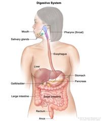
View /Download :
An official website of the United States government
Here’s how you know
Official websites use .gov A .gov website belongs to an official government organization in the United States.
Secure .gov websites use HTTPS A lock ( Lock Locked padlock icon ) or https:// means you’ve safely connected to the .gov website. Share sensitive information only on official, secure websites.
- Entire Site
- Research & Funding
- Health Information
- About NIDDK
- Media Library
- Media Asset Details
Digestive tract - shaded

Description
Drawing of the digestive tract within an outline of the human body. The stomach, small intestine, large intestine, rectum, and anus not labeled.
Alternate Text
Diseases or conditions.
308 KB | 1039 x 1727
Related Keywords

Human Digestive System
HUMAN DIGESTIVE SYSTEM Ever wondered how your body deals with food? Ever wanted to know what happens between the first bite and the number two? You are about to find out... DIGESTION: the way your body turns food into energy and byproducts (excrement). TONGUE WHEN DOES THE DIGESTION PROCESS START?? SALIVA GLANDS Digestion can start well before food reaches your mouth. Saliva production, the first step in digestion, can be activated by smells, sights, or even thoughts. gland is the substance produced in the saliva SALIVA: PHARYNX mouths of humans and most animals, contains 98% water and 2% ESOPHAGUS enzymes, electrolytes, antibacterial compounds, etc. LIVER Around .5 to 1.5 liters of saliva is produced in your mouth a day. STOMACH PANCREAS Eww Saliva stones can form in the saliva glands, causing a great GALLBADDER deal of mouth pain. DUODENUM MOUTH/TONGUE: While saliva gets the food ready, JEJUNUM the mouth begins the first of SMALL INTESTINE many steps of separation. Your tongue, along with tasting the food, mixes saliva with your food while chewing. It is the first stage of mechanical separation. This along with the lubrication from saliva assists getting the food down to your ILEUM LARGE INTESTINE stomach. COLON Like fingerprints, the imprint of the tongue is unique to the RECTUM individual. ANUS The tongue is one of the strongest and most flexible muscles for its size as well as being the fastest healing. PHARYNX & ESOPHAGUS: The pharynx is your CAN YOU SWALLOW FOOD UPSIDE DOWN? throat, which receives the food that has been chewed. The food is sent to your Yes, you can through your body's autonomic peristalsis response. How did you think Spiderman eats his lunch?! stomach via peristalsis. This is a type of wave muscle contraction similar to how worms propel themselves. STOMACH THE BOLUS! THE PANCREAS: In addition to insulin regulation, the Once the chewed food (bolus) reaches the stomach, gastric acid goes to work breaking everything down. So the stomach separates food both mechanically (stomach churning) and chemically (gastric acid). The slurry of food, called chime (yummy!), is released through the pyloric valve into the duodenum (the beginning of the small intestine). pancreas also produces enzymes that reak down carbohydrates, fats, and proteins. The acid in the stomach is strong enough to dissolve razor blades. Hell yeah! Don't eat any blades though to check. THE LIVER: GALLBLADDER: Bile produced by the liver is then stored in the gallbladder and released into the duodenum in varying concentrations, depending upon the foods eaten. While the liver performs over 500 functions, for digestion, a primary function is the creation of bile, which assists in dissolving fats. The largest gallstone ever removed from someone was in 1952, Eww removed from an 80 year old woman and weighed over 13.84 lbs. THE INTESTINE DUODENUM: Before the journey to the small intestines is LARGE INTESTINE: At this stage in the digestion process, food is no longer being broken down. By the time your meal has reached this point, it is mostly water, plant fiber, electrolytes, and some dead cells from the lining of the intestinal tract. The large intestine absorbs remaining water along with vitamins produced by colonic bacteria, then compacting and segmenting the waste pushing toward the colon and rectum. The large intestine has 3 main parts: the cecum, colon, on the way, chyme is mixed in the duodenum with a final mix of enzymes to make the absorption of proteins, fats, and carbohydrates. Approximately 80% of type 2 diabetic patients who underwent gastric and rectum. bypass (which bypasses the duodenum) were cured of their diabetes. COLON CECUM RECTUM SMALL INTESTINE: This part of the digestion mechanism is the part that absorbs nutrients from the food. The average small intestine is approximately 22.5 feet in men and 23.33 feet in women. Tapeworm lives in the intestine. The largest tapeworms can grow up to 30 feet long. JEJUNUM AND ILEUM: COLON: The lining inside these two is specially designed for absorption of proteins The colon extracts water and salt from whatever is left of your food, before it is eliminated from your body. and carbohydrates. RECTUM: ANUS: Once the walls of the rectum are The anus is the opening opposite to the mouth and functions to control expulsion of excrement as well as inhibit exposure to the environment. stretched, a signal is sent to the brain to pass the bowel movement. Eww INCOMING! A veteran of WWII had been using an unusual method of 'treating' his hemorrhoids, using an artillery shell to push the hemorrhoids back in. Yes, this happened! However on one occasion the shell got stuck 'up there'. Upon surgical removal, the physician had to call in the bomb squad to build a lead box around his bum because the shell was live. Thankfully, the shell was successfully removed and nobody was hurt (unless you count the psychological damage to the medical personnel). Talk about explosive bowel movements! Sources: www.wikipedia.org/wiki/Digestion www.wikipedia.org/wiki/Saliva The American Journal of Forensic Medicine and Pathology Bailey and Love's Short Practice of Surgery, 24th edition Infographic design: www.elance.com/s/nikolam Copyright © 2012 Advanced Physical Medicine | www.advancedphysicalmedicine.org

You may also like...

For hosted site:
For wordpress.com:

IMAGES
VIDEO
COMMENTS
What does the digestive system do? The digestive system is a kind of processing plant inside the body. It pulls in food and pushes it through organs and structures where the processing happens. The fuels we need are extracted, and the digestive system discards the rest. What are the 6 processes of the digestive system?
The digestive system is a complex network of organs that work together to break down food, absorb nutrients, and eliminate waste. Learn about the anatomy, functions, and clinical aspects of the digestive system with Kenhub, a comprehensive online learning platform for anatomy and histology. Kenhub offers interactive quizzes, videos, articles, and atlas images to help you master the digestive ...
The gastrointestinal (GI) tract is a collection of organs that allow for food to be swallowed, digested, absorbed, and removed from the body. The organs that make up the GI tract are the mouth, throat, esophagus, stomach, small intestine, large intestine, rectum, and anus. The GI tract is one part of the digestive system.
The human digestive system is a series of organs that converts food into essential nutrients that are absorbed into the body. The digestive organs also move waste material out of the body.
The digestive system is composed of 2 main components: the gastrointestinal tract, or GI tract, where digestion and absorption take place; and accessory organs which secrete various fluids/enzymes to help with digestion. The GI tract is a continuous chain of hollow organs where food enters at one end and waste gets out from the other. These organs are lined with layers of smooth muscles whose ...
The main organs that make up your digestive system are the organs known as your gastrointestinal tract. They are: your mouth, esophagus, stomach, small intestine, large intestine and anus. Assisting your GI organs along the way are your pancreas, gallbladder and liver. Here's how these organs work together in your digestive system.
Narrow section of the digestive tract, about 6.5 meters long, between the stomach and cecum, where a part of digestion and food absorption occurs. large intestine Last wide section of the digestive tract, about 1.5 meters long, where the final stage of digestion and elimination of waste occurs; it includes the colon and the rectum.
All cadaver images from the University of Michigan and are made available by the regents under CC 4.0. For larger images, right click (PC)/command click (Mac) and click open image in new window. This page titled 21.8: Anatomical Atlas - Digestive System is shared under a CC BY-NC-SA 4.0 license and was authored, remixed, and/or curated by ...
Structures and functions of the human digestive system. The abdominal organs are supported and protected by the bones of the pelvis and ribcage and are covered by the greater omentum, a fold of peritoneum that consists mainly of fat. The digestive tract begins at the lips and ends at the anus. It consists of the mouth, or oral cavity, with its ...
The digestive process starts when you put food in your mouth. Mouth. Food starts to move through your GI tract when you eat. When you swallow, your tongue pushes the food into your throat. A small flap of tissue, called the epiglottis, folds over your windpipe to prevent choking and the food passes into your esophagus.
Abdomen and digestive system anatomy. This e-Anatomy illustrates the gross anatomy of the digestive system. We focused especially on the diagrams of the abdominal digestive system (oesophagus is described on the modules about the thorax and oral cavity/pharynx on the ENT modules). 84 anatomical diagrams and histological images with over 300 ...
Animation: Overview of the Human Digestive System | Channels for Pearson+. Next video. General Biology 39. Digestive System Digestion.
The medical information on this site is provided as an information resource only, and is not to be used or relied on for any diagnostic or treatment purposes.
The esophagus is a hollow muscular organ in the digestive system that connects the throat and the stomach. The esophagus has a histological structure typical of the digestive system. The inner mucosa consists of a multilayered, nonkeratinized squamous epithelium.This special epithelial layer protects the esophagus from mechanical wear and tear caused by the food bolus during swallowing.
The digestive system consists of those parts of the body that work together to turn food into nutrients used by the human body. Understanding the parts of the digestive system and the functions of each part can be quite challenging. A mind map simplifies the understanding of the working of the digestive system.
2. Add the mouth, teeth, and tongue. You can draw the mouth open as a sideways "v" and add a small curve on the bottom for a tongue and some small squares for teeth on the top. Your first step of digestion is now complete! [2] Digestion begins in the mouth with ingestion.
In these topics. Appendicitis Overview of the Digestive System Abdominal Abscesses Overview of Gastrointestinal Emergencies. The BioDigital Human is a virtual 3D body that visualizes human anatomy, disease and treatments in an interactive 3D web platform.
A labeled diagram of the human digestive system provides a visual representation of the organs involved and their respective functions. It allows learners to easily identify and understand the different parts of the digestive system, from the mouth to the anus, and how they work together to ensure proper digestion and absorption.
The digestive system is made up of organs that are important for digesting food and liquids. These include the mouth, pharynx (throat), esophagus, stomach, small intestine, large intestine, rectum, and anus. The digestive system also includes the salivary glands, liver, gallbladder, and pancreas, which make digestive juices and enzymes that ...
Drawing of the digestive tract within an outline of the human body. The stomach, small intestine, large intestine, rectum, and anus not labeled. Caption. Drawing of the digestive tract within an outline of the human body. The stomach, small intestine, large intestine, rectum, and anus not labeled. Diseases or Conditions
The large intestine has 3 main parts: the cecum, colon, on the way, chyme is mixed in the duodenum with a final mix of enzymes to make the absorption of proteins, fats, and carbohydrates. Approximately 80% of type 2 diabetic patients who underwent gastric and rectum. bypass (which bypasses the duodenum) were cured of their diabetes.