Hunter's Woods PH


Montessori Biology
The skeletal system: free online and printable worksheets for elementary students, a quick lesson and free online and printable skeletal system worksheets for elementary students..
- Quick lesson on bones and joints
Worksheet 1: How the Skeletal System Works
Worksheet 2: the protection function of bones, worksheet 3: axial skeleton and appendicular skeleton, worksheet 4: axial vs appendicular skeleton and the functions of the skeletal system, worksheet 5: kinds of joints, worksheet 6: know your bones, worksheet 7: labeling the skeleton.
The skeletal system is made up of the bones and joints in your body, as well as the connective tissue that binds them to each other.
Adults have 206 bones (although the exact number depends on whether certain bones are counted as one are separately — it’s kind of complicated). Children are born with more than 206 bones but some of them fuse as we grow up.
The smallest bone in the body is the stapes , which is only 2 to 3 millimeters long and is found inside your ear.
The longest bone is the femur or thigh bone.
Your bones have many important functions (jobs):
- They give your body its shape and framework. It holds you up — without your skeleton, you would just be a bunch of organs in a weirdly-shaped bag of skin.
- Your bones work with your muscles so you can move.
- Your bones also store important minerals like calcium.
- Your bones — specifically, the part of the bone called the bone marrow — are also the site where your blood cells are produced.
- Last but definitely not the least, your bones protect many of your most important organs, such as your brain, spinal cord, heart, and lungs.
The point where two bones meet is called a joint .
There are many types of joints, depending on what they are made of, how much movement they allow, and what kind of movement takes place in them.
When the bones are joined by dense connective tissue that has a lot of collagen fibers , they are called fibrous joints , and there is no movement that happens in them. An example of fibrous joints are those between the plates of your skull, where they are called sutures.
When the bones are joined by a tissue called cartilage , they are called cartilaginous joints . Cartilage is less stiff and so there is a bit of movement that can happen in cartilaginous joints, such as those between the discs of your backbone.
When there is something like a bag of fluid between two bones, they are called synovial joints . They are also known as freely movable joints because they allow a great amount of movement. The fluid in synovial joints is called synovial fluid and it acts like a lubricant to allow smooth and easy movement in the joint. A lot of the joints in your body where movement takes place — such as your knee or your elbow — are synovial joints.
In turn, there are several types of synovial joints , depending on the kind of movement they allow:
- ball and socket joint – such as in the shoulders and hips
- gliding or plane joint – such as in the wrists and ankles
- hinge joint – such as in the knees and elbows
- pivot joint – such as in the neck
- condyloid joint – such as in the jaw and fingers
- saddle joint – such as at the base of the thumb
Skeletal System Worksheets (Online and Printable)
The worksheets below are interactive “live worksheets” — they can be answered and corrected/submitted right on this page.
Printable (PDF) versions of these worksheets are also available for free download — just click the green-colored link provided just before each worksheet.
Note on the Worksheets
You can reduce the size of the worksheet by zooming out your browser screen. For Windows users, scroll down the mouse wheel while pressing the Ctrl key in your keyboard. If there are any errors/glitches, just refresh and try again.
Fill in the Blanks Worksheet for Grade 1
A printable version of this live worksheet can be downloaded here: HuntersWoodsPH How the Skeletal System Works: Fill in the Blanks Worksheet for Grade 1 PDF
Skeletal System Worksheet for Grade 2
A printable version of this live worksheet can be downloaded here: HuntersWoodsPH Skeletal System Worksheet for Grade 2: Protection Function of Bones PDF
Skeletal System Worksheet for Grade 3
A printable version of this live worksheet can be downloaded here: HuntersWoodsPH Skeletal System Worksheet for Grade 3: Axial Skeleton and Appendicular Skeleton PDF
Skeletal System Worksheet for Grade 4
A printable version of this live worksheet can be downloaded here: HuntersWoodsPH Skeletal System Worksheet for Grade 4: Axial Skeleton, Appendicular Skeleton, and Skeletal System Functions PDF
Skeletal System Worksheet for Grade 5
A printable version of this live worksheet can be downloaded here: HuntersWoodsPH Skeletal System Worksheet for Grade 5: Kinds of Joints PDF
Skeletal System Diagram Labeling Worksheet for Grade 6
A printable version of this live worksheet can be downloaded here: HuntersWoodsPH Know Your Bones: Skeletal System Diagram Labeling Worksheet for Grade 6 PDF
Skeletal System Bones Naming Worksheet
A printable version of this live worksheet can be downloaded here: HuntersWoodsPH Label the Skeletal System: Bones Naming Worksheet PDF
Did you enjoy these skeletal system worksheets? See all our free printable and live worksheets here:
Share this:.
LEARNING AND GROWING
Learning / Education Financial Education for Kids Inspiration for Kids
MONTESSORI EDUCATION Montessori Homeschooling Montessori for Toddlers and Preschoolers Montessori for Elementary School Kids Montessori-Inspired Worksheets Montessori-Inspired Interactive Online Quizzes Why Choose Montessori
LEARNING ABOUT THE WORLD Books Environmental Issues Philippine Heritage and Culture World History, Arts and Culture
LEARNING TO ADULT Career Budgeting and Financial Education
How To [Do Stuff]
Family Adventures
Discover more from Hunter's Woods PH
Subscribe now to keep reading and get access to the full archive.
Type your email…
Continue reading

Book Title: Anatomy & Physiology
Subtitle: OpenStax
Authors: Lindsay M. Biga; Staci Bronson; Sierra Dawson; Amy Harwell; Robin Hopkins; Joel Kaufmann; Mike LeMaster; Philip Matern; Katie Morrison-Graham; Kristen Oja; Devon Quick; Jon Runyeon; OSU OERU; and OpenStax
Download this book
- Digital PDF
- Common Cartridge (Web Links)
- Common Cartridge (LTI Links)
Book Description: This work, Anatomy & Physiology, is adapted from Anatomy & Physiology by OpenStax , licensed under CC BY . This edition, with revised content and artwork, is licensed under CC BY-SA except where otherwise noted. Data dashboard Adoption Form Data Dashboard (through 7/31/23)
Book Information
Book description.
This work, Anatomy & Physiology, is adapted from Anatomy & Physiology by OpenStax , licensed under CC BY . This edition, with revised content and artwork, is licensed under CC BY-SA except where otherwise noted.
Anatomy & Physiology Copyright © 2019 by Lindsay M. Biga, Staci Bronson, Sierra Dawson, Amy Harwell, Robin Hopkins, Joel Kaufmann, Mike LeMaster, Philip Matern, Katie Morrison-Graham, Kristen Oja, Devon Quick, Jon Runyeon, OSU OERU, and OpenStax is licensed under a Creative Commons Attribution-ShareAlike 4.0 International License , except where otherwise noted.
Anatomy & Physiology is an adapted version of Anatomy & Physiology by OpenStax , licensed under CC BY .
Download for free at https://open.oregonstate.education/aandp/
Publication and on-going maintenance of this textbook is possible due to grant support from Oregon State University Ecampus.
Suggest a correction
You must enable JavaScript in order to use this site.

- school Campus Bookshelves
- menu_book Bookshelves
- perm_media Learning Objects
- login Login
- how_to_reg Request Instructor Account
- hub Instructor Commons
- Download Page (PDF)
- Download Full Book (PDF)
- Periodic Table
- Physics Constants
- Scientific Calculator
- Reference & Cite
- Tools expand_more
- Readability
selected template will load here
This action is not available.

14.2: Introduction to the Skeletal System
- Last updated
- Save as PDF
- Page ID 16803

- Suzanne Wakim & Mandeep Grewal
- Butte College
Skull and Cross-Bones
The skull and cross-bones symbol has been used for a very long time to represent death, perhaps because after death and decomposition, bones are all that remain. Many people think of bones as being dead, dry, and brittle. These adjectives may correctly describe the bones of a preserved skeleton, but the bones of a living human being are very much alive. Living bones are also strong and flexible. Bones are the major organs of the skeletal system.
_insignia%252C_1980.png?revision=1)
The skeletal system is the organ system that provides an internal framework for the human body. Why do you need a skeletal system? Try to imagine what you would look like without it. You would be a soft, wobbly pile of skin containing muscles and internal organs but no bones. You might look something like a very large slug. Not that you would be able to see yourself — folds of skin would droop down over your eyes and block your vision because of your lack of skull bones. You could push the skin out of the way if you could only move your arms, but you need bones for that as well!
Components of the Skeletal System
In adults, the skeletal system includes 206 bones, many of which are shown in Figure \(\PageIndex{2}\). Bones are organs made of dense connective tissues, mainly the tough protein collagen. Bones contain blood vessels, nerves, and other tissues. Bones are hard and rigid due to deposits of calcium and other mineral salts within their living tissues. Locations, where two or more bones meet, are called joints. Many joints allow bones to move like levers. For example, your elbow is a joint that allows you to bend and straighten your arm.
Besides bones, the skeletal system includes cartilage and ligaments.
- Cartilage is a type of dense connective tissue, made of tough protein fibers. It is strong but flexible and very smooth. It covers the ends of bones at joints, providing a smooth surface for bones to move over.
- Ligaments are bands of fibrous connective tissue that hold bones together. They keep the bones of the skeleton in place.
Axial and Appendicular Skeletons
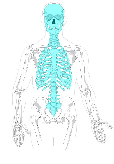
The skeleton is traditionally divided into two major parts: the axial skeleton and the appendicular skeleton, both of which are pictured in Figure \(\PageIndex{3}\).
- The axial skeleton forms the axis of the body. It includes the skull, vertebral column (spine), and rib cage. The bones of the axial skeleton, along with ligaments and muscles, allow the human body to maintain its upright posture. The axial skeleton also transmits weight from the head, trunk, and upper extremities down the back to the lower extremities. In addition, the bones protect the brain and organs in the chest.
- The appendicular skeleton forms the appendages and their attachments to the axial skeleton. It includes the bones of the arms and legs, hands and feet, and shoulder and pelvic girdles. The bones of the appendicular skeleton make possible locomotion and other movements of the appendages. They also protect the major organs of digestion, excretion, and reproduction.
Functions of the Skeletal System
The skeletal system has many different functions that are necessary for human survival. Some of the functions, such as supporting the body, are relatively obvious. Other functions are less obvious but no less important. For example, three tiny bones (hammer, anvil, and stirrup) inside the middle ear transfer sound waves into the inner ear.
Support, Shape, and Protection
The skeleton supports the body and gives it shape. Without the rigid bones of the skeletal system, the human body would be just a bag of soft tissues, as described above. The bones of the skeleton are very hard and provide protection to the delicate tissues of internal organs. For example, the skull encloses and protects the soft tissues of the brain, and the vertebral column protects the nervous tissues of the spinal cord. The vertebral column, ribs, and sternum (breast bone) protect the heart, lungs, and major blood vessels. Providing protection to these latter internal organs requires the bones to be able to expand and contract. The ribs and the cartilage that connects them to the sternum and vertebrae are capable of small shifts that allow breathing and other internal organ movements.
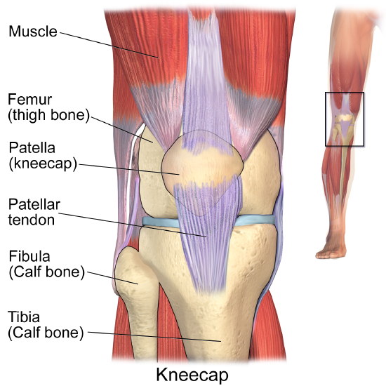
The bones of the skeleton provide attachment surfaces for skeletal muscles. When the muscles contract, they pull on and move the bones. The figure below, for example, shows the muscles attached to the bones at the knee. They help stabilize the joint and allow the leg to bend at the knee. The bones at joints act like levers moving at a fulcrum point, and the muscles attached to the bones apply the force needed for movement.
Hematopoiesis
Hematopoiesis is the process in which blood cells are produced. This process occurs in a tissue called red marrow, which is found inside some bones, including the pelvis, ribs, and vertebrae. Red marrow synthesizes red blood cells, white blood cells, and platelets. Billions of these blood cells are produced inside the bones every day.
Mineral Storage and Homeostasis
Another function of the skeletal system is storing minerals, especially calcium and phosphorus. This storage function is related to the role of bones in maintaining mineral homeostasis. Just the right levels of calcium and other minerals are needed in the blood for the normal functioning of the body. When mineral levels in the blood are too high, bones absorb some of the minerals and store them as mineral salts, which is why bones are so hard. When blood levels of minerals are too low, bones release some of the minerals back into the blood. Bone minerals are alkaline (basic), so their release into the blood buffers the blood against excessive acidity (low pH), whereas their absorption back into bones buffers the blood against excessive alkalinity (high pH). In this way, bones help maintain acid-base homeostasis in the blood.
Another way bones help to maintain homeostasis is by acting as an endocrine organ. One endocrine hormone secreted by bone cells is osteocalcin, which helps regulate blood glucose and fat deposition. It increases insulin secretion and also the sensitivity of cells to insulin. In addition, it boosts the number of insulin-producing cells and reduces fat stores.
- What is the skeletal system? How many bones are there in the adult skeleton?
- Describe the composition of bones.
- Besides bones, what other organs are included in the skeletal system?
- Identify the two major divisions of the skeleton.
- List several functions of the skeletal system.
- Discuss sexual dimorphism in the human skeleton.
- Bones, cartilage, and ligaments are all made of types of ____________ tissue.
- True or False. Bones contain living tissue and can affect processes in other parts of the body.
- True or False. Bone cells contract to pull on muscles in order to initiate a movement.
- If a person has a problem with blood cell production, what type of bone tissue is most likely involved? Explain your answer.
- Are the pelvic girdles part of the axial or appendicular skeleton?
- What are three forms of homeostasis that the skeletal system regulates? Briefly explain how each one is regulated by the skeletal system.
- What do you think would happen to us if we did not have ligaments? Explain your answer.
b. How is cartilage related to joints?
c. Identify one joint in the human body and describe its function.
Explore More
Attributions.
- Fighter squadron 84 by US Navy , public domain via Wikimedia Commons
- Human skeleton front by LadyofHats Mariana Ruiz Villarreal, public domain via Wikimedia Commons
- Axial skeleton by LadyofHats Mariana Ruiz Villarreal, public domain via Wikimedia Commons
- Appendicular skeleton by LadyofHats Mariana Ruiz Villarreal, public domain via Wikimedia Commons
- Knee anatomy by Blausen.com staff (2014). " Medical gallery of Blausen Medical 2014 ". WikiJournal of Medicine 1 (2). DOI : 10.15347/wjm/2014.010 . ISSN 2002-4436 . CC BY 3.0 via Wikimedia Commons
- Text adapted from Human Biology by CK-12 licensed CC BY-NC 3.0

Bones teaches students all about many of the major bones in the human body. Students will discover where these bones are located and learn to identify and label them. They will likewise be able to define other parts of the skeleton and ways to protect bones.
There are several suggestions listed in the “Options for Lesson” section that you can use in your lesson if you want to. One such suggestion is to have students trace their bodies on large pieces of paper and then label the bones of the body. If you can access a model skeleton, you could have students use it to identify each of the bones.
Description
Additional information, what our bones lesson plan includes.
Lesson Objectives and Overview: Bones explores several basic areas of the human skeleton. Students will discover where certain bones are on a skeleton and how to identify them. In addition, they will learn about different ways to help protect their bones from injury. This lesson is for students in 1st grade, 2nd grade, and 3rd grade.
Classroom Procedure
Every lesson plan provides you with a classroom procedure page that outlines a step-by-step guide to follow. You do not have to follow the guide exactly. The guide helps you organize the lesson and details when to hand out worksheets. It also lists information in the yellow box that you might find useful. You will find the lesson objectives, state standards, and number of class sessions the lesson should take to complete in this area. In addition, it describes the supplies you will need as well as what and how you need to prepare beforehand.
Options for Lesson
There are several suggestions in the “Options for Lesson” section that you can add to or incorporate into your lesson. One such suggestion is to allow students to work in pairs for the activity so they can help each other as they complete the worksheet. You could also have students trace their bodies on large paper and then label the bones they learned about. If you can gain access to a model skeleton, students could use to identify each of the bones as you go through the lesson. Another suggestion is to contact a doctor or hospital for X-rays to display to the class. For older students, you could also introduce the scientific names of each bone.
Teacher Notes
This page provides you some extra ideas or guidance on the lesson. The intent of this lesson is to introduce students to the human skeleton at an elementary level. They will only learn about a few major bones. However, you can gauge the class’s level of comprehension and adjust wherever you see fit. There are several blank lines for you to write any more ideas or notes you have before teaching the class this lesson.
BONES LESSON PLAN CONTENT PAGES
Types of bones.
There are two pages of content in this lesson. The first page displays a skeleton and labels several of the specific bones that students should recognize. The lesson starts by asking students why they don’t simply fall down when they stand up. It explains that we can stand, walk, and run because of our bones, which support our bodies. They provide the structure on which the skin hangs and the support and protection for every other part of the body. The skull, for instance, protects the brain and the eyes, face, jaw, nose, and ears.
This lesson continues by explaining several other major bones. The arm bones connect to the collarbone and shoulder bone at the top and then the hand at the bottom. The rib cage covers and protects the organs in the chest and back, such as the heart, lungs, and stomach. The human body typically has 12 pairs of ribs, and they all come in different sizes. The sternum, or breastbone, is part of the rib cage. It is a very hard bone in the center of the chest. It also helps protect the heart and lungs, as well as major blood vessels. The spine that runs down the back protects the spinal cord, which is the path the brain uses to send messages throughout the body.
Students will also learn about some bones in the lower body. The bones in the legs attach to the bottom of the spine by a special group of bones: the pelvis. The upper part of the leg is the thigh bone, which connects to the knee. The lower leg has two bones that attach to the knee and down to the foot.
The Skeleton
The whole bone structure of the body is the skeleton. When a baby is born, it has about 300 bones. However, as people age, some of the bones join together. Generally speaking, adults have a total of 206 bones. About 52 of them make up just the feet and ankles! There are several other parts of the body that work with the skeleton to keep bodies healthy and active.
The bones work with muscles and joints to allow body movement. A joint is the place where two bones come together. The largest and strongest joint in the human body is the knee joint. Ligaments link bones together at the joints. Muscles attach to bones with tendons. Students will discover that they would not be able to move without the muscles, joints, tendons, and ligaments working together with their bones.
These important skeletal structures are necessary to the body. They are strong and can carry heavy weights. However, students will learn that just as their bones protect other parts of their bodies, they should protect their bones. The lesson lists several examples of precautions students should take to help them protect their bodies. They would wear helmets when riding bikes, skateboards, or scooters. If they play sports such as football, soccer, lacrosse, or hockey, they need to wear the proper equipment to protect themselves from injury. They can also strengthen their bones by drinking milk and other dairy products. And finally, they should move. Exercise, sports, running, jumping, dancing, and whatever else they can think of will all help them remain strong.
Here is a list of the vocabulary words students will learn in this lesson plan:
- Bone: a structure of the body on which skin hangs that help provide support and protection for the rest of the body
- Spinal cord: the path by which the brain can send messages throughout the body
- Rib cage (ribs): the bones that protect the organs in the chest region of the body
- Pelvis: a group of bones that attach the leg bones to the bottom of the spine
- Skeleton: the whole bone structure of the body
- Joint: a place where two bones come together
- Ligament: the tissue that links bones together at the joints
- Tendon: a tissue that connects muscles with the bones
BONES LESSON PLAN WORKSHEETS
There are three worksheets in the Bones lesson plan: an activity worksheet, a practice worksheet, and a homework assignment. The goal is to help students grasp the concepts in the lesson in different ways. You can use the classroom procedure guide to know when to hand out each worksheet.
COLOR THE SKELETON ACTIVITY
For the activity, students will cut out each bone from the first worksheet. They will then glue them all onto a separate sheet of paper in the right places. They can color them if they want to or keep them white. The second worksheet page shows 12 bones. Students will have to figure out which area of the skeleton the specific bone represents and color it a unique color.
If you prefer, students can work with a partner for the activity. However, they will need to complete their own skeletons. As an alternative, the activity in the “Options for Lesson” section describes having students trace themselves on large pieces of paper. Then they can draw and label the different bones there. You could also do all three activities if time permits.
LABEL THE SKELETON PRACTICE WORKSHEET
Students must use a word bank to correctly label a skeleton. They may need to refer back to the information from the content pages for some clues. It is also possible that they will need to do some research to complete the worksheet. The lesson does not describe every single part of the skeleton that this worksheet lists.
BONES HOMEWORK ASSIGNMENT
The homework assignment requires students to fill in the blanks on 20 statements. The statements relate to what they learned during the lesson. Students may need to refer back to the content pages to find their answers. The word bank provides the 20 options, and students will use each option only once.
Worksheet Answer Keys
The final pages of the Bones lesson plan document provides answer keys for the worksheets. This includes an answer for the activity since there are specific correct answers. The answer key for the activity shows a skeleton with different colors matching the correct bones. The answer key for the practice worksheet provides the answers in red. As an option, you may choose to display this page for a set amount of time while students study it. Then you can put it away while they try to remember where all the bones were. The answer key for the homework assignment likewise provides the correct answers in red. If you choose to administer the lesson pages to your students via PDF, you will need to save a new file that omits these pages. Otherwise, you can simply print out the applicable pages and keep these as reference for yourself when grading assignments.
Thank you for submitting a review!
Your input is very much appreciated. Share it with your friends so they can enjoy it too!

Great coloring activity
I am using this in my year long science class. The kids did a great job coloring the different bones. I hope I don't start getting spammed after giving my email address.
Wonderful resource
I look forward to using more of these resources with my grandchildren
Very understandable and engaging for kids - love it.
Wonderful learning for the children.
Dem Bones are Great!
This lesson has a lot of information useful for my year long science class. Thank you!
Amazing resource for teacher!
Fantastic material! I recommend to ALL teachers.
Related products
Careers: Electrician

Killer Bees

Man o’ Wars
Make your life easier with our lesson plans, stay up-to-date with new lessons.

- Lesson Plans
- For Teachers
© 2024 Learn Bright. All rights reserved. Terms and Conditions. Privacy Policy.
- Sign Up for Free

Elementary School Science
The Skeletal System Lesson Plan: No More Skeletons in the Closet
This lesson contains affiliate links to products I have used and personally recommend. At no cost to you, I make a commission for purchases made through the links or advertisements. These commissions help to pay for the costs of the site and enable it to remain free for anyone who wants to use it.
Objectives:
The students will learn how many bones are in the body.
The students will be able to identify the name and location of major bones in the body.
The students will be able to describe how our bones protect our body.
The students will be able to explain how bone growth occurs.
The students will be able to name ways to keep our bones safe and healthy.
Questions that encompasses the objective:
Have you ever broken a bone or do you know someone who has?
What was the experience like?
How was your bone repaired?
Prepare the Learner: Activating Prior Knowledge.
How will students prior knowledge be activated?
Warm up by asking students:
What do you know about the skeletal system?
How many bones do you think we have in our body?
Common Core State Standards:
CCSS.ELA-LITERACY.SL.2.1
CCSS.ELA-LITERACY.SL.2.1 B
CCSS.ELA-LITERACY.SL.2.4
CCSS.ELA-LITERACY.W.2.2
CCSS.ELA-LITERACY.W.3.2
CCSS.ELA-LITERACY.W.3.2 B
CCSS.ELA-LITERACY.SL.3.1
CCSS.ELA-LITERACY.SL.3.4
Materials and Free Resources to Download for this Lesson:
Video: “What If We Didn’t Have Bones—What Would Happen?” by Suggested By You - Amazing Facts
Picture of a Skeleton
**If you have access to a model skeleton that should be used instead. Using realia helps the students to better understand the topic being taught, as well as helps to make the lesson more interesting for the students.**
“Bones in Our Body” worksheet
“Take Care of Your Bones!” worksheet
“What is Wrong with Mr. Bones?” activity materials:
“Poor Mr. Bones!” cards
“What is Wrong with Mr. Bones?” worksheet
"What Do You Think? Assessment


Digestive System Lesson Plan: How It All Goes Down
Input: What is the most important content in this lesson? To reach this lesson’s objective, students need to understand:
The name and location of the major bones in the body.
How many bones are in the body.
The importance of the skeletal system and its role in protecting the body.
How bones grow.
The ways we can keep our bones safe and healthy.
How will the learning of this content be facilitated?
The class will begin with the teacher showing the YouTube clip “What If We Didn’t Have Bones—What Would Happen?” (source: https://www.youtube.com/watch?v=_hyqlKVm2ks) The video is about 4 minutes long. After the video is shown, a discussion should begin about what the students just learned. The teacher should ask the students their thoughts about the content.
The teacher should begin explaining the skeletal system more in depth to the students. Show the students the picture of the skeleton or model skeleton.
The skeletal system is a very important component of our body. The skeletal system:
Provides protection for our internal organs
Give us a framework for our body
Allows us to move around (with the help of our joints).
The skeletal system consists of 206 bones, which are constantly changing. When a baby is born, their skeletal system contains 300 bones. But as they grow, some bones begin to fuse/ grow together. Many bones in a child’s body are made of cartilage. As the child grows, with the help of calcium, the cartilage begins to turn into bone. By the time you are 25 years old, bone growth stops and the size your bones are is what they are going to be.
Without our skeletal system, we would not be able to stand upright, walk, move our arms, or turn our heads. All of the bones work together to make movement possible.
Our skeletal system also helps to protect our internal organs from harm. For example, your ribs protect your heart, lungs, and liver; your skull protects your brain.
Information Source: https://kidshealth.org/en/kids/bones.html#
After the discussion, the teacher should hand out the “Bones in Our Body” worksheet. If it is possible, project the “Bones in Our Body” worksheet onto the board using a projector or put into a PowerPoint document and project so that the teacher can point to each bone while they explain. As the teacher explains each bone, the students should write the name in the box. From this activity, the students will learn about the major bones of the body, the location of each bone, and the function of each bone.
Bones and their Functions:
Cranium : the skull; encloses/ protects the brain.
Clavicle : also called the collarbone; allows your arms to hang freely.
Ribs : act as a protective cage for the heart, lungs, and liver; there are 12 pairs of ribs.
Radius : lateral, shorter bone of the forearm.
Ulna : inner, larger bone of the forearm.
Metacarpals : the five bones of the metacarpus, located between the wrist and the fingers.
Femur : the thighbone; the longest and strongest bone in the body.
Tibia : the inner, larger bone of the lower leg.
Tarsals : the seven bones of the ankle joint.
Scapula : also called the shoulder blade; provides a foundation for joint function.
Mandible : the jawbone; holds the lower teeth in place; it is the strongest bone in the face.
Stapes : located in the middle ear; the smallest bone in the body.
Humerus : upper arm bone; supports arm functions, such as lifting.
Vertebrae : any of the 33 bones of the spinal column.
Pelvis : located near the base of the spine where the hind limbs/legs are attached; this bone is separated in children and fused together in adults.
Carpals : any of the 8 bones of the carpus/ wrist.
Phalanges : any of the bones of the fingers.
Patella : the kneecap; allows for knee extension.
Fibula : the outer, smaller bone located between the knee and the ankle.
Metatarsals : any of the bones located between the ankle and the toes.
Information Source: http://medical-dictionary.thefreedictionary.com
Once the bones are explained, the teacher will instruct the students to stand up. Tell the students that they will be playing a game, similar to “Simon Says”, where they will be touching the different bones of their body. Play the game for about 5-10 minutes, incorporating most or all of the bones that were just taught. Begin the game by saying: “[insert your name- Mr./Ms./Mrs….] says, touch your patella.”
**This provides the students with some time to stand and move around before resuming the final discussion of the lesson**
After the game, the teacher should begin a discussion about ways to keep bones healthy and safe. The teacher should hand out the “Take Care of Your Bones!” worksheet. If it is possible, project the “Take Care of Your Bones!” worksheet onto the board using a projector or put into a PowerPoint document and project. The teacher should begin a discussion about ways to keep the bones safe and healthy. The students should write the four ways to keep bones healthy on their worksheet
Always wear a helmet, kneepads, and elbow pads when roller blading, skateboarding, riding a bicycle, or riding a scooter.
If you play sports like football, soccer, or hockey, always listen to your coach and wear the proper safety gear.
Eat foods that contain calcium. These foods include milk, cheese, and yogurt.
Exercise often—playing outside, riding your bicycle, jumping, and dancing all help strengthen your bones.
Once the discussion on how to keep bones safe and healthy is finished, the students will break into groups of three or four. Each student will be given a “What’s Wrong With Mr. Bones?” worksheet. On desks throughout the room will be “Poor Mr. Bones!” cards. On each card will be a description of something that Mr. Bones did that caused him to get hurt. The students will read each card and identify which bone Mr. Bones injured. Allow the students about 15 minutes to go around the room and read each card. Reconvene when 15 minutes is over and review the worksheet/ activity.
The final assessment will be for the students to answer the question:
Think about what you learned in class today about the skeletal system. Why is our skeleton important? What are the benefits of having a skeleton? When we are born, how many bones do we have? Do we still have the same amount of bones when we reach adulthood? If not, what happens to those bones? What are some ways we can keep our bones safe and healthy?

Endocrine System Lesson Plan: A Look at the Endocrine System

Circulatory System Lesson Plan: A Look at the Heart (Cardiovascular System)

Immune System Lesson Plan: Staying Healthy

Integumentary System Lesson Plan: All About Our Skin
Image by Brucel Blaus blausen.com on Wikipedia with a CC 3.0 License

Lymphatic System Lesson Plan: A Look at Our Lymphatic System

Muscular System Lesson Plan: Getting Stronger With Muscles

Nervous System Lesson Plan: How Did My Body Do That?
Time/Application 3-5 minutes Guided Introduction
Review the class/ agenda with the students:
Introductory Activity (video)
Skeletal System Discussion
Skeleton Diagram
Game: “Simon Says”
Taking Care of Our Bones Discussion
Group Activity: “What’s Wrong With Mr. Bones?”
Discussion of Group Activity
Independent Assessment
Introductory Activity:
Have the students sit at their desks.
Show the students the video: “What If We Didn’t Have Bones—What Would Happen?” by Suggested By You - Amazing Facts
Once the video is over, discuss it with the students.
25 Minutes
Skeletal System Discussion | Bones in Our Body | Simon Says | Taking Care of Our Bones
Expand on the video and discuss the skeletal system more in depth.
Give each student the “Bones in Our Body” worksheet.
Project the diagram onto the board either through a projector or PowerPoint presentation.
Tell the students that as each part is explained, they should write the name of the part in the box.
Play “Simon Says” with the students; using the names of the bones they just learned.
Begin a discussion about keeping bones safe and healthy.
Group Activity: “What’s Wrong with Mr. Bones?”
Give each student a “What’s Wrong with Mr. Bones?” worksheet.
Instruct the students to break into groups of three or four.
Set up the “Poor Mr. Bones!” cards on desks around the room. Have the students circulate around the room, read the cards, and decide which bone Mr. Bones injured.
At the end of 15 minutes, have the students return to their desks and discuss their observations.
Closure/Assessment 10 minutes
Independent Assessment:
Appropriate answers should include (but will vary):
Our skeleton is important because it is the framework of our body. Our skeleton helps us to stand upright and move our arms and legs. Our skeleton also helps to protect our internal organs, such as our heart, lungs, and brain. Without our skeletal system we would be like a lump of jelly. When we are born, we have 300 bones, but our bones fuse together as we grow. By the time we are adults we have 206 bones. We can keep our bones healthy and safe by eating foods that contain calcium, exercising, and wearing protective gear when riding out bicycles or playing sports.
If there is additional time, discuss any additional questions the students may have.
Individualized Instruction/Scaffolding
English Language Learners will be supported in this lesson through data-based heterogeneous grouping, verbal and written repetition of new vocabulary words, and multiple representation of vocabulary words through printed images and video.

Reproductive System Lesson Plan: I'm Pregnant!

Respiratory System Lesson Plan: Breathe In, Breathe Out

Skeletal System Lesson Plan: No More Skeletons in the Closet

Urinary System Lesson Plan: Urine Luck!

Human Body Lesson Plans Home Page
Academia.edu no longer supports Internet Explorer.
To browse Academia.edu and the wider internet faster and more securely, please take a few seconds to upgrade your browser .
Enter the email address you signed up with and we'll email you a reset link.
- We're Hiring!
- Help Center

Skeletal System.ppt

Related Papers
Nhật Nguyễn
vinoth kalaiselvan
DIANA MARCELA ESCOBAR MONTOYA
Louiza Aoudia
Skeleton 3A-1 at Aïoun Bériche (also known as Site 12), a Capsian escargotière in eastern Algeria testifies to a complex treatment prior to burial. A study of the skeleton and the field records through the lens of archaeothanatology allows a more detailed interpretation of the burial than previously published. Osteological analysis revealed the presence of cutmarks on different bones of the skeleton. The location of these cutmarks near major joints (neck, elbow, and knee) shows that the intention of the cutting operation was to partition off the body into pieces. Archaeothanatological analysis provides evidence that these operations were conducted promptly after death as well as the deposit in earth of the partitioned body into "anatomical dislocated blocks". This very specific treatment is not a unique case and appears, on the contrary, to play an important part in the Capsian funerary identity.
David Gómez Nieto
Steven Kuehn
Archaeological investigations at the late prehistoric Janey B. Goode site (11S1232) in southwestern Illinois resulted in the recovery of over 5,400 domestic dog (Canis familiaris) remains, representing over 100 individual animals. The substantial size of this well-preserved faunal assemblage allows for a detailed study of Native American dogs during the Late Woodland Patrick phase (A.D. 650-900), Terminal Late Woodland (A.D. 900-1050), and Mississippian (A.D. 1050-1400) periods in the American Bottom. One aspect of this on-going, multifaceted research project is the documentation, analysis, and interpretation of prehistoric trauma and pathologies. This article presents a preliminary summary of the dog paleopathology evidence obtained thus far from the Janey B. Goode site.
CaMi Pineda Correa
Opuscula Annual of the Swedish Institutes at Athens and Rome 12 2019 STOCKHOLM SVENSKA INSTITUTEN I ATHEN OCH ROM INSTITUTUM ATHENIENSE ATQUE INSTITUTUM ROMANUM REGNI SUECIAE
Ylva A E Bäckström , Anne Ingvarsson , Stella Chryssoulaki , Anna Linderholm , Vendela Kempe Lagerholm
P. David Polly
The more than 4,000 living species of mammal have infiltrated almost every habitat in the world. From alpine mountaintops to plains grasslands, from aerial heights to the depths of the ocean, from slender forest branches to narrow subterranean burrows, mammals occupy an extraordinary variety of substrates. Some are capable of running, swimming, or swinging at great speeds, while others creep slowly along limbs or push laboriously through the soil. The limbs of mammals reflect the diversity of their habitats (fig. 15.1). The long, slender legs and two-toed feet of the antelope allow it to survey the plains for predators and bound off at their approach. The short, muscled arms and broad, thick-clawed hands of the mole scrape through soil as the animal searches for worms and grubs. Spidery, elongated hands and fingers with webs of skin between them propel bats on their erratic chases after night-flying insects. Dense, paddlelike fins steer whales through the watery course on which they are propelled by their massive tail flukes. Limbs are crucial for mammalian locomotion, social behavior, and feeding. The functional diversity of mammal limbs is facilitated by sometimes subtle structural differences. A small discrepancy in the proportion of one limb segment and its distal neighbor can translate into significant disparity in running speed. The position of a muscle insertion along a long bone shaft can make the difference between a mammal that can tear its way through thick turf and one that cannot. The fusion of two bones in the forearm or wrist may mean that one animal cannot turn its palm to grasp a limb as it tries to climb, while another species can easily wrap its forelimbs around a tree trunk and scamper to safety. A large part of a mammal's lifestyle can, therefore, be read in the structure of its limbs. This chapter first reviews the anatomical structure of the mammalian limb. Some brief mention of comparative differences is made in relation to structure, but those are reserved for the most striking ones in which the number of elements differs or the structure of homologous elements is particularly diverse. Special emphasis is given to the structures that are most obviously related to function. The chapter then reviews the functional diversity of the mammalian limb from the perspective of gross ecomorphological categories, groups that are primarily locomotory in nature. The chapter then reviews aspects of variation, genetics, and development of the mammalian limb. Finally, the early evolution of the mammalian limb is reviewed.
Loading Preview
Sorry, preview is currently unavailable. You can download the paper by clicking the button above.
RELATED TOPICS
- We're Hiring!
- Help Center
- Find new research papers in:
- Health Sciences
- Earth Sciences
- Cognitive Science
- Mathematics
- Computer Science
- Academia ©2024

IMAGES
VIDEO
COMMENTS
Providing support for the body. Storing minerals (calcium, phosphate) Producing red blood cells. Protecting the organs and tissues. Allowing movement (the bones act as levers) The skeleton can be subcategorized into two divisions: The Axial Skeleton (left, in blue) Includes: Bones of the skull, vertebrae, sternum, ribs, and sacrum.
Abstract The skeletal system is formed of bones and cartilage, which are connected by ligaments to form a framework for the remainder of the body tissues. This article, the first in a two-part series on the structure and function of the skeletal system, reviews the anatomy and physiology of bone. Understanding the structure and
Get free human anatomy worksheets and study guides to download and print. This is a collection of free human anatomy worksheets. The completed worksheets make great study guides for learning bones, muscles, organ systems, etc. The worksheets come in a variety of formats for downloading and printing. In most cases, the PDF worksheets print the best.
The skeletal system includes Cartilage and ligament. The cartilage is soft, gel-like padding between the bones that protect the joints and facilitates movement while the ligament is a rigid muscle group that connects bone to bone and provides joint elasticity. Types of bones there are 5 types of bone that create the skeleton 1.
each individual bone is a separate organ of the skeletal system. ~270 bones (organs) of the Skeletal System. with age the number decreases as bones fuse. by adulthood the number is 206 (typical) even this number varies due to varying numbers of minor bones: sesamoid bones - small rounded bones that form within tendons in response to stress.
Home - University of Cincinnati | University Of Cincinnati
A quick lesson and free online and printable skeletal system worksheets for elementary students. Contents: Quick lesson on bones and joints. Worksheets. Worksheet 1: How the Skeletal System Works. Worksheet 2: The Protection Function of Bones. Worksheet 3: Axial Skeleton and Appendicular Skeleton. Worksheet 4: Axial vs Appendicular Skeleton and ...
Digital PDF Print PDF Common Cartridge (Web Links) Common Cartridge (LTI Links) ... Bone Tissue and the Skeletal System. 6.0 Introduction. 6.1 The Functions of the Skeletal System. 6.2 Bone Classification. 6.3 Bone Structure. ... Download for free at https://open.oregonstate.education/aandp/
Summary. Anatomy and Physiology 2e is developed to meet the scope and sequence for a two-semester human anatomy and physiology course for life science and allied health majors. The book is organized by body systems. The revision focuses on inclusive and equitable instruction and includes new student support. Illustrations have been extensively ...
In adults, the skeletal system includes 206 bones, many of which are shown in Figure 14.2.2 14.2. 2. Bones are organs made of dense connective tissues, mainly the tough protein collagen. Bones contain blood vessels, nerves, and other tissues. Bones are hard and rigid due to deposits of calcium and other mineral salts within their living tissues.
Eder, et al.: Laboratory Atlas of Anatomy and Physiology, Third Edition 2. Human Skeletal Anatomy Text © The McGraw−Hill Companies, 2001 45 2 Human Skeletal Anatomy
Projections where muscles, tendons and ligaments attach. Tubercle - small rounded projection. Tuberosity - rounded projection. Crest - narrow, prominent ridge of bone. Trochanter - large, blunt, irregular surface. Line - narrow ridge of bone. Epicondyle - raised area above a condyle. Spine - sharp, slender projection.
skeletal muscles for body movement, produces blood cells, and stores minerals. Organs Here are the primary structures that comprise the skeletal system: bones joints Word Parts Here are the most common word parts (with their meanings) used to build skeletal system terms. For a more comprehensive list, refer to the Terminology section of this ...
hinge The scientific name of the kneecap. patella A type of fracture where there is a complete break of the bone and the skin. Compound/open The scientific name of the collarbone. clavicle When a bone moves out of position. dislocation The scientific name of the shoulder blades.
Assignment 1 Skeletal System - Free download as Word Doc (.doc / .docx), PDF File (.pdf), Text File (.txt) or read online for free. The axial skeleton, making up 80 of the 206 bones, includes all upper body bones. The bones of the skull can be categorised into two groups, the cranium and face. The thorax (chest) is the sternum and ribs forming the structure of the chest.
Lesson Objectives and Overview: Human Skeleton STEM teaches students about the major bones in the human body. Students will discover that bones, muscles, and tissue work together to help the body move and function properly. They will learn that the skeletal system is not independent of other body systems. This lesson is for students in 4th ...
Model of the Human Skeleton Have students read about the skeletal system below and take the two short answer quizzes. They can study the labeled skeleton and then try to label a whole skeleton themselves. Now they are ready to build their own labeled skeletons. 1. Print out the pdf bones of the body; hands, feet, arms, forearms legs, shins, skull,
A typical myofiber is 2-3 centimeters ( 3/4-1 1/5 in) long and 0.05millimeters (1/500 inch) in diameter and is composed of narrower structures - myofibrils. These contain thick and thin myofilaments made up mainly of the proteins actin and myosin. Numerous capillaries keep the muscle supplied with the oxygen and glucose needed to fuel ...
Rib cage (ribs): the bones that protect the organs in the chest region of the body. Pelvis: a group of bones that attach the leg bones to the bottom of the spine. Skeleton: the whole bone structure of the body. Joint: a place where two bones come together. Ligament: the tissue that links bones together at the joints.
BONES CAN BE CLASSIFIED, INTO FIVE TYPES, Bones of the human skeletal, system are categorized by their, shape and function into five types., The femur is an example of a long, bone., The frontal bone is a flat bone., The patella, also called the knee, cap, is a sesamoid bone., Carpals (in the hand) and tarsals, (in the feet) are examples of ...
Allows us to move around (with the help of our joints). The skeletal system consists of 206 bones, which are constantly changing. When a baby is born, their skeletal system contains 300 bones. But as they grow, some bones begin to fuse/ grow together. Many bones in a child's body are made of cartilage.
The Muscular System Manual: The Skeletal Muscles of the Human Body, 4th edition, is meant to be the most thorough atlas of muscle function that is available. Instead of simply listing muscle attachments and actions that are typically taught, The Muscular System Manual comprehensively covers all muscle functions of each muscle.
A study of the skeleton and the field records through the lens of archaeothanatology allows a more detailed interpretation of the burial than previously published. Osteological analysis revealed the presence of cutmarks on different bones of the skeleton. The location of these cutmarks near major joints (neck, elbow, and knee) shows that the ...