Have a language expert improve your writing
Run a free plagiarism check in 10 minutes, generate accurate citations for free.
- Knowledge Base
Methodology
- How to Write a Literature Review | Guide, Examples, & Templates

How to Write a Literature Review | Guide, Examples, & Templates
Published on January 2, 2023 by Shona McCombes . Revised on September 11, 2023.
What is a literature review? A literature review is a survey of scholarly sources on a specific topic. It provides an overview of current knowledge, allowing you to identify relevant theories, methods, and gaps in the existing research that you can later apply to your paper, thesis, or dissertation topic .
There are five key steps to writing a literature review:
- Search for relevant literature
- Evaluate sources
- Identify themes, debates, and gaps
- Outline the structure
- Write your literature review
A good literature review doesn’t just summarize sources—it analyzes, synthesizes , and critically evaluates to give a clear picture of the state of knowledge on the subject.
Instantly correct all language mistakes in your text
Upload your document to correct all your mistakes in minutes

Table of contents
What is the purpose of a literature review, examples of literature reviews, step 1 – search for relevant literature, step 2 – evaluate and select sources, step 3 – identify themes, debates, and gaps, step 4 – outline your literature review’s structure, step 5 – write your literature review, free lecture slides, other interesting articles, frequently asked questions, introduction.
- Quick Run-through
- Step 1 & 2
When you write a thesis , dissertation , or research paper , you will likely have to conduct a literature review to situate your research within existing knowledge. The literature review gives you a chance to:
- Demonstrate your familiarity with the topic and its scholarly context
- Develop a theoretical framework and methodology for your research
- Position your work in relation to other researchers and theorists
- Show how your research addresses a gap or contributes to a debate
- Evaluate the current state of research and demonstrate your knowledge of the scholarly debates around your topic.
Writing literature reviews is a particularly important skill if you want to apply for graduate school or pursue a career in research. We’ve written a step-by-step guide that you can follow below.

Prevent plagiarism. Run a free check.
Writing literature reviews can be quite challenging! A good starting point could be to look at some examples, depending on what kind of literature review you’d like to write.
- Example literature review #1: “Why Do People Migrate? A Review of the Theoretical Literature” ( Theoretical literature review about the development of economic migration theory from the 1950s to today.)
- Example literature review #2: “Literature review as a research methodology: An overview and guidelines” ( Methodological literature review about interdisciplinary knowledge acquisition and production.)
- Example literature review #3: “The Use of Technology in English Language Learning: A Literature Review” ( Thematic literature review about the effects of technology on language acquisition.)
- Example literature review #4: “Learners’ Listening Comprehension Difficulties in English Language Learning: A Literature Review” ( Chronological literature review about how the concept of listening skills has changed over time.)
You can also check out our templates with literature review examples and sample outlines at the links below.
Download Word doc Download Google doc
Before you begin searching for literature, you need a clearly defined topic .
If you are writing the literature review section of a dissertation or research paper, you will search for literature related to your research problem and questions .
Make a list of keywords
Start by creating a list of keywords related to your research question. Include each of the key concepts or variables you’re interested in, and list any synonyms and related terms. You can add to this list as you discover new keywords in the process of your literature search.
- Social media, Facebook, Instagram, Twitter, Snapchat, TikTok
- Body image, self-perception, self-esteem, mental health
- Generation Z, teenagers, adolescents, youth
Search for relevant sources
Use your keywords to begin searching for sources. Some useful databases to search for journals and articles include:
- Your university’s library catalogue
- Google Scholar
- Project Muse (humanities and social sciences)
- Medline (life sciences and biomedicine)
- EconLit (economics)
- Inspec (physics, engineering and computer science)
You can also use boolean operators to help narrow down your search.
Make sure to read the abstract to find out whether an article is relevant to your question. When you find a useful book or article, you can check the bibliography to find other relevant sources.
You likely won’t be able to read absolutely everything that has been written on your topic, so it will be necessary to evaluate which sources are most relevant to your research question.
For each publication, ask yourself:
- What question or problem is the author addressing?
- What are the key concepts and how are they defined?
- What are the key theories, models, and methods?
- Does the research use established frameworks or take an innovative approach?
- What are the results and conclusions of the study?
- How does the publication relate to other literature in the field? Does it confirm, add to, or challenge established knowledge?
- What are the strengths and weaknesses of the research?
Make sure the sources you use are credible , and make sure you read any landmark studies and major theories in your field of research.
You can use our template to summarize and evaluate sources you’re thinking about using. Click on either button below to download.
Take notes and cite your sources
As you read, you should also begin the writing process. Take notes that you can later incorporate into the text of your literature review.
It is important to keep track of your sources with citations to avoid plagiarism . It can be helpful to make an annotated bibliography , where you compile full citation information and write a paragraph of summary and analysis for each source. This helps you remember what you read and saves time later in the process.
To begin organizing your literature review’s argument and structure, be sure you understand the connections and relationships between the sources you’ve read. Based on your reading and notes, you can look for:
- Trends and patterns (in theory, method or results): do certain approaches become more or less popular over time?
- Themes: what questions or concepts recur across the literature?
- Debates, conflicts and contradictions: where do sources disagree?
- Pivotal publications: are there any influential theories or studies that changed the direction of the field?
- Gaps: what is missing from the literature? Are there weaknesses that need to be addressed?
This step will help you work out the structure of your literature review and (if applicable) show how your own research will contribute to existing knowledge.
- Most research has focused on young women.
- There is an increasing interest in the visual aspects of social media.
- But there is still a lack of robust research on highly visual platforms like Instagram and Snapchat—this is a gap that you could address in your own research.
There are various approaches to organizing the body of a literature review. Depending on the length of your literature review, you can combine several of these strategies (for example, your overall structure might be thematic, but each theme is discussed chronologically).
Chronological
The simplest approach is to trace the development of the topic over time. However, if you choose this strategy, be careful to avoid simply listing and summarizing sources in order.
Try to analyze patterns, turning points and key debates that have shaped the direction of the field. Give your interpretation of how and why certain developments occurred.
If you have found some recurring central themes, you can organize your literature review into subsections that address different aspects of the topic.
For example, if you are reviewing literature about inequalities in migrant health outcomes, key themes might include healthcare policy, language barriers, cultural attitudes, legal status, and economic access.
Methodological
If you draw your sources from different disciplines or fields that use a variety of research methods , you might want to compare the results and conclusions that emerge from different approaches. For example:
- Look at what results have emerged in qualitative versus quantitative research
- Discuss how the topic has been approached by empirical versus theoretical scholarship
- Divide the literature into sociological, historical, and cultural sources
Theoretical
A literature review is often the foundation for a theoretical framework . You can use it to discuss various theories, models, and definitions of key concepts.
You might argue for the relevance of a specific theoretical approach, or combine various theoretical concepts to create a framework for your research.
Like any other academic text , your literature review should have an introduction , a main body, and a conclusion . What you include in each depends on the objective of your literature review.
The introduction should clearly establish the focus and purpose of the literature review.
Depending on the length of your literature review, you might want to divide the body into subsections. You can use a subheading for each theme, time period, or methodological approach.
As you write, you can follow these tips:
- Summarize and synthesize: give an overview of the main points of each source and combine them into a coherent whole
- Analyze and interpret: don’t just paraphrase other researchers — add your own interpretations where possible, discussing the significance of findings in relation to the literature as a whole
- Critically evaluate: mention the strengths and weaknesses of your sources
- Write in well-structured paragraphs: use transition words and topic sentences to draw connections, comparisons and contrasts
In the conclusion, you should summarize the key findings you have taken from the literature and emphasize their significance.
When you’ve finished writing and revising your literature review, don’t forget to proofread thoroughly before submitting. Not a language expert? Check out Scribbr’s professional proofreading services !
This article has been adapted into lecture slides that you can use to teach your students about writing a literature review.
Scribbr slides are free to use, customize, and distribute for educational purposes.
Open Google Slides Download PowerPoint
If you want to know more about the research process , methodology , research bias , or statistics , make sure to check out some of our other articles with explanations and examples.
- Sampling methods
- Simple random sampling
- Stratified sampling
- Cluster sampling
- Likert scales
- Reproducibility
Statistics
- Null hypothesis
- Statistical power
- Probability distribution
- Effect size
- Poisson distribution
Research bias
- Optimism bias
- Cognitive bias
- Implicit bias
- Hawthorne effect
- Anchoring bias
- Explicit bias
A literature review is a survey of scholarly sources (such as books, journal articles, and theses) related to a specific topic or research question .
It is often written as part of a thesis, dissertation , or research paper , in order to situate your work in relation to existing knowledge.
There are several reasons to conduct a literature review at the beginning of a research project:
- To familiarize yourself with the current state of knowledge on your topic
- To ensure that you’re not just repeating what others have already done
- To identify gaps in knowledge and unresolved problems that your research can address
- To develop your theoretical framework and methodology
- To provide an overview of the key findings and debates on the topic
Writing the literature review shows your reader how your work relates to existing research and what new insights it will contribute.
The literature review usually comes near the beginning of your thesis or dissertation . After the introduction , it grounds your research in a scholarly field and leads directly to your theoretical framework or methodology .
A literature review is a survey of credible sources on a topic, often used in dissertations , theses, and research papers . Literature reviews give an overview of knowledge on a subject, helping you identify relevant theories and methods, as well as gaps in existing research. Literature reviews are set up similarly to other academic texts , with an introduction , a main body, and a conclusion .
An annotated bibliography is a list of source references that has a short description (called an annotation ) for each of the sources. It is often assigned as part of the research process for a paper .
Cite this Scribbr article
If you want to cite this source, you can copy and paste the citation or click the “Cite this Scribbr article” button to automatically add the citation to our free Citation Generator.
McCombes, S. (2023, September 11). How to Write a Literature Review | Guide, Examples, & Templates. Scribbr. Retrieved March 20, 2024, from https://www.scribbr.com/dissertation/literature-review/
Is this article helpful?
Shona McCombes
Other students also liked, what is a theoretical framework | guide to organizing, what is a research methodology | steps & tips, how to write a research proposal | examples & templates, unlimited academic ai-proofreading.
✔ Document error-free in 5minutes ✔ Unlimited document corrections ✔ Specialized in correcting academic texts
- - Google Chrome
Intended for healthcare professionals
- Access provided by Google Indexer
- My email alerts
- BMA member login
- Username * Password * Forgot your log in details? Need to activate BMA Member Log In Log in via OpenAthens Log in via your institution

Search form
- Advanced search
- Search responses
- Search blogs
- Writing a case report...
Writing a case report in 10 steps
- Related content
- Peer review
- Victoria Stokes , foundation year 2 doctor, trauma and orthopaedics, Basildon Hospital ,
- Caroline Fertleman , paediatrics consultant, The Whittington Hospital NHS Trust
- victoria.stokes1{at}nhs.net
Victoria Stokes and Caroline Fertleman explain how to turn an interesting case or unusual presentation into an educational report
It is common practice in medicine that when we come across an interesting case with an unusual presentation or a surprise twist, we must tell the rest of the medical world. This is how we continue our lifelong learning and aid faster diagnosis and treatment for patients.
It usually falls to the junior to write up the case, so here are a few simple tips to get you started.
First steps
Begin by sitting down with your medical team to discuss the interesting aspects of the case and the learning points to highlight. Ideally, a registrar or middle grade will mentor you and give you guidance. Another junior doctor or medical student may also be keen to be involved. Allocate jobs to split the workload, set a deadline and work timeframe, and discuss the order in which the authors will be listed. All listed authors should contribute substantially, with the person doing most of the work put first and the guarantor (usually the most senior team member) at the end.
Getting consent
Gain permission and written consent to write up the case from the patient or parents, if your patient is a child, and keep a copy because you will need it later for submission to journals.
Information gathering
Gather all the information from the medical notes and the hospital’s electronic systems, including copies of blood results and imaging, as medical notes often disappear when the patient is discharged and are notoriously difficult to find again. Remember to anonymise the data according to your local hospital policy.
Write up the case emphasising the interesting points of the presentation, investigations leading to diagnosis, and management of the disease/pathology. Get input on the case from all members of the team, highlighting their involvement. Also include the prognosis of the patient, if known, as the reader will want to know the outcome.
Coming up with a title
Discuss a title with your supervisor and other members of the team, as this provides the focus for your article. The title should be concise and interesting but should also enable people to find it in medical literature search engines. Also think about how you will present your case study—for example, a poster presentation or scientific paper—and consider potential journals or conferences, as you may need to write in a particular style or format.
Background research
Research the disease/pathology that is the focus of your article and write a background paragraph or two, highlighting the relevance of your case report in relation to this. If you are struggling, seek the opinion of a specialist who may know of relevant articles or texts. Another good resource is your hospital library, where staff are often more than happy to help with literature searches.
How your case is different
Move on to explore how the case presented differently to the admitting team. Alternatively, if your report is focused on management, explore the difficulties the team came across and alternative options for treatment.
Finish by explaining why your case report adds to the medical literature and highlight any learning points.
Writing an abstract
The abstract should be no longer than 100-200 words and should highlight all your key points concisely. This can be harder than writing the full article and needs special care as it will be used to judge whether your case is accepted for presentation or publication.
Discuss with your supervisor or team about options for presenting or publishing your case report. At the very least, you should present your article locally within a departmental or team meeting or at a hospital grand round. Well done!
Competing interests: We have read and understood BMJ’s policy on declaration of interests and declare that we have no competing interests.

Organizing Your Social Sciences Research Paper: Writing a Case Study
- Purpose of Guide
- Design Flaws to Avoid
- Independent and Dependent Variables
- Glossary of Research Terms
- Narrowing a Topic Idea
- Broadening a Topic Idea
- Extending the Timeliness of a Topic Idea
- Academic Writing Style
- Choosing a Title
- Making an Outline
- Paragraph Development
- Executive Summary
- The C.A.R.S. Model
- Background Information
- The Research Problem/Question
- Theoretical Framework
- Citation Tracking
- Content Alert Services
- Evaluating Sources
- Reading Research Effectively
- Primary Sources
- Secondary Sources
- Tiertiary Sources
- What Is Scholarly vs. Popular?
- Qualitative Methods
- Quantitative Methods
- Using Non-Textual Elements
- Limitations of the Study
- Common Grammar Mistakes
- Writing Concisely
- Avoiding Plagiarism
- Footnotes or Endnotes?
- Further Readings
- Annotated Bibliography
- Dealing with Nervousness
- Using Visual Aids
- Grading Someone Else's Paper
- Types of Structured Group Activities
- Group Project Survival Skills
- Multiple Book Review Essay
- Reviewing Collected Essays
- Writing a Case Study
- About Informed Consent
- Writing Field Notes
- Writing a Policy Memo
- Writing a Research Proposal
- Bibliography
The term case study refers to both a method of analysis and a specific research design for examining a problem, both of which are used in most circumstances to generalize across populations. This tab focuses on the latter--how to design and organize a research paper in the social sciences that analyzes a specific case.
A case study research paper examines a person, place, event, phenomenon, or other type of subject of analysis in order to extrapolate key themes and results that help predict future trends, illuminate previously hidden issues that can be applied to practice, and/or provide a means for understanding an important research problem with greater clarity. A case study paper usually examines a single subject of analysis, but case study papers can also be designed as a comparative investigation that shows relationships between two or among more than two subjects. The methods used to study a case can rest within a quantitative, qualitative, or mixed-method investigative paradigm.
Case Studies . Writing@CSU. Colorado State University; Mills, Albert J. , Gabrielle Durepos, and Eiden Wiebe, editors. Encyclopedia of Case Study Research . Thousand Oaks, CA: SAGE Publications, 2010 ; “What is a Case Study?” In Swanborn, Peter G. Case Study Research: What, Why and How? London: SAGE, 2010.
How to Approach Writing a Case Study Research Paper
General information about how to choose a topic to investigate can be found under the " Choosing a Research Problem " tab in this writing guide. Review this page because it may help you identify a subject of analysis that can be investigated using a single case study design.
However, identifying a case to investigate involves more than choosing the research problem . A case study encompasses a problem contextualized around the application of in-depth analysis, interpretation, and discussion, often resulting in specific recommendations for action or for improving existing conditions. As Seawright and Gerring note, practical considerations such as time and access to information can influence case selection, but these issues should not be the sole factors used in describing the methodological justification for identifying a particular case to study. Given this, selecting a case includes considering the following:
- Does the case represent an unusual or atypical example of a research problem that requires more in-depth analysis? Cases often represent a topic that rests on the fringes of prior investigations because the case may provide new ways of understanding the research problem. For example, if the research problem is to identify strategies to improve policies that support girl's access to secondary education in predominantly Muslim nations, you could consider using Azerbaijan as a case study rather than selecting a more obvious nation in the Middle East. Doing so may reveal important new insights into recommending how governments in other predominantly Muslim nations can formulate policies that support improved access to education for girls.
- Does the case provide important insight or illuminate a previously hidden problem? In-depth analysis of a case can be based on the hypothesis that the case study will reveal trends or issues that have not been exposed in prior research or will reveal new and important implications for practice. For example, anecdotal evidence may suggest drug use among homeless veterans is related to their patterns of travel throughout the day. Assuming prior studies have not looked at individual travel choices as a way to study access to illicit drug use, a case study that observes a homeless veteran could reveal how issues of personal mobility choices facilitate regular access to illicit drugs. Note that it is important to conduct a thorough literature review to ensure that your assumption about the need to reveal new insights or previously hidden problems is valid and evidence-based.
- Does the case challenge and offer a counter-point to prevailing assumptions? Over time, research on any given topic can fall into a trap of developing assumptions based on outdated studies that are still applied to new or changing conditions or the idea that something should simply be accepted as "common sense," even though the issue has not been thoroughly tested in practice. A case may offer you an opportunity to gather evidence that challenges prevailing assumptions about a research problem and provide a new set of recommendations applied to practice that have not been tested previously. For example, perhaps there has been a long practice among scholars to apply a particular theory in explaining the relationship between two subjects of analysis. Your case could challenge this assumption by applying an innovative theoretical framework [perhaps borrowed from another discipline] to the study a case in order to explore whether this approach offers new ways of understanding the research problem. Taking a contrarian stance is one of the most important ways that new knowledge and understanding develops from existing literature.
- Does the case provide an opportunity to pursue action leading to the resolution of a problem? Another way to think about choosing a case to study is to consider how the results from investigating a particular case may result in findings that reveal ways in which to resolve an existing or emerging problem. For example, studying the case of an unforeseen incident, such as a fatal accident at a railroad crossing, can reveal hidden issues that could be applied to preventative measures that contribute to reducing the chance of accidents in the future. In this example, a case study investigating the accident could lead to a better understanding of where to strategically locate additional signals at other railroad crossings in order to better warn drivers of an approaching train, particularly when visibility is hindered by heavy rain, fog, or at night.
- Does the case offer a new direction in future research? A case study can be used as a tool for exploratory research that points to a need for further examination of the research problem. A case can be used when there are few studies that help predict an outcome or that establish a clear understanding about how best to proceed in addressing a problem. For example, after conducting a thorough literature review [very important!], you discover that little research exists showing the ways in which women contribute to promoting water conservation in rural communities of Uganda. A case study of how women contribute to saving water in a particular village can lay the foundation for understanding the need for more thorough research that documents how women in their roles as cooks and family caregivers think about water as a valuable resource within their community throughout rural regions of east Africa. The case could also point to the need for scholars to apply feminist theories of work and family to the issue of water conservation.
Eisenhardt, Kathleen M. “Building Theories from Case Study Research.” Academy of Management Review 14 (October 1989): 532-550; Emmel, Nick. Sampling and Choosing Cases in Qualitative Research: A Realist Approach . Thousand Oaks, CA: SAGE Publications, 2013; Gerring, John. “What Is a Case Study and What Is It Good for?” American Political Science Review 98 (May 2004): 341-354; Mills, Albert J. , Gabrielle Durepos, and Eiden Wiebe, editors. Encyclopedia of Case Study Research . Thousand Oaks, CA: SAGE Publications, 2010; Seawright, Jason and John Gerring. "Case Selection Techniques in Case Study Research." Political Research Quarterly 61 (June 2008): 294-308.
Structure and Writing Style
The purpose of a paper in the social sciences designed around a case study is to thoroughly investigate a subject of analysis in order to reveal a new understanding about the research problem and, in so doing, contributing new knowledge to what is already known from previous studies. In applied social sciences disciplines [e.g., education, social work, public administration, etc.], case studies may also be used to reveal best practices, highlight key programs, or investigate interesting aspects of professional work. In general, the structure of a case study research paper is not all that different from a standard college-level research paper. However, there are subtle differences you should be aware of. Here are the key elements to organizing and writing a case study research paper.
I. Introduction
As with any research paper, your introduction should serve as a roadmap for your readers to ascertain the scope and purpose of your study . The introduction to a case study research paper, however, should not only describe the research problem and its significance, but you should also succinctly describe why the case is being used and how it relates to addressing the problem. The two elements should be linked. With this in mind, a good introduction answers these four questions:
- What was I studying? Describe the research problem and describe the subject of analysis you have chosen to address the problem. Explain how they are linked and what elements of the case will help to expand knowledge and understanding about the problem.
- Why was this topic important to investigate? Describe the significance of the research problem and state why a case study design and the subject of analysis that the paper is designed around is appropriate in addressing the problem.
- What did we know about this topic before I did this study? Provide background that helps lead the reader into the more in-depth literature review to follow. If applicable, summarize prior case study research applied to the research problem and why it fails to adequately address the research problem. Describe why your case will be useful. If no prior case studies have been used to address the research problem, explain why you have selected this subject of analysis.
- How will this study advance new knowledge or new ways of understanding? Explain why your case study will be suitable in helping to expand knowledge and understanding about the research problem.
Each of these questions should be addressed in no more than a few paragraphs. Exceptions to this can be when you are addressing a complex research problem or subject of analysis that requires more in-depth background information.
II. Literature Review
The literature review for a case study research paper is generally structured the same as it is for any college-level research paper. The difference, however, is that the literature review is focused on providing background information and enabling historical interpretation of the subject of analysis in relation to the research problem the case is intended to address . This includes synthesizing studies that help to:
- Place relevant works in the context of their contribution to understanding the case study being investigated . This would include summarizing studies that have used a similar subject of analysis to investigate the research problem. If there is literature using the same or a very similar case to study, you need to explain why duplicating past research is important [e.g., conditions have changed; prior studies were conducted long ago, etc.].
- Describe the relationship each work has to the others under consideration that informs the reader why this case is applicable . Your literature review should include a description of any works that support using the case to study the research problem and the underlying research questions.
- Identify new ways to interpret prior research using the case study . If applicable, review any research that has examined the research problem using a different research design. Explain how your case study design may reveal new knowledge or a new perspective or that can redirect research in an important new direction.
- Resolve conflicts amongst seemingly contradictory previous studies . This refers to synthesizing any literature that points to unresolved issues of concern about the research problem and describing how the subject of analysis that forms the case study can help resolve these existing contradictions.
- Point the way in fulfilling a need for additional research . Your review should examine any literature that lays a foundation for understanding why your case study design and the subject of analysis around which you have designed your study may reveal a new way of approaching the research problem or offer a perspective that points to the need for additional research.
- Expose any gaps that exist in the literature that the case study could help to fill . Summarize any literature that not only shows how your subject of analysis contributes to understanding the research problem, but how your case contributes to a new way of understanding the problem that prior research has failed to do.
- Locate your own research within the context of existing literature [very important!] . Collectively, your literature review should always place your case study within the larger domain of prior research about the problem. The overarching purpose of reviewing pertinent literature in a case study paper is to demonstrate that you have thoroughly identified and synthesized prior studies in the context of explaining the relevance of the case in addressing the research problem.
III. Method
In this section, you explain why you selected a particular subject of analysis to study and the strategy you used to identify and ultimately decide that your case was appropriate in addressing the research problem. The way you describe the methods used varies depending on the type of subject of analysis that frames your case study.
If your subject of analysis is an incident or event . In the social and behavioral sciences, the event or incident that represents the case to be studied is usually bounded by time and place, with a clear beginning and end and with an identifiable location or position relative to its surroundings. The subject of analysis can be a rare or critical event or it can focus on a typical or regular event. The purpose of studying a rare event is to illuminate new ways of thinking about the broader research problem or to test a hypothesis. Critical incident case studies must describe the method by which you identified the event and explain the process by which you determined the validity of this case to inform broader perspectives about the research problem or to reveal new findings. However, the event does not have to be a rare or uniquely significant to support new thinking about the research problem or to challenge an existing hypothesis. For example, Walo, Bull, and Breen conducted a case study to identify and evaluate the direct and indirect economic benefits and costs of a local sports event in the City of Lismore, New South Wales, Australia. The purpose of their study was to provide new insights from measuring the impact of a typical local sports event that prior studies could not measure well because they focused on large "mega-events." Whether the event is rare or not, the methods section should include an explanation of the following characteristics of the event: a) when did it take place; b) what were the underlying circumstances leading to the event; c) what were the consequences of the event.
If your subject of analysis is a person. Explain why you selected this particular individual to be studied and describe what experience he or she has had that provides an opportunity to advance new understandings about the research problem. Mention any background about this person which might help the reader understand the significance of his/her experiences that make them worthy of study. This includes describing the relationships this person has had with other people, institutions, and/or events that support using him or her as the subject for a case study research paper. It is particularly important to differentiate the person as the subject of analysis from others and to succinctly explain how the person relates to examining the research problem.
If your subject of analysis is a place. In general, a case study that investigates a place suggests a subject of analysis that is unique or special in some way and that this uniqueness can be used to build new understanding or knowledge about the research problem. A case study of a place must not only describe its various attributes relevant to the research problem [e.g., physical, social, cultural, economic, political, etc.], but you must state the method by which you determined that this place will illuminate new understandings about the research problem. It is also important to articulate why a particular place as the case for study is being used if similar places also exist [i.e., if you are studying patterns of homeless encampments of veterans in open spaces, why study Echo Park in Los Angeles rather than Griffith Park?]. If applicable, describe what type of human activity involving this place makes it a good choice to study [e.g., prior research reveals Echo Park has more homeless veterans].
If your subject of analysis is a phenomenon. A phenomenon refers to a fact, occurrence, or circumstance that can be studied or observed but with the cause or explanation to be in question. In this sense, a phenomenon that forms your subject of analysis can encompass anything that can be observed or presumed to exist but is not fully understood. In the social and behavioral sciences, the case usually focuses on human interaction within a complex physical, social, economic, cultural, or political system. For example, the phenomenon could be the observation that many vehicles used by ISIS fighters are small trucks with English language advertisements on them. The research problem could be that ISIS fighters are difficult to combat because they are highly mobile. The research questions could be how and by what means are these vehicles used by ISIS being supplied to the militants and how might supply lines to these vehicles be cut? How might knowing the suppliers of these trucks from overseas reveal larger networks of collaborators and financial support? A case study of a phenomenon most often encompasses an in-depth analysis of a cause and effect that is grounded in an interactive relationship between people and their environment in some way.
NOTE: The choice of the case or set of cases to study cannot appear random. Evidence that supports the method by which you identified and chose your subject of analysis should be linked to the findings from the literature review. Be sure to cite any prior studies that helped you determine that the case you chose was appropriate for investigating the research problem.
IV. Discussion
The main elements of your discussion section are generally the same as any research paper, but centered around interpreting and drawing conclusions about the key findings from your case study. Note that a general social sciences research paper may contain a separate section to report findings. However, in a paper designed around a case study, it is more common to combine a description of the findings with the discussion about their implications. The objectives of your discussion section should include the following:
Reiterate the Research Problem/State the Major Findings Briefly reiterate the research problem you are investigating and explain why the subject of analysis around which you designed the case study were used. You should then describe the findings revealed from your study of the case using direct, declarative, and succinct proclamation of the study results. Highlight any findings that were unexpected or especially profound.
Explain the Meaning of the Findings and Why They are Important Systematically explain the meaning of your case study findings and why you believe they are important. Begin this part of the section by repeating what you consider to be your most important or surprising finding first, then systematically review each finding. Be sure to thoroughly extrapolate what your analysis of the case can tell the reader about situations or conditions beyond the actual case that was studied while, at the same time, being careful not to misconstrue or conflate a finding that undermines the external validity of your conclusions.
Relate the Findings to Similar Studies No study in the social sciences is so novel or possesses such a restricted focus that it has absolutely no relation to previously published research. The discussion section should relate your case study results to those found in other studies, particularly if questions raised from prior studies served as the motivation for choosing your subject of analysis. This is important because comparing and contrasting the findings of other studies helps to support the overall importance of your results and it highlights how and in what ways your case study design and the subject of analysis differs from prior research about the topic.
Consider Alternative Explanations of the Findings It is important to remember that the purpose of social science research is to discover and not to prove. When writing the discussion section, you should carefully consider all possible explanations for the case study results, rather than just those that fit your hypothesis or prior assumptions and biases. Be alert to what the in-depth analysis of the case may reveal about the research problem, including offering a contrarian perspective to what scholars have stated in prior research.
Acknowledge the Study's Limitations You can state the study's limitations in the conclusion section of your paper but describing the limitations of your subject of analysis in the discussion section provides an opportunity to identify the limitations and explain why they are not significant. This part of the discussion section should also note any unanswered questions or issues your case study could not address. More detailed information about how to document any limitations to your research can be found here .
Suggest Areas for Further Research Although your case study may offer important insights about the research problem, there are likely additional questions related to the problem that remain unanswered or findings that unexpectedly revealed themselves as a result of your in-depth analysis of the case. Be sure that the recommendations for further research are linked to the research problem and that you explain why your recommendations are valid in other contexts and based on the original assumptions of your study.
V. Conclusion
As with any research paper, you should summarize your conclusion in clear, simple language; emphasize how the findings from your case study differs from or supports prior research and why. Do not simply reiterate the discussion section. Provide a synthesis of key findings presented in the paper to show how these converge to address the research problem. If you haven't already done so in the discussion section, be sure to document the limitations of your case study and needs for further research.
The function of your paper's conclusion is to: 1) restate the main argument supported by the findings from the analysis of your case; 2) clearly state the context, background, and necessity of pursuing the research problem using a case study design in relation to an issue, controversy, or a gap found from reviewing the literature; and, 3) provide a place for you to persuasively and succinctly restate the significance of your research problem, given that the reader has now been presented with in-depth information about the topic.
Consider the following points to help ensure your conclusion is appropriate:
- If the argument or purpose of your paper is complex, you may need to summarize these points for your reader.
- If prior to your conclusion, you have not yet explained the significance of your findings or if you are proceeding inductively, use the conclusion of your paper to describe your main points and explain their significance.
- Move from a detailed to a general level of consideration of the case study's findings that returns the topic to the context provided by the introduction or within a new context that emerges from your case study findings.
Note that, depending on the discipline you are writing in and your professor's preferences, the concluding paragraph may contain your final reflections on the evidence presented applied to practice or on the essay's central research problem. However, the nature of being introspective about the subject of analysis you have investigated will depend on whether you are explicitly asked to express your observations in this way.
Problems to Avoid
Overgeneralization One of the goals of a case study is to lay a foundation for understanding broader trends and issues applied to similar circumstances. However, be careful when drawing conclusions from your case study. They must be evidence-based and grounded in the results of the study; otherwise, it is merely speculation. Looking at a prior example, it would be incorrect to state that a factor in improving girls access to education in Azerbaijan and the policy implications this may have for improving access in other Muslim nations is due to girls access to social media if there is no documentary evidence from your case study to indicate this. There may be anecdotal evidence that retention rates were better for girls who were on social media, but this observation would only point to the need for further research and would not be a definitive finding if this was not a part of your original research agenda.
Failure to Document Limitations No case is going to reveal all that needs to be understood about a research problem. Therefore, just as you have to clearly state the limitations of a general research study , you must describe the specific limitations inherent in the subject of analysis. For example, the case of studying how women conceptualize the need for water conservation in a village in Uganda could have limited application in other cultural contexts or in areas where fresh water from rivers or lakes is plentiful and, therefore, conservation is understood differently than preserving access to a scarce resource.
Failure to Extrapolate All Possible Implications Just as you don't want to over-generalize from your case study findings, you also have to be thorough in the consideration of all possible outcomes or recommendations derived from your findings. If you do not, your reader may question the validity of your analysis, particularly if you failed to document an obvious outcome from your case study research. For example, in the case of studying the accident at the railroad crossing to evaluate where and what types of warning signals should be located, you failed to take into consideration speed limit signage as well as warning signals. When designing your case study, be sure you have thoroughly addressed all aspects of the problem and do not leave gaps in your analysis.
Case Studies . Writing@CSU. Colorado State University; Gerring, John. Case Study Research: Principles and Practices . New York: Cambridge University Press, 2007; Merriam, Sharan B. Qualitative Research and Case Study Applications in Education . Rev. ed. San Francisco, CA: Jossey-Bass, 1998; Miller, Lisa L. “The Use of Case Studies in Law and Social Science Research.” Annual Review of Law and Social Science 14 (2018): TBD; Mills, Albert J., Gabrielle Durepos, and Eiden Wiebe, editors. Encyclopedia of Case Study Research . Thousand Oaks, CA: SAGE Publications, 2010; Putney, LeAnn Grogan. "Case Study." In Encyclopedia of Research Design , Neil J. Salkind, editor. (Thousand Oaks, CA: SAGE Publications, 2010), pp. 116-120; Simons, Helen. Case Study Research in Practice . London: SAGE Publications, 2009; Kratochwill, Thomas R. and Joel R. Levin, editors. Single-Case Research Design and Analysis: New Development for Psychology and Education . Hilldsale, NJ: Lawrence Erlbaum Associates, 1992; Swanborn, Peter G. Case Study Research: What, Why and How? London : SAGE, 2010; Yin, Robert K. Case Study Research: Design and Methods . 6th edition. Los Angeles, CA, SAGE Publications, 2014; Walo, Maree, Adrian Bull, and Helen Breen. “Achieving Economic Benefits at Local Events: A Case Study of a Local Sports Event.” Festival Management and Event Tourism 4 (1996): 95-106.
Writing Tip
At Least Five Misconceptions about Case Study Research
Social science case studies are often perceived as limited in their ability to create new knowledge because they are not randomly selected and findings cannot be generalized to larger populations. Flyvbjerg examines five misunderstandings about case study research and systematically "corrects" each one. To quote, these are:
Misunderstanding 1 : General, theoretical [context-independent knowledge is more valuable than concrete, practical (context-dependent) knowledge. Misunderstanding 2 : One cannot generalize on the basis of an individual case; therefore, the case study cannot contribute to scientific development. Misunderstanding 3 : The case study is most useful for generating hypotheses; that is, in the first stage of a total research process, whereas other methods are more suitable for hypotheses testing and theory building. Misunderstanding 4 : The case study contains a bias toward verification, that is, a tendency to confirm the researcher’s preconceived notions. Misunderstanding 5 : It is often difficult to summarize and develop general propositions and theories on the basis of specific case studies [p. 221].
While writing your paper, think introspectively about how you addressed these misconceptions because to do so can help you strengthen the validity and reliability of your research by clarifying issues of case selection, the testing and challenging of existing assumptions, the interpretation of key findings, and the summation of case outcomes. Think of a case study research paper as a complete, in-depth narrative about the specific properties and key characteristics of your subject of analysis applied to the research problem.
Flyvbjerg, Bent. “Five Misunderstandings About Case-Study Research.” Qualitative Inquiry 12 (April 2006): 219-245.
- << Previous: Reviewing Collected Essays
- Next: Writing a Field Report >>
- Last Updated: Jan 17, 2023 10:50 AM
- URL: https://libguides.pointloma.edu/ResearchPaper
Advertisement
Reactive arthritis following COVID-19: clinical case presentation and literature review
- Case Based Review
- Published: 06 October 2023
- Volume 44 , pages 191–195, ( 2024 )
Cite this article
- Dana Bekaryssova ORCID: orcid.org/0000-0002-9651-7295 1 ,
- Marlen Yessirkepov ORCID: orcid.org/0000-0003-2511-6918 1 &
- Sholpan Bekarissova ORCID: orcid.org/0009-0007-1359-9859 2
224 Accesses
4 Altmetric
Explore all metrics
Reactive arthritis (ReA) is a clinical condition typically triggered by extra-articular bacterial infections and often associated with the presence of HLA-B27. While ReA has traditionally been associated with gastrointestinal and genitourinary infections, its pathogenesis involves immune and inflammatory responses that lead to joint affections. The emergence of COVID-19, caused by SARS-CoV-2, has prompted studies of plausible associations of the virus with ReA. We present a case of ReA in a patient who survived COVID-19 and presented with joint affections. The patient, a 31-year-old man, presented with lower limb joints pain. SARS-CoV-2 was confirmed by PCR testing during COVID-19-associated pneumonia. Following a thorough examination and exclusion of all ReA-associated infections, a diagnosis of ReA after COVID-19 was confirmed. In addition, this article encompasses a study of similar clinical cases of ReA following COVID-19 reported worldwide.
This is a preview of subscription content, log in via an institution to check access.
Access this article
Price includes VAT (Russian Federation)
Instant access to the full article PDF.
Rent this article via DeepDyve
Institutional subscriptions
Reactive arthritis occurring after COVID-19 infection: a narrative review
Maroua Slouma, Maissa Abbes, … Bassem Louzir
Reactive arthritis following COVID-19 current evidence, diagnosis, and management strategies
Filippo Migliorini, Andreas Bell, … Nicola Maffulli
Reactive arthritis after COVID-19: a case-based review
Burhan Fatih Kocyigit & Ahmet Akyol
Zeidler H, Hudson AP (2021) Reactive arthritis update: spotlight on new and rare infectious agents implicated as pathogens. Curr Rheumatol Rep 23(7):53. https://doi.org/10.1007/s11926-021-01018-6
Article PubMed PubMed Central CAS Google Scholar
Medical Subject Headings Dictionary (MESH) Joint Diseases. https://www.ncbi.nlm.nih.gov/mesh/?term=reactive+arthritis . Accessed 27 June 2023
Leirisalo-Repo M (2005) Reactive arthritis. Scand J Rheumatol 34(4):251–259. https://doi.org/10.1080/03009740500202540
Article PubMed CAS Google Scholar
Gravano DM, Hoyer KK (2013) Promotion and prevention of autoimmune disease by CD8+ T cells. J Autoimmun 45:68–79. https://doi.org/10.1016/j.jaut.2013.06.004
Selmi C, Gershwin ME (2014) Diagnosis and classification of reactive arthritis. Autoimmun Rev 13(4–5):546–549. https://doi.org/10.1016/j.autrev.2014.01.005
Stavropoulos PG, Soura E, Kanelleas A, Katsambas A, Antoniou C (2015) Reactive arthritis. J Eur Acad Dermatol Venereol 29(3):415–424. https://doi.org/10.1111/jdv.12741
Pal A, Roongta R, Mondal S, Sinha D, Sinhamahapatra P, Ghosh A, Chattopadhyay A (2023) Does post-COVID reactive arthritis exist? Experience of a tertiary care centre with a review of the literature. Reumatol Clin(Engl Ed) 19(2):67–73. https://doi.org/10.1016/j.reumae.2022.03.005
Jubber A, Moorthy A (2021) Reactive arthritis: a clinical review. J R Coll Physicians Edinb 51(3):288–297. https://doi.org/10.4997/JRCPE.2021.319
Article PubMed Google Scholar
Bekaryssova D, Joshi M, Gupta L, Yessirkepov M, Gupta P, Zimba O, Gasparyan AY, Ahmed S, Kitas GD, Agarwal V (2022) Knowledge and perceptions of reactive arthritis diagnosis and management among healthcare workers during the COVID-19 pandemic: online survey. J Korean Med Sci 37(50):e355. https://doi.org/10.3346/jkms.2022.37.e355
Article PubMed PubMed Central Google Scholar
Hannu T (2011) Reactive arthritis. Best Pract Res Clin Rheumatol 25(3):347–357. https://doi.org/10.1016/j.berh.2011.01.018
Leirisalo-Repo M, Hannu T, Mattila L (2003) Microbial factors in spondyloarthropathies: insights from population studies. Curr Opin Rheumatol 15(4):408–412. https://doi.org/10.1097/00002281-200307000-00006
Bremell T, Bjelle A, Svedhem A (1991) Rheumatic symptoms following an outbreak of campylobacter enteritis: a five year follow up. Ann Rheum Dis 50(12):934–938. https://doi.org/10.1136/ard.50.12.934
Bekaryssova D, Yessirkepov M, Zimba O, Gasparyan AY, Ahmed S (2022) Reactive arthritis before and after the onset of the COVID-19 pandemic. Clin Rheumatol 41(6):1641–1652. https://doi.org/10.1007/s10067-022-06120-3
Danssaert Z, Raum G, Hemtasilpa S (2020) Reactive arthritis in a 37-year-old female with SARS-CoV2 infection. Cureus 12(8):e9698. https://doi.org/10.7759/cureus.9698
Migliorini F, Karlsson J, Maffulli N (2023) Reactive arthritis following COVID-19: cause for concern. Knee Surg Sports Traumatol Arthrosc 31(6):2068–2070. https://doi.org/10.1007/s00167-023-07332-z
Seet D, Yan G, Cho J (2023) Reactive arthritis in a patient with COVID-19 infection and pleural tuberculosis. Singapore Med J. https://doi.org/10.4103/singaporemedj.SMJ-2021-132
Wendling D, Verhoeven F, Chouk M, Prati C (2021) Can SARS-CoV-2 trigger reactive arthritis? Joint Bone Spine 88(1):105086. https://doi.org/10.1016/j.jbspin.2020.105086
Dombret S, Skapenko A, Schulze-Koops H (2022) Reactive arthritis after SARS-CoV-2 infection. RMD Open 8(2):e002519. https://doi.org/10.1136/rmdopen-2022-002519
Bekaryssova D, Yessirkepov M, Mahmudov K (2023) Structure, demography, and medico-social characteristics of articular syndrome in rheumatic diseases: a retrospective monocentric analysis of 2019–2021 data. Rheumatol Int. https://doi.org/10.1007/s00296-023-05435-x
Soy M, Keser G, Atagündüz P, Tabak F, Atagündüz I, Kayhan S (2020) Cytokine storm in COVID-19: pathogenesis and overview of anti-inflammatory agents used in treatment. Clin Rheumatol 39(7):2085–2094. https://doi.org/10.1007/s10067-020-05190-5
Yadav S, Bonnes SL, Gilman EA, Mueller MR, Collins NM, Hurt RT, Ganesh R (2023) Inflammatory arthritis After COVID-19: A Case Series. Am J Case Rep 24:e939870. https://doi.org/10.12659/AJCR.939870
Brahem M, Jomaa O, Arfa S, Sarraj R, Tekaya R, Berriche O, Hachfi H, Younes M (2023) Acute arthritis following SARS-CoV-2 infection: about two cases. Clin Case Rep 11(5):e7334. https://doi.org/10.1002/ccr3.7334
Gasparyan AY, Ayvazyan L, Blackmore H, Kitas GD (2011) Writing a narrative biomedical review: considerations for authors, peer reviewers, and editors. Rheumatol Int 31(11):1409–1417. https://doi.org/10.1007/s00296-011-1999-3
Cincinelli G, Di Taranto R, Orsini F, Rindone A, Murgo A, Caporali R (2021) A case report of monoarthritis in a COVID-19 patient and literature review: simple actions for complex times. Medicine (Baltimore) 100(23):e26089. https://doi.org/10.1097/MD.0000000000026089
Saricaoglu EM, Hasanoglu I, Guner R (2021) The first reactive arthritis case associated with COVID-19. J Med Virol 93(1):192–193. https://doi.org/10.1002/jmv.26296
Gasparotto M, Framba V, Piovella C, Doria A, Iaccarino L (2021) Post-COVID-19 arthritis: a case report and literature review. Clin Rheumatol 40(8):3357–3362. https://doi.org/10.1007/s10067-020-05550-1
Basheikh M (2022) Reactive arthritis after COVID-19: a case report. Cureus 14(4):e24096. https://doi.org/10.7759/cureus.24096
Shokraee K, Moradi S, Eftekhari T, Shajari R, Masoumi M (2021) Reactive arthritis in the right hip following COVID-19 infection: a case report. Trop Dis Travel Med Vaccines 7(1):18. https://doi.org/10.1186/s40794-021-00142-6
Ouedraogo F, Navara R, Thapa R, Patel KG (2021) Reactive arthritis post-SARS-CoV-2. Cureus 13(9):e18139. https://doi.org/10.7759/cureus.18139
Coath FL, Mackay J, Gaffney JK (2021) Axial presentation of reactive arthritis secondary to COVID-19 infection. Rheumatology (Oxford) 60(7):e232–e233. https://doi.org/10.1093/rheumatology/keab009
Hønge BL, Hermansen MF, Storgaard M (2021) Reactive arthritis after COVID-19. BMJ Case Rep 14(3):e241375. https://doi.org/10.1136/bcr-2020-241375
Kocyigit BF, Akyol A (2021) Reactive arthritis after COVID-19: a case-based review. Rheumatol Int 41(11):2031–2039. https://doi.org/10.1007/s00296-021-04998-x
Sureja NP, Nandamuri D (2021) Reactive arthritis after SARS-CoV-2 infection. Rheumatol Adv Pract 5(1):rkab001. https://doi.org/10.1093/rap/rkab001
Farisogullari B, Pinto AS, Machado PM (2022) COVID-19-associated arthritis: an emerging new entity? RMD Open 8(2):e002026. https://doi.org/10.1136/rmdopen-2021-002026
Leirisalo M, Skylv G, Kousa M, Voipio-Pulkki LM, Suoranta H, Nissilä M, Hvidman L, Nielsen ED, Svejgaard A, Tilikainen A, Laitinen O (1982) Followup study on patients with Reiter’s disease and reactive arthritis, with special reference to HLA-B27. Arthritis Rheum 25(3):249–259. https://doi.org/10.1002/art.1780250302
Daher J, Nammour M, Nammour AG, Tannoury E, Sisco-Wise L (2023) Reactive arthritis following coronavirus 2019 infection in a pediatric patient: a rare case report. J Hand Surg Glob Online 5(4):483–487. https://doi.org/10.1016/j.jhsg.2023.04.012
Jali I (2020) Reactive arthritis after COVID-19 infection. Cureus 12(11):e11761. https://doi.org/10.7759/cureus.11761
Bekaryssova D, Yessirkepov M, Zimba O, Gasparyan AY, Ahmed S (2022) Revisiting reactive arthritis during the COVID-19 pandemic. Clin Rheumatol 41(8):2611–2612. https://doi.org/10.1007/s10067-022-06252-6
Ono K, Kishimoto M, Shimasaki T, Uchida H, Kurai D, Deshpande GA, Komagata Y, Kaname S (2020) Reactive arthritis after COVID-19 infection. RMD Open 6(2):e001350. https://doi.org/10.1136/rmdopen-2020-001350
Yokogawa N, Minematsu N, Katano H, Suzuki T (2021) Case of acute arthritis following SARS-CoV-2 infection. Ann Rheum Dis 80(6):e101. https://doi.org/10.1136/annrheumdis-2020-218281
Migliorini F, Bell A, Vaishya R, Eschweiler J, Hildebrand F, Maffulli N (2023) Reactive arthritis following COVID-19 current evidence, diagnosis, and management strategies. J Orthop Surg Res 18(1):205. https://doi.org/10.1186/s13018-023-03651-6
Ergözen S, Kaya E (2021) Avascular necrosis due to corticosteroid therapy in covid-19 as a syndemic. Cent Asian J Med Hypotheses Ethics 2(2):91–95. https://doi.org/10.47316/cajmhe.2021.2.2.03
Baimukhamedov C, Botabekova A, Lessova Z, Abshenov B, Kurmanali N (2023) Osteonecrosis amid the COVID-19 pandemic. Rheumatol Int 43(7):1377–1378. https://doi.org/10.1007/s00296-023-05332-3
Download references
Author information
Authors and affiliations.
Department of Biology and Biochemistry, South Kazakhstan Medical Academy, Shymkent, Kazakhstan
Dana Bekaryssova & Marlen Yessirkepov
Astana Medical University, Astana, Kazakhstan
Sholpan Bekarissova
You can also search for this author in PubMed Google Scholar
Contributions
All co-authors have contributed substantially to the concept, case description, searches of relevant articles, and revisions. They approved the final version of the manuscript and take full responsibility for all aspects of the work.
Corresponding author
Correspondence to Dana Bekaryssova .
Ethics declarations
Conflicts of interest.
The authors have no conflict of interest to declare.
Informed consent
Written informed consent was obtained from the patient for publication of this report.
Additional information
Publisher's note.
Springer Nature remains neutral with regard to jurisdictional claims in published maps and institutional affiliations.
Rights and permissions
Springer Nature or its licensor (e.g. a society or other partner) holds exclusive rights to this article under a publishing agreement with the author(s) or other rightsholder(s); author self-archiving of the accepted manuscript version of this article is solely governed by the terms of such publishing agreement and applicable law.
Reprints and permissions
About this article
Bekaryssova, D., Yessirkepov, M. & Bekarissova, S. Reactive arthritis following COVID-19: clinical case presentation and literature review. Rheumatol Int 44 , 191–195 (2024). https://doi.org/10.1007/s00296-023-05480-6
Download citation
Received : 10 September 2023
Accepted : 22 September 2023
Published : 06 October 2023
Issue Date : January 2024
DOI : https://doi.org/10.1007/s00296-023-05480-6
Share this article
Anyone you share the following link with will be able to read this content:
Sorry, a shareable link is not currently available for this article.
Provided by the Springer Nature SharedIt content-sharing initiative
- Case reports
- Reactive arthritis
- Find a journal
- Publish with us
- Track your research

Researched by Consultants from Top-Tier Management Companies

Powerpoint Templates
Icon Bundle
Kpi Dashboard
Professional
Business Plans
Swot Analysis
Gantt Chart
Business Proposal
Marketing Plan
Project Management
Business Case
Business Model
Cyber Security
Business PPT
Digital Marketing
Digital Transformation
Human Resources
Product Management
Artificial Intelligence
Company Profile
Acknowledgement PPT
PPT Presentation
Reports Brochures
One Page Pitch
Interview PPT
All Categories
10 Best Literature Review Templates for Scholars and Researchers [Free PDF Attached]
Imagine being in a new country and taking a road trip without GPS. You would be so lost. Right? Similarly, think about delving into a topic without having a clue or proper understanding of the reason behind studying it.
That’s when a well-written literature review comes to the rescue. It provides a proper direction to the topic being studied.
The literature review furnishes a descriptive overview of the existing knowledge relevant to the research statement. It is a crucial step in the research process as it enables you to establish the theoretical roots of your field of interest, elucidate your ideas, and develop a suitable methodology. A literature review can include information from various sources, such as journals, books, documents, and other academic materials. This promotes in-depth understanding and analytical thinking, thereby helping in critical evaluation.
Regardless of the type of literature review — evaluative, exploratory, instrumental, systematic, and meta-analysis, a well-written article consists of three basic elements: introduction, body, and conclusion. Also its essence blooms in creating new knowledge through the process of review, critique, and synthesis.
But writing a literature review can be difficult. Right?
Relax, our collection of professionally designed templates will leave no room for mistakes or anxious feelings as they will help you present background information concisely.
10 Designs to Rethink Your Literature Reviews
These designs are fully customizable to help you establish links between your proposition and already existing literature. Our PowerPoint infographics are of the highest quality and contain relevant content. Whether you want to write a short summary or review consisting of several pages, these exclusive layouts will serve the purpose.
Let’s get started.
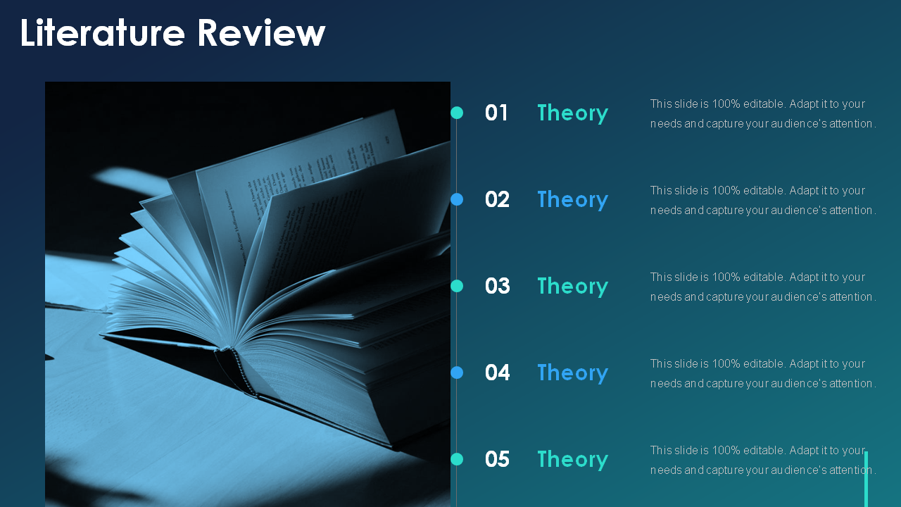
Template 1: Literature Review PPT Template
This literature review design is a perfect tool for any student looking to present a summary and critique of knowledge on their research statement. Using this layout, you can discuss theoretical and methodological contributions in the related field. You can also talk about past works, books, study materials, etc. The given PPT design is concise, easy to use, and will help develop a strong framework for problem-solving. Download it today.

Download this template
Template 2: Literature Review PowerPoint Slide
Looking to synthesize your latest findings and present them in a persuasive manner? Our literature review theme will help you narrow relevant information and design a framework for rational investigation. The given PPT design will enable you to present your ideas concisely. From summary details to strengths and shortcomings, this template covers it all. Grab it now.

Template 3: Literature Review Template
Craft a literature review that is both informative and persuasive with this amazing PPT slide. This predesigned layout will help you in presenting the summary of information in an engaging manner. Our themes are specifically designed to aid you in demonstrating your critical thinking and objective evaluation. So don't wait any longer – download our literature review template today.
Template 4: Comprehensive Literature Review PPT Slide
Download this tried-and-true literature review template to present a descriptive summary of your research topic statement. The given PPT layout is replete with relevant content to help you strike a balance between supporting and opposing aspects of an argument. This predesigned slide covers components such as strengths, defects, and methodology. It will assist you in cutting the clutter and focus on what's important. Grab it today.

Template 5: Literature Review for Research Project Proposal PPT
Writing a literature review can be overwhelming and time-consuming, but our project proposal PPT slides make the process much easier. This exclusive graphic will help you gather all the information you need by depicting strengths and weaknesses. It will also assist you in identifying and analyzing the most important aspects of your knowledge sources. With our helpful design, writing a literature review is easy and done. Download it now.
Template 6: Literature Review for Research Project Proposal Template
Present a comprehensive and cohesive overview of the information related to your topic with this stunning PPT slide. The given layout will enable you to put forward the facts and logic to develop a new hypothesis for testing. With this high-quality design, you can enumerate different books and study materials taken into consideration. You can also analyze and emphasize the technique opted for inquiry. Get this literature review PowerPoint presentation template now.

Template 7: Literature Review for Research Paper Proposal PowerPoint Slide
Lay a strong foundation for your research topic with this impressive PowerPoint presentation layout. It is easy to use and fully customizable. This design will help you describe the previous research done. Moreover, you can enlist the strengths and weaknesses of the study clearly. Therefore, grab it now.
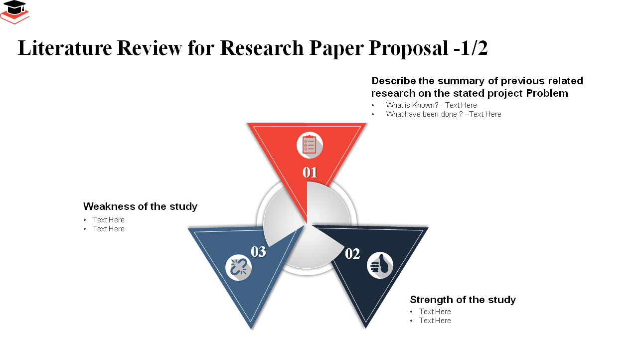
Template 8: Literature Review for Research Paper Proposal PPT
Download this high-quality PPT template and write a well-formatted literature review. The given layout is professionally designed and easy to follow. It will enable you to emphasize various elements, such as materials referred to, past work, the list of books, approach for analysis, and more. So why wait? Download this PowerPoint design immediately.

Template 9: Literature Review for Academic Student Research Proposal PPT
With this exclusive graphic, you'll have everything you need to create a well-structured and convincing literature review. The given design is well-suited for students and researchers who wish to mention reliable information sources, such as books and journals, and draw inferences from them. You can even focus on the strong points of your study, thereby making an impactful research statement. Therefore, grab this PPT slide today.

Template 10: Literature Review Overview for Research Process PPT
Demonstrate your analytical skills and understanding of the topic with this predesigned PowerPoint graphic. The given research overview PPT theme is perfect for explaining what has been done in the area of your topic of interest. Using this impressive design, you can provide an accurate comparison showcasing the connections between the different works being reviewed. Get it right away.
Creating an effective literature review requires discipline, study, and patience. Our collection of templates will assist you in presenting an extensive and cohesive summary of the relevant works. These PPT layouts are professionally designed, fully editable, and visually appealing. You can modify them and create perfect presentations according to your needs. So download them now!
P.S. Are you looking for a way to communicate your individual story? Save your time with these predesigned book report templates featured in this guide .
Download the free Literature Review Template PDF .
Related posts:
- How to Design the Perfect Service Launch Presentation [Custom Launch Deck Included]
- Quarterly Business Review Presentation: All the Essential Slides You Need in Your Deck
- [Updated 2023] How to Design The Perfect Product Launch Presentation [Best Templates Included]
- 99% of the Pitches Fail! Find Out What Makes Any Startup a Success
Liked this blog? Please recommend us

Top 11 Book Report Templates to Tell Your Inspirational Story [Free PDF Attached]
6 thoughts on “10 best literature review templates for scholars and researchers [free pdf attached]”.
This form is protected by reCAPTCHA - the Google Privacy Policy and Terms of Service apply.

Digital revolution powerpoint presentation slides

Sales funnel results presentation layouts
3d men joinning circular jigsaw puzzles ppt graphics icons

Business Strategic Planning Template For Organizations Powerpoint Presentation Slides

Future plan powerpoint template slide

Project Management Team Powerpoint Presentation Slides

Brand marketing powerpoint presentation slides


Launching a new service powerpoint presentation with slides go to market

Agenda powerpoint slide show
Four key metrics donut chart with percentage

Engineering and technology ppt inspiration example introduction continuous process improvement

Meet our team representing in circular format

- Search Menu
- Volume 2024, Issue 3, March 2024 (In Progress)
- Volume 2024, Issue 2, February 2024
- Bariatric Surgery
- Breast Surgery
- Cardiothoracic Surgery
- Colorectal Surgery
- Colorectal Surgery, Upper GI Surgery
- Gynaecology
- Hepatobiliary Surgery
- Interventional Radiology
- Neurosurgery
- Ophthalmology
- Oral and Maxillofacial Surgery
- Otorhinolaryngology - Head & Neck Surgery
- Paediatric Surgery
- Plastic Surgery
- Transplant Surgery
- Trauma & Orthopaedic Surgery
- Upper GI Surgery
- Vascular Surgery
- Author Guidelines
- Submission Site
- Open Access
- Reasons to Submit
- About Journal of Surgical Case Reports
- Editorial Board
- Advertising and Corporate Services
- Journals Career Network
- Self-Archiving Policy
- Journals on Oxford Academic
- Books on Oxford Academic

Article Contents
Introduction, case report, conflict of interest statement.
- < Previous
A young female patient with multiple unilateral low-grade oncocytic renal tumors and angiomyolipoma: a case report and literature review
- Article contents
- Figures & tables
- Supplementary Data
Yi Xu, Xuewen Jiang, Hui Meng, A young female patient with multiple unilateral low-grade oncocytic renal tumors and angiomyolipoma: a case report and literature review, Journal of Surgical Case Reports , Volume 2024, Issue 3, March 2024, rjae125, https://doi.org/10.1093/jscr/rjae125
- Permissions Icon Permissions
We identified a young female patient admitted for suspected renal malignancy. Partial nephrectomy was performed after imaging evaluation and discussion. Postoperative biopsy pathology reported multiple low-grade eosinophilic renal tumors (LOTs) with angiomyolipoma growth. After reviewing the data, we found that LOT was mostly solitary and occurred in middle-aged and elderly patients. This case is unique and we share it to improve the understanding of this disease.
There are many types of eosinophilic renal tumors, ranging from benign to malignant. Low-grade eosinophilic renal tumors (LOTs) have long been classified into the category of other eosinophilic tumors because of their morphological similarity to renal eosinophilic tumors (RO) and eosinophilic smoky cell carcinoma (eCRCC). To further understand their immunohistochemistry and genetics, LOTs were listed as a separate category for the first time in the 2022 classification by the World Health Organization (WHO). According to previous reports, LOTs usually occur in middle-aged and elderly patients and are primarily unilateral solitary tumors that do not grow with other tumors. We encountered a young female patient with multiple LOTs that grew simultaneously with renal angiomyolipoma (AML). We believe that the LOTs in this patient are unique, and report it here with the hope of improving our understanding of this tumor.
A 29-year-old female was found to have abnormal echoes in her left kidney during a physical examination. The patient had no symptoms (frequent urination, urgency, dysuria, hematuria, low back pain, abdominal pain, fever, or fatigue). She had no history of chronic diseases such as hypertension and diabetes and was married with children. There was no history of food or drug allergies, smoking, alcohol consumption, or family history of hereditary diseases.
No obvious positive signs were found on general physical examination upon admission, and no signs of tuberous sclerosis were observed. No lumbar mass, percussion pain in the lumbar region, pressure pain in the ureteral tract, or abnormalities in the vulva were detected during specialized examination.
To further clarify the diagnosis, an intensive computed tomography (CT) examination of the abdominopelvic region was performed, revealing a soft tissue mass and renal tumor in the left kidney, multiple small cysts in both kidneys, and multiple high-density foci in the pelvis, lumbosacral vertebral body, and adnexa ( Fig. 1 ).

CT shows uneven density and disordered structure in the upper pole of the left kidney, with turbidity and increased density in the surrounding fat spaces.
Following a comprehensive discussion under the multidisciplinary team model, a decision was made to proceed with surgical excision of the tumor. The preoperative examination revealed no contraindications to surgery. The procedure used involved da Vinci robot-assisted laparoscopic partial left nephrectomy. Intraoperatively, a tumor measuring ~3 × 4 cm was identified at the upper pole of the left kidney, and additional satellite tumors ~0.8 cm in size were detected near the primary lesion. All tumors were excised and sent for pathological examination. Rapid intraoperative pathological analysis indicated an eosinophilic cell tumor, prompting a localized excision. The surgery progressed smoothly, and postoperative recovery was satisfactory. The patient refused further genetic testing, and a 3-month post-discharge follow-up examination revealed no abnormalities in the surgical area. The patient is currently undergoing regular follow-up.
Four nodular tissue samples were examined postoperatively. Nodules 1 and 2 were identified as low-grade oncocytic renal tumors (LOTs). Microscopic examination revealed a distinct boundary between the tumor cells and normal renal tissue. The tumor cells exhibited a sheet-like arrangement with an eosinophilic cytoplasm ( Fig. 2A ); the nuclei were round or oval, with visible nucleoli. Some regions showed inconspicuous perinuclear halos ( Fig. 2B ). Immunohistochemical staining indicated positive expression of CK7 ( Fig. 2C ), partial PAX-8, and SDHB in the tumor cells, with no CD117 ( Fig. 2D ), RCC, CD10, or CA-IX expression. The Ki-67 proliferation index was ~1–2%.

(A) (HE,×4). Eosinophilic cell tumor with well-defined tumor boundaries and acidophilic tumor cells in solid tubular, nested, and microcystic arrangements; (B) (HE,×10). Eosinophilic cell tumor, homogeneous eosinophilic or eosinophilic granular cytoplasm with no vacuoles in the cytoplasm, round to oval nuclei with some perinuclear halos and sparse binuclei with slender perinuclear vacuoles; (C&D) (×10). Immunohistochemistry showed tumor cells positive for CK7 but negative for CD117.
Nodule 3 contained both LOT and AML tissue. Microscopically, a clear demarcation was observed between the LOT, AML, and normal renal tissues ( Fig. 3A ). Immunohistochemistry revealed positive staining for smooth muscle actin (SMA) ( Fig. 3B ) in the AML as well as Desmin, HMB45, and Melan-A (occasionally), while CD117 was negative. Nodule 4 consisted exclusively of AML tissue.

(A) (HE,×4). LOT, AML, and normal kidney tissue boundaries, where region a is AML, region b is normal kidney tissue, and region c is LOT; (B) (×10). Immunohistochemistry showing AML cells positive for SMA.
Renal tumors that are eosinophilic, or that have eosinophilic characteristics, range from benign to malignant and often occur sporadically; multiple tumors have also been reported [ 1 ]. In 2019, Trpkov et al. reported 28 cases of tumors with low nuclear grade and eosinophilic cytoplasm that were CD117-negative/CK7-positive, naming them LOTs [ 2 ]. This term was then adopted as a new classification entity in the 2022 edition of the WHO classification of urological and male genital tumors [ 3 ].
In current reports, LOTs are sporadic solitary tumors that are more common in the elderly population. The average age of patients with LOTs is 63 years and they are more commonly found in women. The median tumor size is 2–3 cm, and there are typically no complications [ 2 ]. Microscopically, the tumors have round-to-oval-shaped nuclei and focal perinuclear halos, as well as areas of edema with scattered or irregularly distributed cells. Growing evidence suggests that they have characteristic immunohistochemical profiles (diffusely positive for CK7 and negative for CD117/KIT) [ 4 ].
Other immunohistochemical indicators in LOTs in addition to CK7 and CD117 were also studied. Chen et al. found that GATA3, E-cadherin, Pax-8, succinate dehydrogenase B (SDHB), and fumarate hydratase (FH) were all positive in five cases of LOT, while vimentin, carbonic anhydrase 9 (CA9), CD10, P504s, CK20, TFE3, TFEB, HMB45, ALK, and forkhead box protein I1 (FOXI1) were negative [ 1 ]. Williamson et al. found that GATA3 was positive in a study of 16 LOT tumors [ 5 ]. The immunohistochemical results of the patient presented here partially support this view. The detection of GATA3, PAX-8, SDHB, Pax-8, CD10, and other immune indicators may be significant for accurate clinical and differential diagnoses.
As molecular techniques such as next-generation sequencing are increasingly being used to evaluate LOTs, many case series have reported new findings. Ricci et al. found that MTOR, TCS1, and TSC2 mutations in the mTOR pathway were closely related to the pathogenesis of this tumor. Other mutated genes, including PIK3CA, NF2, and PTEN, were also identified that can act as both upstream and downstream effectors [ 6 ]. Williamson et al. also confirms the above conclusion [ 5 ]. Mutations in the mTOR pathway may play an important role in LOT diagnosis; however, these gene changes are nonspecific and are also common in several other eosinophilic tumors such as eosinophilic solid and cystic renal cell carcinoma (ESC RCC), renal eosinophilic vacuolated tumors (EVT), and renal cell carcinoma with vascular leiomyoma matrix (RCC FMS) [ 7 ]. For instance, Xia et al. recently found that LOTs, ESC RCC, EVT, and a group of similar tumors that do not fully meet these criteria all show TSC/mTOR mutations and have different RNA clustering expression profiles [ 8 ].
For LOTs, surgical resection is sufficient to achieve a satisfactory prognosis; no progression or metastasis has been reported with local resection or unilateral total nephrectomy. The key to the clinical diagnosis and treatment of this type of disease should therefore be the former. The prehospital diagnosis of the patient in this case was suspected to be malignant. The nature of the tumor was determined by rapid pathological examination during operation, and partial resection was performed. Clinical diagnosis requires knowledge of the pathological features of LOTs, timely puncture biopsy, or intraoperative pathological examination to avoid misdiagnosis and unnecessary treatment.
At present, English literature reports on the close growth of LOTs and AML are rare. In this case, the patient was only 29 years old but had multiple large tumors, which is relatively rare among reported cases. The patient refused to undergo genetic testing; genetic detection may have revealed a relationship between the LOTs and AML. An analysis of the diagnosis and treatment of these patients can enhance our understanding of LOTs.
None declared.
Lerma LA , Schade GR , Tretiakova MS . Co-existence of ESC-RCC, EVT, and LOT as synchronous and metachronous tumors in six patients with multifocal neoplasia but without clinical features of tuberous sclerosis complex . Hum Pathol 2021 ; 116 : 1 – 11 .
Google Scholar
Trpkov K , Williamson SR , Gao Y , et al. Low-grade oncocytic tumour of kidney (CD117-negative, cytokeratin 7-positive): a distinct entity? Histopathology 2019 ; 75 : 174 – 84 .
Hes O , Michalová K , Pivovarčíková K . New insights in the new WHO classification of adult renal tumors . Cesk Patol 2022 ; 67 : 187 – 91 .
Trpkov K , Williamson SR , Gill AJ , et al. Novel, emerging and provisional renal entities: the Genitourinary Pathology Society (GUPS) update on renal neoplasia . Mod Pathol 2021 ; 34 : 1167 – 84 .
Williamson SR , Hes O , Trpkov K , et al. Low-grade oncocytic tumour of the kidney is characterised by genetic alterations of TSC1, TSC2, MTOR or PIK3CA and consistent GATA3 positivity . Histopathology 2023 ; 82 : 296 – 304 .
Ricci C , Ambrosi F , Franceschini T , et al. Evaluation of an institutional series of low-grade oncocytic tumor (LOT) of the kidney and review of the mutational landscape of LOT . Virchows Arch 2023 ; 483 : 687 – 98 .
Trpkov K , Yilmaz A , Uzer D , et al. Renal oncocytoma revisited: a clinicopathological study of 109 cases with emphasis on problematic diagnostic features . Histopathology 2010 ; 57 : 893 – 906 .
Xia Q-Y , Wang X-T , Zhao M , et al. TSC/MTOR-associated eosinophilic renal tumors exhibit a heterogeneous clinicopathologic spectrum : a targeted next-generation sequencing and gene expression profiling study . Am J Surg Pathol 2022 ; 46 : 1562 – 76 .
- angiomyolipoma
- kidney neoplasms
- proto-oncogene protein c-kit
- older adult
Email alerts
Citing articles via, affiliations.
- Online ISSN 2042-8812
- Copyright © 2024 Oxford University Press and JSCR Publishing Ltd
- About Oxford Academic
- Publish journals with us
- University press partners
- What we publish
- New features
- Open access
- Institutional account management
- Rights and permissions
- Get help with access
- Accessibility
- Advertising
- Media enquiries
- Oxford University Press
- Oxford Languages
- University of Oxford
Oxford University Press is a department of the University of Oxford. It furthers the University's objective of excellence in research, scholarship, and education by publishing worldwide
- Copyright © 2024 Oxford University Press
- Cookie settings
- Cookie policy
- Privacy policy
- Legal notice
This Feature Is Available To Subscribers Only
Sign In or Create an Account
This PDF is available to Subscribers Only
For full access to this pdf, sign in to an existing account, or purchase an annual subscription.
- Reference Manager
- Simple TEXT file
People also looked at
Original research article, the “arrhythmic” presentation of peripartum cardiomyopathy: case series and critical review of the literature.

- 1 Cardiac Electrophysiology and Clinical Arrhythmology Department, IRCCS San Raffaele Scientific Institute, Milan, Italy
- 2 Disease Unit for Myocarditis and Arrhythmogenic Cardiomyopathies, IRCCS San Raffaele Scientific Institute, Milan, Italy
- 3 School of Medicine, Vita-Salute San Raffaele University, Milan, Italy
- 4 Department of Cardiac Electrophysiology and Clinical Arrhythmology, IRCCS Policlinico San Donato, Milan, Italy
- 5 Department of Cardiology, IRCCS Fondazione Don Carlo Gnocchi, Parma, Italy
- 6 Department of Obstetrics and Gynecology, IRCCS San Raffaele Scientific Institute, Milan, Italy
- 7 Interventional Cardiology Unit, IRCCS San Raffaele Scientific Institute, Milan, Italy
Peripartum Cardiomyopathy (PPCM) is a polymorphic myocardial disease occurring late during pregnancy or early after delivery. While reduced systolic function and heart failure (HF) symptoms have been widely described, there is still a lack of reports about the arrhythmic manifestations of the disease. Most importantly, a broad range of unidentified pre-existing conditions, which may be missed by general practitioners and gynecologists, must be considered in differential diagnosis. The issue is relevant since some arrhythmias are associated to sudden cardiac death occurring in young patients, and the overall risk does not cease during the early postpartum period. This is why multimodality diagnostic workup and multidisciplinary management are highly suggested for these patients. We reported a series of 16 patients diagnosed with PPCM following arrhythmic clinical presentation. Both inpatients and outpatients were identified retrospectively. We performed several tests to identify the arrhythmic phenomena, inflammation and fibrosis presence. Cardiomyopathies phenotypes were reclassified in compliance with the updated ESC guidelines recommendations. Arrhythmias were documented in all the patients during the first cardiological assessment. PVC were the most common recorder arrhythmias, followed by VF, NSVT, AF, CSD.
Introduction
Peripartum cardiomyopathy (PPCM) is a rare myocardial disease occurring during late pregnancy or early postpartum period ( 1 ). Because of the frequent finding of reduced systolic dysfunction and heart failure (HF), PPCM is currently classified as a variant of dilated cardiomyopathy (DCM) ( 2 ). Consistently, the European Society of Cardiology (ESC) ( 3 , 4 ) has adopted the revised version of the very earliest proposed diagnostic criteria ( 5 , 6 ), namely: (1) development of HF from one month before delivery to the following five months—a narrow timeframe, which has been subsequently extended ( 1 ); (2) absence of an evident alternative cause other than pregnancy; (3) absence of known heart diseases diagnosed before the pregnancy; (4) LV ejection fraction (LVEF) < 45%, as defined by transthoracic echocardiogram.
While the DCM phenotype and associated mechanical manifestations are widely characterized in PPCM, to date the arrhythmic presentation of the disease is still under-investigated. In fact, although a broad range of tachy- and brady-arrhythmias have been described in patients with PPCM ( 1 ), most of the current knowledge relies on case reports and small-sized studies. The aim of the current review is: (1) to describe a series of patients evaluated for clinically-suspected PPCM following arrhythmic presentation; (2) to summarize the status of the art about the arrhythmic manifestations of PPCM.
Case series
We present a series of n = 16 consecutive patients evaluated for clinically-suspected PPCM at two centers specialized in arrhythmia management. Both inpatients and outpatients were identified retrospectively, based on the following screening criteria: 1) female sex in childbearing age (15 to 45 years); 2) first clinical presentation with arrhythmias (including bradyarrhythmias and either supraventricular or ventricular tachyarrhythmias), as documented either during pregnancy or in the 6 months after delivery; and: 3) lack of known cardiological history beforhead. In addition, in keeping with the local standard of care, multimodal diagnostic workup and multidisciplinary management were applied, respectively, to clarify the underlying diagnosis and enable patient-tailored treatment choices. In detail, on top of laboratory exams, transthoracic echocardiogram, 12-lead ECG and inhospital telemonitoring/outpatient Holter ECG monitoring, advanced diagnostic workup included one or more of the following exams: cardiac magnetic resonance (CMR) with late gadolinium enhancement (LGE) and additional sequences to investigate structural diseases (T2-weighted sequences, fat-sat sequences, parametric mapping whenever applicable); genetic test by next-generation sequencing to screen for cardiomyopathic gene variants (CGVs); histology exams, including hematoxylin-eosin and trichrome assays to detect myocardial inflammation and fibrosis, as well as immunohistochemistry analysis to further characterize the inflammatory infiltrates; 18-F fluorodeoxyglucose positron emission tomography (FDG-PET) scan, to screen for cardiac sarcoidosis in suspected cases; and electroanatomical map (EAM), to characterize the arrhythmogenic substrates in patients with clinical indication to catheter ablation. Cardiomyopathic phenotypes were reclassified in compliance with the updated (2023) ESC guideline recommendations ( 7 ).
But for the restrictions applied for pregnancy and lactation timeframes, all patients were offered optimal guideline-based medical therapy. Implantation of cardiac devices, as well as catheter ablation of arrhythmias, were in keeping with the current recommendations. At both centers, regular follow-up took place at dedicated outpatient settings for cardiomyopathy. The content of this report is fully compliant with the Declaration of Helsinki, and all patients signed informed consent to be enrolled in a research registry.
SPSS Version 20 (IBM Corp., Armonk, New York) was used for statistical analysis. Continuous variables were expressed as mean or median with standard deviation (SD) or range, depending on the distribution of data, as assessed by the Shapiro-Wilk's test. Categorical variables are reported as counts and percentages. Because of the small sample size and the absence of a prespecified study design, no statistical models were introduced for risk stratification, and no p -values were presented for comparison between groups.
Results: clinical presentation
The series includes 16 women (mean age 31 years, range 24–36; 88% Caucasian), of whom 14 (88%) presented with symptoms, and were managed as inpatients. In detail, their clinical presentation was: cardiocirculatory arrest ( n = 1), syncope ( n = 1), palpitation ( n = 5), dyspnea ( n = 4), asthenia ( n = 2), and chest pain ( n = 1). The first clinical manifestation occurred during the third trimester of pregnancy in n = 3 cases (19%), and after delivery (median 4, range 1–6 months) in the remaining 13 (81%).
The key clinical features of the case series are show in Table 1 . Obstetric history was unremarkable, except for two cases of twin pregnancy (13%, including one case occurring following in-vitro fertilization). No other patients had infertility or history of radiation exposure. The cardiovascular risk profile of the sample was generally low: in particular, there were no diabetic patients, and hypertension with criteria for preeclampsia was found in one single case (6%). Also, only two patients (13%) reported family history of sudden cardiac death (SCD) or cardiomyopathy.
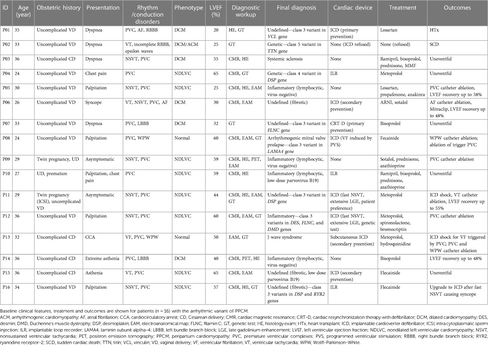
Table 1 . Key clinical features of the case series ( n = 16).
Arrhythmias were documented in all patients at the time of first cardiological assessment after clinical presentation. In detail, premature ventricular complexes (PVC) were the most commonly recorded arrhythmia (median daily burden 1,128, range 322–21,960; short-coupled in two cases only), and showed dominant right bundle branch block morphology suggesting LV origin in 11/16; cases (69%). Other arrhythmias included ventricular fibrillation (VF) causing out-of-hospital cardiocirculatory arrest ( n = 1), sustained ventricular tachycardia (VT; n = 2), nonsustained ventricular tachycardias (NSVT; n = 7), atrial fibrillation (AF; n = 1), Wolff-Parkinson-White syndrome (WPW; n = 2, incidental diagnosis), and conduction system disorders (CSD; n = 4). By the end of the baseline workup, most patients (88%) had more than one arrhythmia type documented.
Results: diagnostic workup and clinical management
At presentation, the mean LVEF was 47% (range 20%–65%), and phenotype was consistent with DCM in 6 patients (38%), non-dilated LV cardiomyopathy (NDLVC) in 8 (50%), and no criteria for structural disease in n = 2 (13%). Multimodality diagnostic workup included CMR ( n = 13; 81%), genetic test ( n = 9; 56%), histology ( n = 8; 50%), FDG-PET scan ( n = 2; 13%), and EAM ( n = 8; 50%). Overall, a mean of 2.5 exams per patient on top of baseline echocardiogram were required to identify the final diagnosis, which was: defined genetic cardiomyopathy ( n = 2), myocarditis ( n = 5), systemic sclerosis ( n = 1), arrhythmogenic mitral valve prolapse ( n = 1), and J-wave syndrome ( n = 1). Representative examples of the diagnostic workup are shown in Figure 1 . The median time from clinical onset of final diagnosis was 18 (range 9–42) months, with no patients being diagnosed during pregnancy.
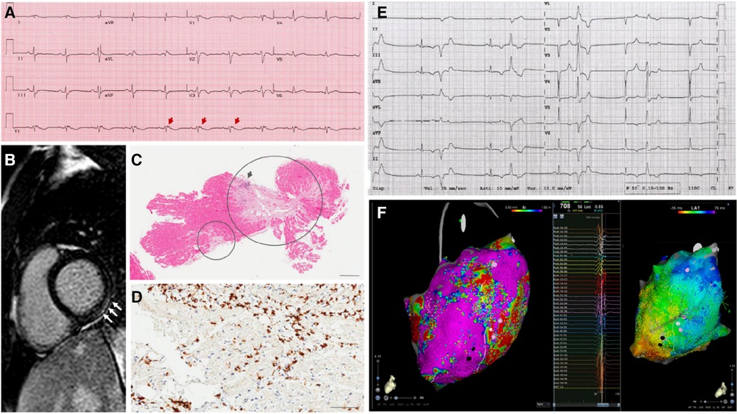
Figure 1 . Representative examples of the diagnostic workup in the case series. The main results of diagnostic findings are shown. ( A ) 12-lead electrocardiogram in a patient (P02) with signs of arrhthmogenic cardiomyopathy, including negative T-waves in anterior precordial leads, and epsilon waves (arrows). ( B ) Strial of late gadolinium enhancement involving the basal segments of the inferolateral left ventricular wall (arrows), in a patient (P15) with nondilated phenotype. ( C ) Endomyocardial biopsy findings in a patient (03) with subsequent diagnosis of systemic sclerosis. Extensive areas of replacement fibrosis are shown (circles) on hematoxylin-eosin assay. ( D ) Immunohistochemical analysis on endomyocardial biopsy show >7/mm 2 CD3-positive T-lymphocytes, meeting the diagnostic criteria for active-phase myocarditis in a patient (P09) with arrhythmic presentation. ( E ) 12-lead recording of polymorphic premature ventricular complexes in a patient (P04) with underlying mitral valve prolapse an nonischemic scar in the left ventricle. ( F ) High-density electroanatomical maps of the left ventricular epicardium (CARTO system, Biosense Webster; Octaray multielectrode catheter), including low-voltage areas (voltage map—on the left) and late potentials (activation map during sinus rhythm—on the right) involving the inferolateral wall, in a patient (P11) undergoing catheter ablation of a drug-refractory ventricular tachycardia.
On top of standard medical treatment, including betablockers and renin-angiontensin-aldosterone-inhibitors in the postpartum period, antiarrhythmic agents were used in 6 patients (38%). Out of 7 women choosing breastfeeding, 4 had medical treatment temporarily interrupted during lactation. One patient (6%) received bromocriptine, and 4 (25%) underwent immunosuppressive therapy to target myocardial inflammation. Before discharge, implantable devices were placed in 11 patients (69%), including cardioverter defibrillators (ICD; n = 6, of whom 1 subcutaneous) and loop recorders in (ILR; n = 5). As per local standard practice, no patients received wearable cardioverter defibrillators (WCD). Because of uncontrolled psychiatric comorbidity, one patient refused any kind of therapy, including ICD implant.
Results: outcomes
All patients had uncomplicated pregnancy, including n = 1 preterm (6%) and n = 4 caesarean deliveries (25%). No health issues were reported in children. By a median follow-up of 7 (range 2–34) years, n = 4 patients (25%) experienced major adverse outcomes including SCD from cardiocirculatory arrest ( n = 1), appropriate ICD shocks ( n = 2), and end-stage heart failure requiring heart transplantation ( n = 1). In addition, 6 patients (38%) requires catheter ablation of arrhythmias (PVC, n = 4; VT, n = 1; AF, n = 1, WPW, n = 2). Follow-up was uneventful in the remaining patients, and was remarkable for left ventricular reverse remodelling (LVRR) with improvement in LVEF in four of the six cases with DCM (67%). Three patients (19%) subsequently underwent new uncomplicated pregnancy and delivery, without LVEF decrease. Four patients underwent exercise stress test late after delivery without complications.
The relationships between baseline features and outcomes are summarized in Table 2 . The variales showing better association with the occurrence of major adverse outcomes included clinical presentation with sustained VT or VF, and presence of notable ECG abnormalities as epsilon- and J-waves (incidence of major adverse outcomes: 2/2 vs. 2/14, p = 0.05, for both variables). To be noted that the adverse outcomes (VF for P08, ICD shock for P13, see Table 1 ) were not related to WPW, which was previously treated via catheter ablation. A weaker association ( p < 0.30) was found with LVEF < 50%, CSD, supraventricular arrhythmias, and abnormal T -waves ( Table 2 ). For other clinically-relevant variables, such as genotypes and LGE, any reliable association analyses were prevented by the very small sample size. It should be highlighted that no adverse events occurred in patients receiving either immunomodulatory or prolactin-inhibitory therapy (0/5 vs. 4/11, p = 0.24).
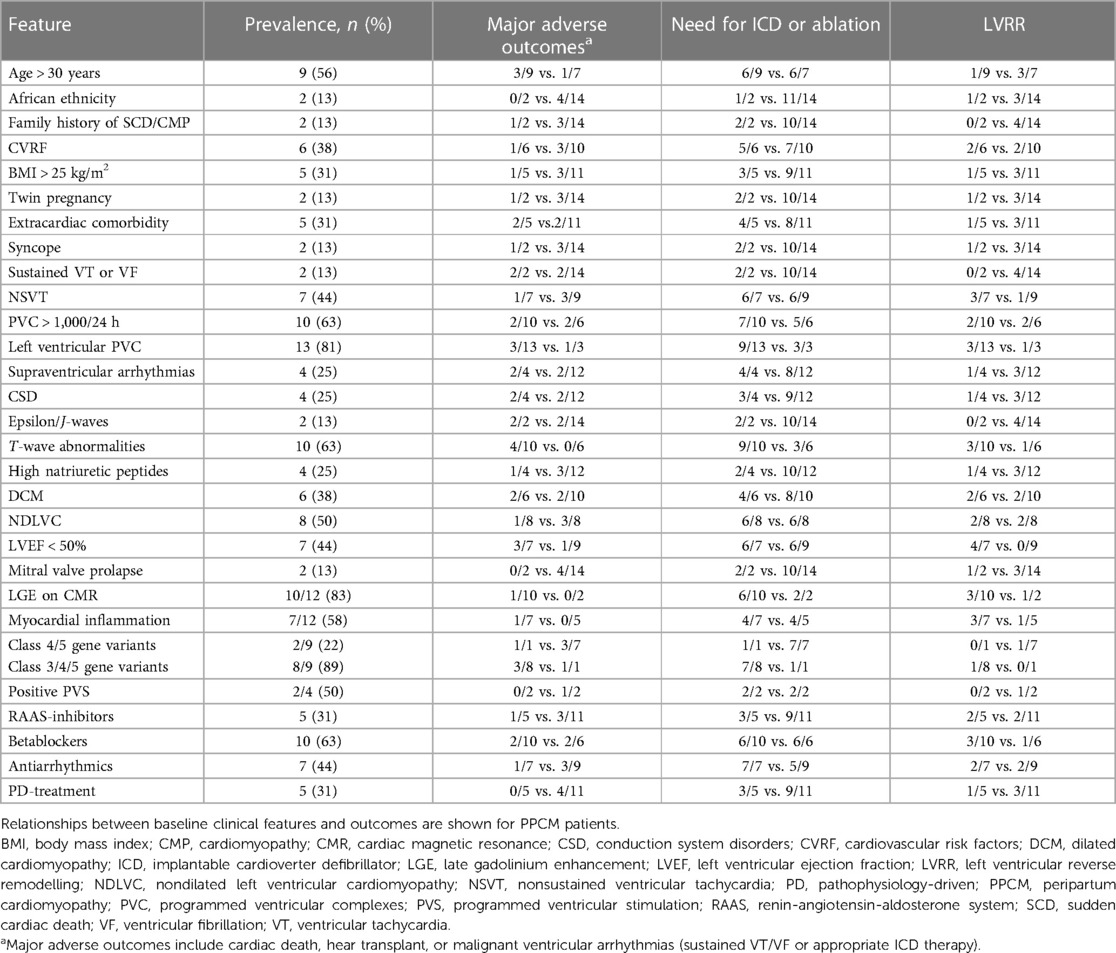
Table 2 . Relationships between clinical features and outcomes.
Critical review of the literature
Epidemiology.
The global incidence of PPCM is 1 in 1,000 worldwide, with peak values in northern Nigeria (1:100) and Haiti (1:300) ( 8 ). Recognized risk factors for PPCM include African American ethnicity, maternal age over 30 years, chronic hypertension, pregnancy-associated-hypertensive conditions as preeclampsia, anemia, and prolonged use of beta-agonist tocolytics during threatened preterm labor ( 2 , 8 , 9 ).
Our report was notable for including women with no prior cardiological history, the majority of whom being Caucasian (88%). In addition, we hereby provided extensive characterization of patients with arrhythmias recorded during baseline workup, either at clinical presentation or immediately after. Remarkably, DCM phenotype accounted for <50% of our cohort, so that an “arrhythmic variant” of PPCM was hereby described. In the largest study on a population of 9,841 patients with classically-defined PPCM, the overall prevalence of arrhythmias was 19% ( 10 ). Among them, ventricular arrhythmias were the most common ones (4% for VT, 1% for VF), followed by supraventricular arrhythmias (1.3% for AF, 0.5% each for atrial flutter and atrial tachycardia, 0.3% for paroxysmal reentry tachycardia including WPW) and 2.5% of CSD mainly including left bundle branch blocks ( 10 , 11 ). No conflicting data emerged from our series, except for PVC, which was the most common arrhythmia in our experience (15/16) in contrast with the 0.1% prevalence reported so far for both atrial and ventricular ectopies ( 10 , 11 ). In this setting, we attempt to bridge a knowledge gap ( 10 , 11 ), by providing data about daily burden (widely variable in range 322–21,960), and morphology (mainly right bundle branch block, suggesting LV origin, as expected in PPCM).
Pathophysiology
The pathophysiology of arrhythmias in PPCM reflects the multifactorial nature of the disease, whose dominant mechanisms are summarized in Table 3 . Briefly, hemodynamic changes, autonomic dysregulation, electrolyte imbalances, systemic inflammation, metabolic and hormonal effects have been described, either as a substrate or triggering events for HF and arrhythmias related to PPCM ( 2 , 14 , 15 ).
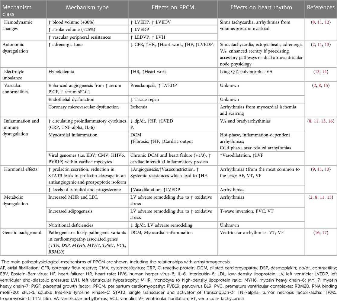
Table 3 . Pathophysiological mechanisms of PPCM.
As an alternative to the multisystemic dysregulation hypothesis, it has been suggested that latent preexisting myocardial diseases, including but not limited to myocarditis and primary cardiomyopathies, may retain a primary role in the disease pathophysiology ( 2 , 12 ). In this setting, the current definition of PPCM ( 2 , 3 ) is challenging, since a preexisting undiagnosed disease may be simply unmasked during pregnancy or after delivery. In a study ( 9 ), almost one third of PPCM patients showed biopsy-proven cardiotropic viral genomes, suggesting that DCM and HF may occur as late manifestations of chronic myocarditis. An increased risk of preeclampsia has been reported also in association with COVID-19 infection ( 18 ). Autoimmune virus-negative myocarditis has also been described as a driver mechanism in PPCM ( 2 ), also because of the microchimerism from fetus-derived cells during the immune-suppressed pregnant state ( 19 ). As known, myocarditis may account for a broad range of arrhythmias, complicating both the inflammatory and the postinflammatory phases of the disease, even in the patients with preserved LVEF ( 20 , 21 ). In our series, myocarditis was detected either by CMR or EMB in 7 of 16 patients (44%). As the only viral genome found in the myocardium, parvovirus B19 (load < 500 copies/mcg) was infrequently found ( Table 1 ). While prior studies failed in demonstrating higher rates of EMB-proven myocarditis among PPCM cases ( 22 , 23 ), the role of myocardial inflammation as an arrhythmogenic substrate is still to be investigated.
In turn, the genetic basis of PPCM has been recently revealed ( 16 , 24 ). Historically, PPCM has been differentiated from primary DCM because of its idiopathic, non-familial, non-genetic substrate ( 3 , 4 , 9 ). For instance, distinct cellular pathways downstream prolactin have been described as cardiomyopathic susbstrate specific of PPCM ( 25 ). However, in a recent study on 172 women with PPCM ( 17 ), truncating variants in genes predisposing to DCM were identified in 26 cases (15%). In this setting, volume overload and other systemic changes associated with pregnancy, may act as accelerating factors in sensitive genotypes. The main reports involved genes encoding titin ( TTN ), desmoplakin ( DSP ), alpha myosin heavy chain protein ( MYH6 ), tropomyosin ( TPM1 ), vinculin ( VCL ) and lamin A/C ( LMNA ), which constitute key structural and functional components for the cytoskeleton organization ( 7 , 17 , 26 ). Consistently, we detected CGVs in 8 of the 9 gentoyped patients (89%). While two patients only (13%) carried CGVs with a compelling pathogenic role (class 4/5), the hemodynamic changes associated with physiological pregnancy may have unmasked a concealed cardiomyopathic substrate even in the remaining subjects. Similar effects have been described for women carrying TTN truncating variants, where pregnancy has been described as a “second hit” for the classic PPCM presentation ( 17 ). In this setting, the presence and type of arrhythmias may strongly depend on the genotype. For instance, cytoskeletal genes may predispose to maladaptive evolution towards DCM, whereas desmosomal genes towards ventricular arrhythmias and myocardial inflammation ( 17 , 27 ). In turn, mutations in the LMNA gene may account for both brady- and tachyarrhythmias, much earlier than overt LV systolic dysfunction occurs ( 28 ). Preliminary evidence suggests that life-threatening arrhythmias in the peripartum may be associated even with Brugada syndrome or long QT syndrome ( 29 – 31 ). Dedicated studies are needed to add confirmatory evidence in this setting.
Multimodality diagnostic workup
In compliance with the current standards, PPCM should be suspected every time signs or symptoms of cardiac disease are found for the first time in a pregnant woman ( 2 , 3 ). In the “classic” DCM phenotype, PPCM may be easily detected by routine transthoracic echocardiogram ( 8 ). In particular, a number of parameters may differentiate pregnancy-associated physiological findings from maladaptive PPCM changes by ultrasounds ( 32 – 35 ). However, diagnosis may be more challenging following clinical onset of arrhythmias: as noted above, many of the patients included in our report (63%) had either NDLVC or normal phenotype. While the finding of LVEF < 45% was uncommon, and the diagnosis of the classic variant of PPCM was subsequently not met, all patients in our series had documented arrhythmias with or without signs of associated muscle disease ( Table 1 ). To be noted, only two patients in our series (13%) had relatives known for SCD or cardiomyopathy, in line with the 15% prevalence reported in a published German registry ( 36 ). While a number of arrhythmogenic conditions, such as WPW, were likely preexisting and simply unmasked by pregnancy, diagnostic assessment was challenging for most women. As a uniform tract, arrhythmias were documented, and diagnosed for the first time either during pregnancy or by 6 months after delivery, so that an “arrhythmic” variant of PPCM is hereby proposed. Importantly, arrhythmic manifestations occurred irrespectively of LVEF values ( Table 1 ). In this context, the overlap with arrhythmogenic cardiomyopathy, channelopathies and inflammatory heart diseases is more demanding as compared with DCM. As currently suggested for many arrhythmogenic cardiomyopathies ( 7 , 37 ), even in our experience a multimodality diagnostic approach was useful in characterizing the disease. Table 4 summarizes the spectrum of diagnostic techniques available to detect cardiomyopathic substrates in PPCM.
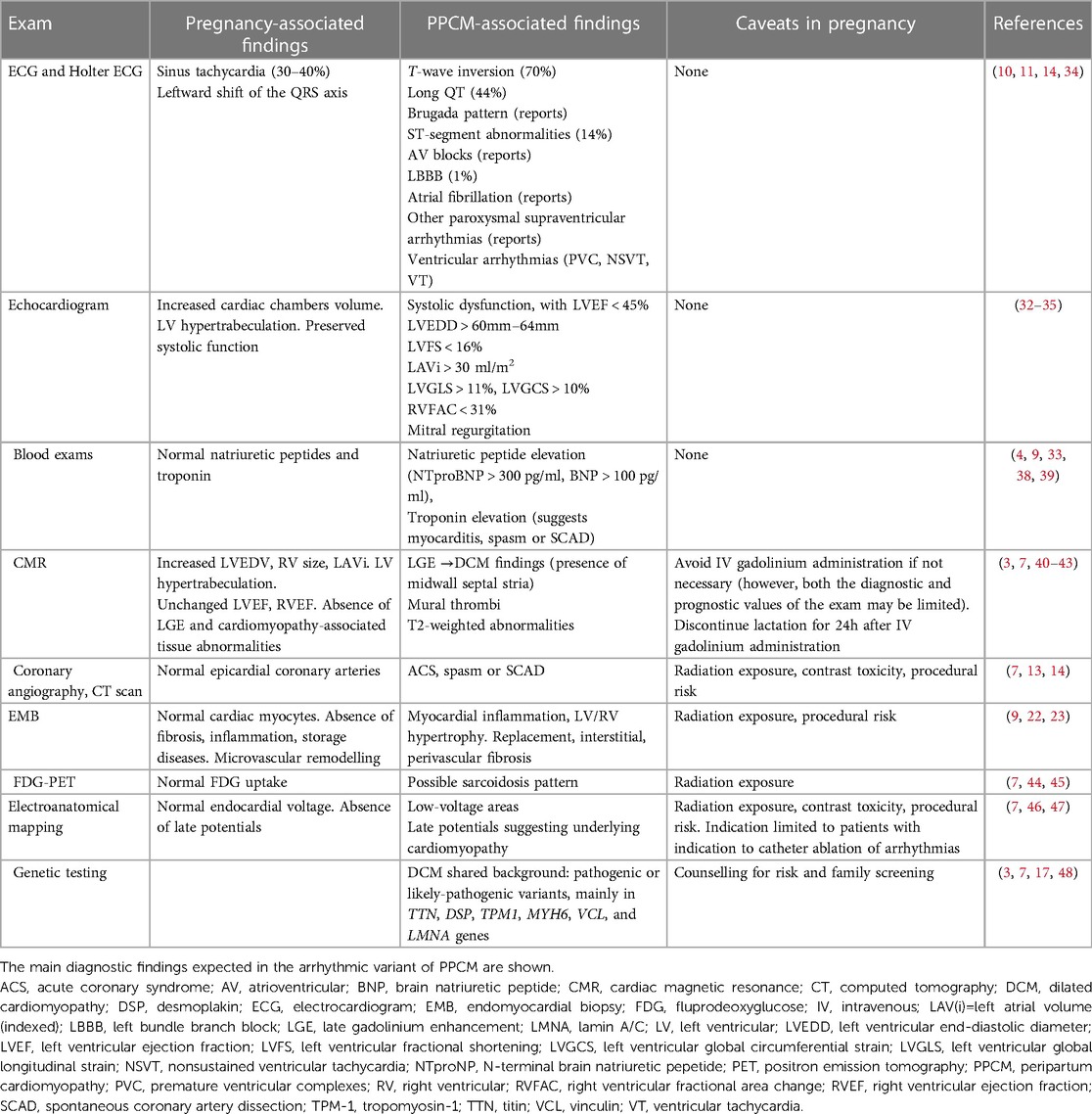
Table 4 . Diagnostic workup and findings in pregnancy and PPCM.
In classic PPCM, sinus rhythm ECG may reveal signs suggestive for PPCM, like T -wave inversion in up to 70% of patients ( 14 , 49 ). Cardiac biomarkers, such as natriuretic peptides BNP and NT-proBNP, are frequently elevated ( 9 , 38 , 39 ). Beyond hypokynesis, echocardiogram may show extensive remodeling of cardiac chambers and diastolic dysfunction ( 3 , 34 , 50 , 51 ). Not infrequently, LV hypertrabeculation exceeding the degree expected during pregnancy is observed ( 8 , 52 ). While functional mitral valve regurgitation in classic PPCM may occur secondarily to LV dilation, thickened leaflets and specific signs should call for mitral valve prolapse as an alternative source of arrhythmias ( 53 ).
Among second-level imaging techniques, CMR is currently considered as the gold standard in cardiomyopathies ( 7 ), and it is proven safe in pregnancy ( 3 ). Although no specific diagnostic criteria for PPCM have been described at CMR, most patients with classic PPCM phenotype had no evidence of LGE ( 40 , 41 ). As opposed, we documented nonischemic LGE in almost all women with the arrhythmic variant of PPCM ( Table 2 ). In this setting, distinct patterns of LGE may also point to specific diagnoses, such as primary DCM in the presence of midwall septal stria ( 7 ), myocarditis in association with subepicardial involvement of the inferolateral wall ( 42 ), and distinct variants of NDLVC in the presence of a ring-like appearance ( 7 , 43 ). In addition, abnormalities on T2-weighted sequences enforce the suspicion of myocardial inflammation ( 54 ), which frequently deserves confirmation and further etiological characterization by EMB, as recommended in patients with myocarditis ( 42 ). Histology may also reveal tissue remodeling, fibrosis, and associated viral genomes ( 9 , 22 , 23 ). As an alternative to histology, FDG-PET may be particularly useful whenever cardiac sarcoidosis is clinically suspected, or implantable device-related artifacts prevent the interpretation of CMR ( 7 , 44 – 55 ). Finally, in patients with clinical indication to catheter ablation, EAM may help identifying low-voltage areas or electrogram abnormalities suggestive for a cardiomyopathic substrate ( 46 , 47 ). Whenever familial disease is suspected, or upstream workup suggests signs of a genetic disease, wide-spectrum genetic test should be strongly considered ( 3 , 7 , 48 ). Even in the absence of macroscopic substrate abnormalities, long QT syndrome, Brugada syndrome, catecholamine-related VT syndromes may account for concealed arrhythmogenic substrates ( 56 , 57 ). In such heterogeneous scenarios, genotyping may help in reaching a definite diagnosis. In partial agreement with the published literature ( 16 , 17 ), 50% of patients in our report had CGVs detected by genetic test. Nonetheless, because of the frequent finding of variants of unknown significance, diagnosing a genetically-proven cardiomyopathy was challenging in the majority of cases.
It is worth noting that, in our series, the average number of the above-mentioned second level exams was 2.5 per patient, thus allowing to reach the diagnosis by a median follow-up of 18 months from clinical onset, i.e., late after delivery. Our data indicate that diagnostic characterization for clinically-suspected PPCM may be complex and lengthy, and relies on multimodality workup.
One last critical point concerns the detection of arrhythmic episodes. Since arrhythmias may arise suddenly, discontinuous monitoring by means of repeated Holter ECG of 24 or 48h registration, may result in significant underdetection of rhythm disorders ( 58 ). In our series, most patients had arrhythmias detected because of inhospital setting and continuous telemonitoring. For instance, NSVT episodes were detected in up to 7 of 16 patients (44%), in contrast to the 21% detected by Holter ECG in a published series ( 58 ). Given that continuous electrical monitoring techniques had demonstrated superiority to discontinuous monitoring in similar clinical scenarios ( 59 ), ILR may find application in selected cases considered at lower risk of SCD and no indication to ICD ( 60 ). In our series, one patient carrying ILR subsequently underwent upgrade to ICD because of fast NSVT episodes missed by Holter ECG. The proposed diagnostic algorithm for arrhythmia detection in PPCM is shown in Figure 2 .
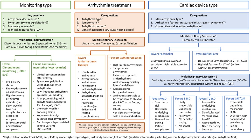
Figure 2 . Clinical scenarios in the arrhythmic variant of PPCM. The main clinical challenges for multidisciplinary heathcare teams to manage patients with clinically-suspected peripartum cardiomyopathy and either proven or suspected arrhythmias are shown. AF, atrial fibrillation; ATP, anti-tachycardia pacing; AV, atrioventricular; CCA, cardiocirculatory arrest; CMR, cardiac magnetic resonance; CRT/CSP, cariac resynchronization therapy/conduction systema pacing; ECG, electrocardiogram; ICD, implantable cardioverter defibrillator (S, suubcutaneous; TV, transvenous); LBBB, left bundle branch block; LGE, late gadolinium enhancement; LTVA, life-threatening ventricular arrhythmias; LVEF, left ventricular ejection fraction; MVP, mitral valve prolapse, NSVT, nonsustained ventricular tachycardia; PSVT, paroxysmal supraventricular tachycardia; SVA, supraventricular arrhythmias; VF, ventricular fibrillation; VT, ventricular tachycardia; WCD, wearable cardioverter defibrillator; WPW, Wolff-Parkinson-White syndrome.
Risk stratification
PPCM has an increasing incidence ( 8 ), and has been reported as the leading cause of maternal cardiovascular death ( 61 ), with mortality rates ranging from 1.3% inhospital, to 16% at 7 years ( 62 , 63 ). Overall, VA are the most threatening manifestations of classic PPCM, accounting for up to 1 out of 4 cases of SCDs ( 13 ). Mortality of PPCM-patients experiencing arrhythmia is 2.1%, three-fold higher than without arrhythmias ( 11 ).
Instead, bradyarrhythmias are reported benign and self-limited in most cases ( 10 ). In fact, unless accompanied by ventricular arrhythmias, underlying diseases with adverse prognostic significance such as sarcoidosis and LMNA cardiomyopathy are unlikely ( 60 ).
As for the mechanical manifestations of the disease, LV reverse remodeling and full recovery of LVEF have been described in many patients during the postpartum period, with LVEF normalization rates of up to 71% by 6 months after delivery ( 64 ). Also in our series, LVEF at presentation was associated with higher recovery rates ( Table 2 ), confirming the published data ( 8 ). In classic PPCM, additional prognostic factors for heart failure include LVEF below 45%, increased LV end-diastolic diameter, reduced LV strain parameters, right ventricular or biventricular dysfunction, and increased left atrial volume ( 32 , 33 , 65 , 66 ). Also, women whose LV ejection fraction failed to return within the normal range after their first episode of PPCM showed an increased risk of PPCM recurrence in a subsequent pregnancy ( 67 , 68 ). In this setting, NT-proBNP values ≥900 pg/ml were found as negative predictors of LV reverse remodeling ( 38 ), whereas a BNP value <100 pg/ml was found accurate in ruling out adverse events related to PPCM ( 39 ).
Even in the absence of an overt DCM phenotype, the identification of LGE on CMR, especially with a septal distribution pattern, can be predictive of both for SCD and end-stage heart failure ( 60 , 69 , 70 ). Advanced myocardial imaging may also identify mitral annular disjunction and additional prognostic signs for arrhythmogenic mitral valve prolapse ( 71 ). In this setting, the genetic test has a major impact on patient prognosis: in fact, in compliance with the updated guideline recommendations ( 7 ), the identification of “high risk” genotypes may significantly contribute to both the arrhythmic risk stratification and the clinician's decision of ICD implant. In our experience, while both medical treatment and ICD were refused by the patient, the only case of SCD occurred in a patient with overt DCM who harbored a pathogenic TTN truncating mutation (as reported in Table 1 ).
Remarkably, prognostic evaluation by disease-specific risk factors cannot be applied in the absence of a specific etiology ( 7 , 60 ). As far as no comprehensive risk score calculators become available for PPCM from large multicenter studies, a multimodal and patient-tailored arrhythmic risk stratification strategy is strongly advised.
Personalized treatment strategies
An evidence-based overview of the available treatment options to manage arrhythmias in PPCM is presented in Table 5 . The traditional RAAS-inhibitors, as well as angiotensin receptor-neprilysin inhibitors and mineralocorticoid receptor antagonists, are contraindicated during pregnancy and can be only used in the post-partum period ( 2 – 4 , 73 ), as occurred in our cases. Episodes of acute HF are managed by oxygen administration, fluid restriction, loop diuretics, nitrates and vasodilators as hydralazine ( 8 , 9 , 72 ). In severe cases, inotropes and mechanical circulatory support are needed ( 2 , 8 ). Anticoagulants can be administered according to the current recommendations in patients with LVEF < 30%–35% ( 76 , 77 ), in particular in the presence of risk factors for thromboembolic events, as in AF or LV hypertrabeculation ( 52 ).
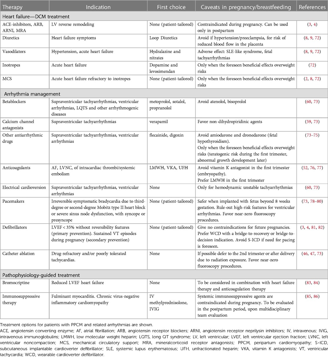
Table 5 . Treatment options for PPCM and related arrhythmias.
Among antiarrhythmic drugs, the use of amiodarone is restricted during pregnancy, since it can induce fetal hypothyroidism, growth retardation, and prematurity ( 73 ). Most betablockers, including sotalol, and central calcium-channel antagonists as verapamil, are well tolerated during pregnancy ( 60 , 73 ). Instead, digoxin or flecainide should be used when benefits overwhelm risks ( 73 – 75 ), such as in the event of fetal arrhythmias ( 87 ). For unstable arrhythmias, electrical cardioversion is a suitable and safe option during pregnancy, but the presence of an obstetrician in advisable in light of the risk of increased intrauterine activity ( 11 , 73 ). In our experience, betablocker and antiarrhythmic agents were employed in 63% and 44% of patients, respectively, without safety issues when used during pregnancy.
Among cardiac devices, pacemakers are indicated in case of severe bradycardia or CSD ( 78 – 80 ). Importantly, underlying arrhythmogenic diseases and/or risk factors for malignant ventricular arrhythmias should be carefully ruled out, to ensure that ICD are not needed instead. A guide for the clinical decision making is summarized in Figure 2 . Given the transitory nature of PPCM and associated arrhythmias, WCD constitute a reasonable approach to protect pregnant women from arrhythmias in the short term. Women with a severe systolic dysfunction, as in PPCM with a LVEF under 35%, are more likely to manifest SCD from malignant ventricular arrhythmias ( 2 , 8 ). Remarkably, the incidence of appropriate ICD shocks in PPCM was as high as 37% over a mean 3-year follow-up ( 88 ), i.e., at a significant longer term as compared to the postpartum period. Therefore, to avoid unnecessary ICD implant in primary prevention, WCD may be used for a few months with a bridge-to-recovery indication ( 2 , 81 , 82 ). Similar considerations are applied in the context of secondary prevention of SCD in patients presenting their first VT episode in pregnancy ( 4 , 60 ). In this setting, withdrawal of WCD may be more challenging since no temporal cutoffs are available to notify the end-of-risk timing. It should be noted that, reflecting the local standard practice, no patients in our series had WCD. Consistently, in a recent consensus document of the Heart Rhythm Society ( 73 ), it has been reported that the criteria for early ICD placement should be more stringent compared to other cardiac conditions. This particularly applies to patients presenting with LVEF below 30% in conjunction with a LV end-diastolic diameter equal to or exceeding 60 mm, because of the low likelihood of LVRR even in the long term ( 89 ). The ESC guidelines recommend that for women presenting with symptoms and severe LV dysfunction 6 months after initial presentation, despite optimal medical therapy and left bundle branch block-shaped QRS with duration greater than 120 ms, cardiac resynchronization therapy should be strongly considered, because of the reported beneficial effects in classic PPCM ( 90 ). While transvenous devices may be placed even during pregnancy in selected cases, delivery prior to device implantation is advised for most PPCM patients ( 11 ). In fact, although the current reaching the fetus is minimal, transient fetal arrhythmias after electrical resynchronization have been described ( 91 ). Efforts should be also made to minimize fetal radiation exposure by limiting fluoroscopy and using abdominal shielding. Successful ICD implantation using echocardiography without fluoroscopy is a desirable option ( 92 ). Of 16 patients, the clinical indication to ICD implant applied to up to 10 patients (64%) by the end of follow-up.
Catheter ablation is another key therapeutic weapon that applies to a range of arrhythmias in a number of clinical scenarios. In the acute setting, catheter ablation is reserved for women suffering from hemodynamically unstable arrhythmias ( 10 ), as well as for VT persisting in spite of antiarrhythmic therapy ( 46 , 47 ). In our series all patients received ablation late after clinical onset. As for cardiac device implant, also catheter ablation is preferred after delivery or when a pregnancy is planned in case arrhythmias have been already diagnosed ( 4 , 46 ). This particularly applies to non-life threatening arrhythmias such as AF, as well as for reentry circuits likely to be completely abolished by ablation, such as WPW ( 73 ). Before performing catheter ablation in a pregnant woman, risk and benefit of both mother and fetus must be considered, because consequences include fetal radiation exposure, maternal hemodynamic imbalance, impaired placental perfusion ( 33 ).
As a final remark, among pathophysiology-directed strategies, the inhibition of prolactin secretion by means of bromocriptine in addition to standard heart failure therapy has shown promising results in two clinical trials ( 83 , 84 ), but evidence is still contradictory ( 2 , 8 , 12 ), and no data are available about antiarrhythmic effects. Concerns have been raised also about drug-associated maternal adverse vascular events ( 93 ). For classic PPCM, the ESC included a weak recommendation (class II b, level of evidence B) for the use of bromocriptine ( 4 ). In our series on arrhythmic PPCM, only one patient (6%) received bromocriptine, but despite association with metoprolol she still required catheter ablation of PVC ( Table 1 ). In selected patients with PPCM secondary to myocarditis, intravenous immunoglobulin administration has shown an improvement in LVEF ( 85 ). While no role is currently recognized for other etiology-driven therapies, it should be noted that three patients with EMB-proven virus-negative lymphocytic myocarditis underwent safe immunosuppressive therapy ( 86 ) in the postpartum period. All of them had uneventful follow-up, except for the need of PVC ablation in a patient with residual monomorphic PVC: results are consistent with the pleiotropic beneficial effects of immunosuppression in arrhythmic myocarditis ( 94 ), but it should deserve dedicated investigation as pathophysiology-guided therapy in the PPCM population. Our experience showed that 6 patients were ablated in the postpartum (38%), including 50% (2 of 4) of those showing LVRR during follow-up ( Table 1 ).
Given the complex and multifactorial nature of the disease, multidisciplinary healthcare teams should become the gold-standard model of care, in compliance with the current recommendations applying to all cardiomyopathies ( 7 ). Figure 3 summarizes the model proposed based on the literature review and our own experience. On top of HF specialists, cardiac electrophysiologists have a critical role in decision making about management of arrhythmias and the prevention of SCD, both in the short and in the long term. Geneticists have a key contribution in defining clinical indications to genetic tests and enabling family screening. Gynecologists retain a key role, also for defining the mode and optimal timing of delivery. Other specialists may provide relevant contributions, such as immunologists and endocrinologists for the administration of pathophysiology-driven therapies. Also, since one patient in our series underwent arrhythmic SCD after refusing ICD and therapies, psychiatrists should assist in managing either preexisting or peripartum-associated mental comorbidities.
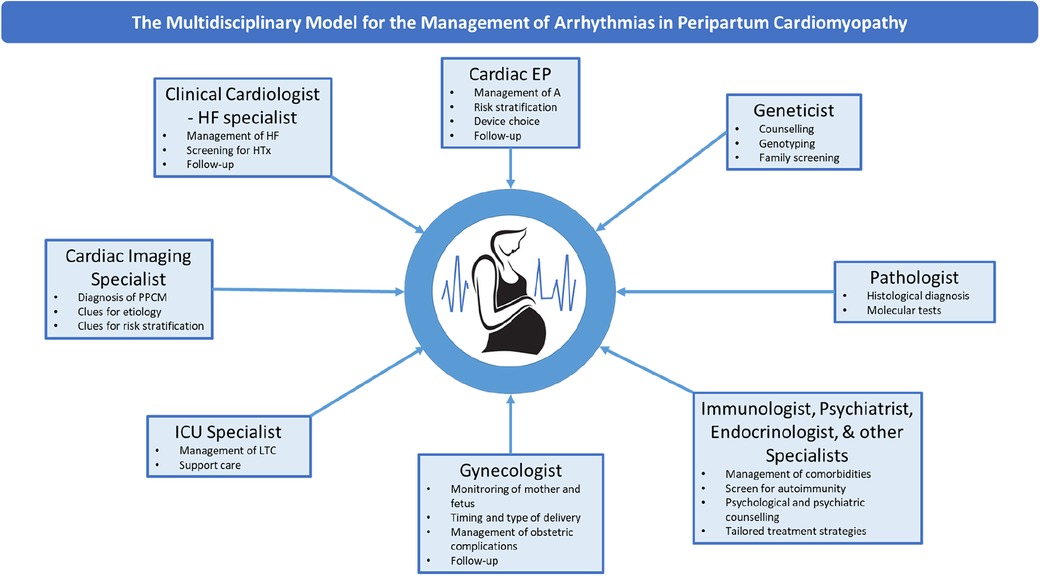
Figure 3 . The multidisciplinary model for the management of arrhythmias in PPCM. The main components of the multidisciplinary healthcare team for the management of PPCM and related arrhythmias are shown. EP, electrophysiologist; HF, heart failure; HTx, heart transplant; LTC, life-threatening conditions; PPCM, peripartum cardiomyopathy.
Conclusions
PPCM is a complex and multifactorial disease, whose arrhythmic manifestations are currently under-investigated. While treatment choices are strongly conditioned by the pregnancy status, an open-minded and patient-tailored diagnostic workup is strongly encouraged to allow optimal treatment options after differential diagnosis is solved.
Efforts are needed to describe and further characterize the “arrhythmic” variant of PPCM, which posed hard clinical challenges for SCD risk assessment, as compared to the classic DCM phenotype with heart failure manifestations. In fact, differentiating bystander vs. PPCM-triggered arrhythmias, as well as revealing a missed preexisting diagnosis are a major issue, as shown in our case series. In these settings, multimodality diagnostic workup and multidisciplinary care models should be promoted. Similarly, regular follow-up is required in the long term to clarify the underlying diagnosis and prevent complications.
Given the association between PPCM and arrhythmic phenomena, which can even result in SCD, efforts are needed to early identify the best candidates to undergo definitive implantation of ICD. Multicentre prospective studies on well selected populations of PPCM patients are advocated, to substantially advance our knowledge in such a hot topic of modern medicine.
Data availability statement
The raw data supporting the conclusions of this article will be made available by the authors, without undue reservation.
Ethics statement
The studies involving humans were approved by AINICM Protocol, IRCCS San Raffaele Hospital. The studies were conducted in accordance with the local legislation and institutional requirements. The participants provided their written informed consent to participate in this study. Written informed consent was obtained from the individual(s) for the publication of any potentially identifiable images or data included in this article.
Author contributions
GP: Writing – original draft, Conceptualization, Data curation, Formal Analysis. EM: Data curation, Formal Analysis, Investigation, Methodology, Writing – original draft. GC: Investigation, Methodology, Resources, Writing – review & editing. MM: Data curation, Investigation, Writing – original draft. ML: Data curation, Investigation, Writing – original draft. MC: Data curation, Investigation, Writing – original draft. AB: Data curation, Investigation, Writing – original draft. DL: Resources, Supervision, Visualization, Writing – review & editing. LA: Investigation, Methodology, Supervision, Validation, Visualization, Writing – review & editing. PC: Investigation, Methodology, Resources, Validation, Visualization, Writing – review & editing. AC: Methodology, Project administration, Resources, Supervision, Validation, Visualization, Writing – review & editing. PD: Project administration, Resources, Validation, Visualization, Writing – review & editing. CP: Project administration, Resources, Supervision, Validation, Visualization, Writing – review & editing.
The author(s) declare financial support was received for the research, authorship, and/or publication of this article.
This study was partially supported by Ricerca Corrente funding from Italian Ministry of Health to IRCCS Policlinico San Donato.
Acknowledgments
This work was performed during GP tenure as the Clinical Research Award in Honor of Mark Josephson and Hein Wellens Fellow of the Heart Rhythm Society.
Conflict of interest
The authors declare that the research was conducted in the absence of any commercial or financial relationships that could be construed as a potential conflict of interest.
The author(s) declared that they were an editorial board member of Frontiers, at the time of submission. This had no impact on the peer review process and the final decision.
Publisher's note
All claims expressed in this article are solely those of the authors and do not necessarily represent those of their affiliated organizations, or those of the publisher, the editors and the reviewers. Any product that may be evaluated in this article, or claim that may be made by its manufacturer, is not guaranteed or endorsed by the publisher.
1. Homans DC. Peripartum cardiomyopathy. N Engl J Med . (1985) 312(22):1432–7. doi: 10.1056/NEJM198505303122206
PubMed Abstract | Crossref Full Text | Google Scholar
2. Davis MB, Arany Z, McNamara DM, Goland S, Elkayam U. Peripartum cardiomyopathy: JACC state-of-the-art review. J Am Coll Cardiol . (2020) 75(2):207–21. doi: 10.1016/j.jacc.2019.11.014
3. Sliwa K, Hilfiker-Kleiner D, Petrie MC, Mebazaa A, Pieske B, Buchmann E. Current state of knowledge on aetiology, diagnosis, management, and therapy of peripartum cardiomyopathy: a position statement from the heart failure association of the European society of cardiology working group on peripartum cardiomyopathy. Eur J Heart Fail . (2010) 12(8):767–78. doi: 10.1093/eurjhf/hfq120
4. Regitz-Zagrosek V, Roos-Hesselink JW, Bauersachs J, Blomström-Lundqvist C, Cífková R, De Bonis M. 2018 ESC guidelines for the management of cardiovascular diseases during pregnancy. Eur Heart J . (2018) 39(34):3165–241. doi: 10.1093/eurheartj/ehy340
5. Demakis JG, Rahimtoola SH. Peripartum cardiomyopathy. Circulation . (1971) 44:964–8. doi: 10.1161/01.CIR.44.5.964
6. Pearson GD, Veille JC, Rahimtoola S, Hsia J, Oakley CM, Hosenpud JD, et al. Peripartum cardiomyopathy: national heart, lung, and blood institute and office of rare diseases (national institutes of health) workshop recommendations and review. JAMA . (2000) 283(9):1183–8. doi: 10.1001/jama.283.9.1183
7. Arbelo E, Protonotarios A, Gimeno JR, Arbustini E, Barriales-Villa R, Basso C. 2023 ESC guidelines for the management of cardiomyopathies. Eur Heart J . (2023) 44(37):3503–626. doi: 10.1093/eurheartj/ehad194
8. Arany Z, Elkayam U. Peripartum cardiomyopathy. Circulation . (2016) 133(14):1397–409. doi: 10.1161/CIRCULATIONAHA.115.020491
9. Shah T, Ather S, Bavishi C, Bambhroliya A, Ma T, Bozkurt B. Peripartum cardiomyopathy: a contemporary review. Methodist Debakey Cardiovasc J . (2013) 9(1):38–41. doi: 10.14797/mdcj-9-1-38
Crossref Full Text | Google Scholar
10. Lindley KJ, Judge N. Arrhythmias in pregnancy. Clin Obstet Gynecol . (2020) 63(4):878–92. doi: 10.1097/GRF.0000000000000567
11. Honigberg MC, Givertz MM. Arrhythmias in peripartum cardiomyopathy. Card Electrophysiol Clin . (2015) 7(2):309–17. doi: 10.1016/j.ccep.2015.03.010
12. Baris L, Cornette J, Johnson MR, Sliwa K, Roos-Hesselink JW. Peripartum cardiomyopathy: disease or syndrome? Heart . (2019) 105(5): 357–62. Published online February 12, 2019. doi: 10.1136/heartjnl-2018-314252
13. Hähnle L, Hähnle J, Sliwa K, Viljoe C. Detection and management of arrhythmias in peripartum cardiomyopathy. Cardiovasc Diagn Ther . (2020) 10(2):325–35. doi: 10.21037/cdt.2019.05.03
14. Dunker D, Pfeffer TJ, Bauersachs J, Veltmann C. ECG and arrhythmias in peripartum cardiomyopathy. Herzschr Elektrophys . (2021) 32:207–13. doi: 10.1007/s00399-021-00760-9
15. Mebazaa A, Séronde M, Gayat E, Tibazarwa K, Anumba D, Akrout N. Imbalanced angiogenesis in peripartum cardiomyopathy- diagnostic value of placenta growth factor. Circ J . (2017) 81(11):1654–61. doi: 10.1253/circj.CJ-16-1193
16. Spracklen TF, Chakafana G, Schwartz PJ, Kotta MC, Shaboodien G, Ntusi NAB. Genetics of peripartum cardiomyopathy: current knowledge, future directions and clinical implications. Genes (Basel) . (2021) 12(1):103. Published online January 15, 2021. doi: 10.3390/genes12010103
17. Ware JS, Li J, Mazaika E, Yasso CM, DeSouza T, Cappola TP. Shared genetic predisposition in peripartum and dilated cardiomyopathies. N Engl J Med . (2016) 374:233–41. doi: 10.1056/nejmoa1505517
18. Papageorghiou AT, Deruelle P, Gunier RB, Rauch S, García-May PK, Mhatre M. Preeclampsia and COVID-19: results from the INTERCOVID prospective longitudinal study. Am J Obstet Gynecol . (2021) 225(3):289.e1–e17. doi: 10.1016/j.ajog.2021.05.014
19. Kinder JM, Stelzer IA, Arck PC, Way SS. Immunological implications of pregnancy-induced microchimerism. Nat Rev Immunol . (2017) 17(8):483–94. doi: 10.1038/nri.2017.38
20. Peretto G, Sala S, Rizzo S, De Luca G, Campochiaro C, Sartorelli S. Arrhythmias in myocarditis: state of the art. Heart Rhythm . (2019) 16(5):793–801. doi: 10.1016/j.hrthm.2018.11.024
21. Peretto G, Sala S, Rizzo S, Palmisano A, Esposito A, De Cobelli F. Ventricular arrhythmias in myocarditis: characterization and relationships with myocardial inflammation. J Am Coll Cardiol . (2020) 75(9):1046–57. doi: 10.1016/j.jacc.2020.01.036
22. Bültmann BD, Klingel K, Näbauer M, Wallwiener D, Kandolf R. High prevalence of viral genomes and inflammation in peripartum cardiomyopathy. Am J Obstet Gynecol . (2005) 193:363–5. doi: 10.1016/j.ajog.2005.01.022
23. Fett JD. Viral particles in endomyocardial biopsy tissue from peripartum cardiomyopathy patients. Am J Obstet Gynecol . (2006) 195:330–1; author reply 331. doi: 10.1016/j.ajog.2005.10.810
24. Goli R, Li J, Brandimarto J, Levine LD, Riis V, McAfee Q. IMAC-2 and IPAC investigators genetic and phenotypic landscape of peripartum cardiomyopathy. Circulation . (2021) 143(19):1852–62. doi: 10.1161/CIRCULATIONAHA.120.052395
25. Bollen IA, Van Deel ED, Kuster DWD, Van Der Velden J. Peripartum cardiomyopathy and dilated cardiomyopathy: different at heart. Front Physiol . (2015) 5:2–6. doi: 10.3389/fphys.2014.00531
26. Micaglio E, Monasky MM, Bernardini A, Mecarocci V, Borrelli V, Ciconte G, et al. Clinical considerations for a family with dilated cardiomyopathy, sudden cardiac death, and a novel TTN frameshift mutation. Int J Mol Sci . (2021) 22(2):670. doi: 10.3390/ijms22020670
27. Peretto G, Sommariva E, Di Resta C, Rabino M, Villatore A, Lazzeroni D. Myocardial inflammation as a manifestation of genetic cardiomyopathies: from bedside to the bench. Biomolecules . (2023) 13(4):646. doi: 10.3390/biom13040646
28. Peretto G, Sala S, Benedetti S, Di Resta C, Gigli L, Ferrari M. Updated clinical overview on cardiac laminopathies: an electrical and mechanical disease. Nucleus . (2018) 9(1):380–91. doi: 10.1080/19491034.2018.1489195
29. Prochnau D, Figulla HR, Surber R. First clinical manifestation of brugada syndrome during pregnancy. Herzschrittmacherther Elektrophysiol . (2013) 24(3):194–6. doi: 10.1007/s00399-013-0287-1
30. Ambardekar AV, Lewkowiez L, Krantz MJ. Mastitis unmasks brugada syndrome. Int J Cardiol . (2009) 132(3):e94–6. doi: 10.1016/j.ijcard.2007.07.154
31. Nakatsukasa T, Ishizu T, Adachi T, Hamada H, Nogami A, Ieda M. Long-QT syndrome with peripartum cardiomyopathy causing fatal ventricular arrhythmia after delivery. Int Heart J . (2023) 64(1):90–4. doi: 10.1536/ihj.22-408
32. Ravi Kiran G, RajKumar C, Chandrasekhar P. Clinical and echocardiographic predictors of outcomes in patients with peripartum cardiomyopathy: a single centre, six month follow-up study. Indian Heart J . (2021) 73(3):319–24. doi: 10.1016/j.ihj.2021.01.009
33. Sanusi M, Momin ES, Mannan V, Kashyap T, Pervaiz MA, Akram A. Using echocardiography and biomarkers to determine prognosis in peripartum cardiomyopathy: a systematic review. Cureus . (2022) 14:5–7. doi: 10.7759/cureus.26130
34. Afari HA, Davis EF, Sarma AA. Echocardiography for the pregnant heart. Curr Treat Options Cardiovasc Med . (2021) 23(8):55. doi: 10.1007/s11936-021-00930-5
35. Khaddash I, Hawatmeh A, Altheeb Z, Hamdan A, Shamoon F. An unusual cause of postpartum heart failure. Ann Card Anaesth . (2017) 20(1):102–3. doi: 10.4103/0971-9784.197846
36. Haghikia A, Podewski E, Libhaber E, Labidi S, Fischer D, Roentgen P. Phenotyping and outcome on contemporary management in a German cohort of patients with peripartum cardiomyopathy. Basic Res Cardiol . (2013) 108(4):366. doi: 10.1007/s00395-013-0366-9
37. Peretto G, De Luca G, Villatore A, Di Resta C, Sala S, Palmisano A. Multimodal detection and targeting of biopsy-proven myocardial inflammation in genetic cardiomyopathies: a pilot report. JACC Basic Transl Sci . (2023) 8(7):755–65. doi: 10.1016/j.jacbts.2023.02.018
38. Hoevelmann J, Muller E, Azibani F, Kraus S, Cirota J, Briton O. Prognostic value of NT-proBNP for myocardial recovery in peripartum cardiomyopathy (PPCM). Clin Res Cardiol . (2021) 110(8):1259–69. doi: 10.1007/s00392-021-01808-z
39. Tanous D, Siu SC, Mason J, Greutmann M, Wald RM, Parker JD, et al. B-type natriuretic peptide in pregnant women with heart disease. J Am Coll Cardiol . (2010) 56(15):1247–53. doi: 10.1016/j.jacc.2010.02.076
40. Schelbert EB, Elkayam U, Cooper LT, Givertz MM, Alexis JD, Briller J. Myocardial damage detected by late gadolinium enhancement cardiac magnetic resonance is uncommon in peripartum cardiomyopathy. J Am Heart Assoc . (2017) 6(4):e005472. doi: 10.1161/JAHA.117.005472
41. Ersbøll AS, Bojer AS, Hauge MG, Johansen M, Damm P, Gustafsson F. Long-term cardiac function after peripartum cardiomyopathy and preeclampsia: a danish nationwide, clinical follow-up study using maximal exercise testing and cardiac magnetic resonance imaging. J Am Heart Assoc . (2018) 7(20):e008991. doi: 10.1161/JAHA.118.008991
42. Caforio AL, Pankuweit S, Arbustini E, Basso C, Gimeno-Blanes J, Felix SB. Current state of knowledge on aetiology, diagnosis, management, and therapy of myocarditis: a position statement of the European Society of Cardiology Working Group on myocardial and pericardial diseases. Eur Heart J . (2013) 34:2636–48. doi: 10.1093/eurheartj/eht210
43. Augusto JB, Eiros R, Nakou E, Moura-Ferreira S, Treibel TA, Captur G. Dilated cardiomyopathy and arrhythmogenic left ventricular cardiomyopathy: a comprehensive genotype-imaging phenotype study. Eur Heart J Cardiovasc Imaging . (2020) 21:326–36. doi: 10.1093/ehjci/jez188
44. Seballos RJ, Mendel SG, Mirmiran-Yazdy A, Khoury W, Marshall JB. Sarcoid cardiomyopathy precipitated by pregnancy with cocaine complications. Chest . (1994) 105(1):303–5. doi: 10.1378/chest.105.1.303
45. DeFilippis EM, Beale A, Martyn T, Agarwal A, Elkayam U, Lam CSP. Heart failure subtypes and cardiomyopathies in women. Circ Res . (2022) 130(4):436–54. doi: 10.1161/CIRCRESAHA.121.319900
46. Cronin EM, Bogun FM, Maury P, Peichl P, Chen M, Namboodiri N. 2019 HRS/EHRA/APHRS/LAHRS expert consensus statement on catheter ablation of ventricular arrhythmias: executive summary. Europace . (2020) 22(3):450–95. doi: 10.1093/europace/euz332
47. Tokuda M, Stevenson WG, Nagashima K, Rubin DA. Electrophysiological mapping and radiofrequency catheter ablation for ventricular tachycardia in a patient with peripartum cardiomyopathy. J Cardiovasc Electrophysiol . (2013) 24(11):1299–301. doi: 10.1111/jce.12250
48. Richards S, Aziz N, Bale S, Bick D, Das S, Gastier-Foster J. Standards and guidelines for the interpretation of sequence variants: a joint consensus recommendation of the American College of Medical Genetics and Genomics and the Association for Molecular Pathology. Genet Med . (2015) 17:405–24. doi: 10.1038/gim.2015.30
49. Mallikethi-Reddy S, Akintoye E, Trehan N, Sharma S, Briasoulis A, Jagadeesh K. Burden of arrhythmias in peripartum cardiomyopathy: analysis of 9841 hospitalizations. Int J Cardiol . (2017) 235:114–7. doi: 10.1016/j.ijcard.2017.02.084
50. Ekizler FA, Cay S. A novel marker of persistent left ventricular systolic dysfunction in patients with peripartum cardiomyopathy: monocyte count- to- HDL cholesterol ratio. BMC Cardiovasc Disord . (2019) 19:114. doi: 10.1186/s12872-019-1100-9
51. Fukumitsu A, Muneuchi J, Watanabe M, Sugitani Y, Kawakami T, Ito K. Echocardiographic assessments for peripartum cardiac events in pregnant women with low-risk congenital heart disease. Int Heart J . (2021) 62:1062–8. doi: 10.1536/ihj.20-807
52. Vergani V, Lazzeroni D, Peretto G. Bridging the gap between hypertrabeculation phenotype, noncompaction phenotype and left ventricular noncompaction cardiomyopathy. J Cardiovasc Med (Hagerstown) . (2020) 21(3):192–9. doi: 10.2459/JCM.0000000000000924
53. Basso C, Iliceto S, Thiene G, Perazzolo Marra M. Mitral valve prolapse, ventricular arrhythmias, and sudden death. Circulation . (2019) 140(11):952–64. doi: 10.1161/CIRCULATIONAHA.118.034075
54. Ferreira VM, Schulz-Menger J, Holmvang G, Kramer CM, Carbone I, Sechtem U. Cardiovascular magnetic resonance in nonischemic myocardial inflammation: expert recommendations. J Am Coll Cardiol . (2018) 72:3158–76. doi: 10.1016/j.jacc.2018.09.072
55. Peretto G, Busnardo E, Ferro P, Palmisano A, Vignale D, Esposito A. Clinical applications of FDG-PET scan in arrhythmic myocarditis. JACC Cardiovasc Imaging . (2022) 15(10):1771–80. doi: 10.1016/j.jcmg.2022.02.029
56. Pappone C, Santinelli V, Mecarocci V, Tondi L, Ciconte G, Manguso F. Brugada syndrome: new insights from cardiac magnetic resonance and electroanatomical imaging. Circ Arrhythm Electrophysiol . (2021) 14(11):e010004. doi: 10.1161/CIRCEP.121.010004
57. Di Resta C, Berg G, Villatore A, Concealed substrates in Brugada syndrome: isolated channelopathy or associated cardiomyopathy? Genes (Basel) . (2022) 13(10):1755. doi: 10.3390/genes13101755
58. Diao M, Diop IB, Kane A, Camara S, Kane A, Sarr M, et al. Electrocardiographic recording of long duration (holter) of 24 h during idiopathic cardiomyopathy of the peripartum [in French]. Arch Mal Coeur Vaiss . (2004) 97:25–30. 15002707
PubMed Abstract | Google Scholar
59. Peretto G, Mazzone P, Paglino G, Marzi A, Tsitsinakis G, Rizzo S. Continuous electrical monitoring in patients with arrhythmic myocarditis: insights from a referral center. J Clin Med . (2021) 10(21):5142. doi: 10.3390/jcm10215142
60. Zeppenfeld K, Tfelt-Hansen J, de Riva M, Gregers Winkel B, Behr ER, Blom NA. 2022 ESC guidelines for the management of patients with ventricular arrhythmias and the prevention of sudden cardiac death. Eur Heart J . (2022) 43(40):3997–4126. doi: 10.1093/eurheartj/ehac262
61. Hameed AB, Lawton ES, McCain CL, Morton CH, Mitchell C, Main EK. Pregnancy-related cardiovascular deaths in California: beyond peripartum cardiomyopathy. Am J Obstet Gynecol . (2015) 213(3):379.e1–10. doi: 10.1016/j.ajog.2015.05.008
62. Kolte D, Khera S, Aronow WS, Palaniswamy C, Mujib M, Ahn C. Temporal trends in incidence and outcomes of peripartum cardiomyopathy in the United States: a nationwide population-based study. J Am Heart Assoc . (2014) 3:e001056. doi: 10.1161/JAHA.114.001056
63. Harper MA, Meyer RE, Berg CJ. Peripartum cardiomyopathy: population-based birth prevalence and 7-year mortality. Obstet Gynecol . (2012) 120:1013–9. doi: 10.1097/AOG.0b013e31826e46a1
64. McNamara DM, Elkayam U, Alharethi R, Damp J, Hsich E, Ewald G, IPAC Investigators. Clinical outcomes for peripartum cardiomyopathy in North America: results of the IPAC study (investigations of pregnancy-associated cardiomyopathy). J Am Coll Cardiol . (2015) 66:905–14. doi: 10.1016/j.jacc.2015.06.1309
65. American College of Obstetricians and Gynecologists’ Presidential Task Force on Pregnancy and Heart Disease and Committee on Practice Bulletins —Obstetrics. ACOG practice bulletin No. 212: pregnancy and heart disease. Obstet Gynecol . (2019) 133:e320–56. doi: 10.1097/AOG.0000000000003243
66. Maish B, Lamparter S, Ristić A, Pankuweit S. Pregnancy, and cardiomyopathies. Herz . (2003) 28:196–208. doi: 10.1007/s00059-003-2468-x
67. Elkayam U, Tummala PP, Rao K, Akhter MW, Karaalp IS, Wani OR. Maternal and fetal outcomes of subsequent pregnancies in women with peripartum cardiomyopathy. N Engl J Med . (2001) 344(21):1567–71. doi: 10.1056/NEJM200105243442101
68. Habli M, O'Brien T, Nowack E, Khoury S, Barton JR, Sibai B. Peripartum cardiomyopathy: prognostic factors for long-term maternal outcome. Am J Obstet Gynecol . (2008) 199(4):415.e1–5. doi: 10.1016/j.ajog.2008.06.087
69. Peretto G, Sala S, Lazzeroni D, Palmisano A, Gigli L, Esposito A. Septal late gadolinium enhancement and arrhythmic risk in genetic and acquired non-ischaemic cardiomyopathies. Heart Lung Circ . (2019) 29(9):1356–65. doi: 10.1016/j.hlc.2019.08.018
70. Elkayam U. Clinical characteristics of peripartum cardiomyopathy in the United States: diagnosis, prognosis, and management. J Am Coll Cardiol . (2011) 58(7):659–70. doi: 10.1016/j.jacc.2011.03.047
71. Miller MA, Dukkipati SR, Turagam M, Liao SL, Adams DH, Reddy VY. Arrhythmic mitral valve prolapse: jACC review topic of the week. J Am Coll Cardiol . (2018) 72(23 Pt A):2904–14. doi: 10.1016/j.jacc.2018.09.048
72. Hilfiker-Kleiner D, Westhoff-Bleck M, Gunter HH, von Kaisenberg CS, Bohnhorst B, Hoeltje M, et al. A management algorithm for acute heart failure in pregnancy. The hannover experience. Eur Heart J . (2015) 36:769–70. doi: 10.1093/eurheartj/ehv009
73. Joglar JA, Kapa S, Saarel EV, Dubin AM, Gorenek B, Hameed AB. 2023 HRS expert consensus statement on the management of arrhythmias during pregnancy. Heart Rhythm . (2023) 20(10):e175–264. doi: 10.1016/j.hrthm.2023.05.017
74. Li JM, Nguyen C, Joglar JA, Hamdan MH, Page RL. Frequency and outcome of arrhythmias complicating admission during pregnancy: experience from a high-volume and ethnically-diverse obstetric service. Clin Cardiol . (2008) 31:538–41. doi: 10.1002/clc.20326
75. Chang SH, Kuo CF, Chou IJ, See LC, Yu KH, Luo SF. Outcomes associated with paroxysmal sup- raventricular tachycardia during pregnancy. Circulation . (2017) 135:616–8. doi: 10.1161/CIRCULATIONAHA.116.025064
76. Bozkurt B, Colvin M, Cook J, Cooper LT, Deswal A, Fonarow GC. Current diagnostic and treatment strategies for specific dilated cardiomyopathies: a scientific statement from the American Heart Association. Circulation . (2016) 134(23):e579–646. doi: 10.1161/CIR.0000000000000455
77. Bauersachs J, Arrigo M, Hilfiker-Kleiner D, Veltmann C, Coats AJS, Crespo-Leiro MG. Current management of patients with severe acute peripartum cardiomyopathy: practical guidance from the Heart Failure Association of the European Society of Cardiology Study Group on peripartum cardiomyopathy. Eur J Heart Fail . (2016) 18:1096–105. doi: 10.1002/ejhf.586
78. Tuzcu V, Elvin Gul E, Erdem A, Kamali H, Saritas T, Karadeniz C. Cardiac interventions in pregnant patients without fluoroscopy. Pediatr Cardiol . (2015) 36:1304–7. doi: 10.1007/s00246-015-1181-x
79. Payne J, Lo M, Paydak H, Maskoun W. Near-zero fluoroscopy implantation of dual-chamber pacemaker in pregnancy using electroanatomic mapping. Heart Rhythm Case Rep . (2017) 3:205–9. doi: 10.1016/j.hrcr.2016.12.008. eCollection.
80. Chua KCM, Lim ETS, Chong DTT, Tan BY, Ho KL, Ching CK. Implantation of a dual-chamber permanent pacemaker in a pregnant patient guided by intracar- diac echocardiography and electroanatomic mapping. Heart Rhythm Case Rep . (2017) 3:542–5. doi: 10.1016/j.hrcr.2017.09.003
81. Bauersachs J, Arrigo M, Hilfiker-Kleiner D, Veltmann C, Coats AJ, Crespo-Leiro MG, et al. Current management of patients with severe acute peripartum cardiomyopathy: practical guidance from the Heart Failure Association of the European Society of Cardiology Study Group on peripartum cardiomyopathy. Eur J Heart Fail . (2016) 18:1096–105. doi: 10.1002/ejhf.586
82. Duncker D, Haghikia A, Konig T, Hohmann S, Gutleben KJ, Westenfeld R, et al. Risk for ventricular fibrillation in peripartum cardiomyopathy with severely reduced left. Ventricular function-value of the wearable cardioverter/defibrillator. Eur J Heart Fail . (2014) 16:1331–6. doi: 10.1002/ejhf.188
83. Sliwa K, Blauwet L, Tibazarwa K, Libhaber E, Smedema JP, Becker A, et al. Evaluation of bromocriptine in the treatment of acute severe peripartum cardiomyopathy: a proof-of-concept pilot study. Circulation . (2010) 121:1465–73. doi: 10.1161/CIRCULATIONAHA.109.901496
84. Hilfiker-Kleiner D, Haghikia A, Berliner D, Vogel-Claussen J, Schwab J, Franke A, et al. Bromocriptine for the treatment of peripartum cardiomyopathy: a multicenter randomized study. Eur Heart J . (2017) 38:2671–9. doi: 10.1093/eurheartj/ehx355
85. Bozkurt B, Villaneuva FS, Holubkov R, Tokarczyk T, Alvarez RJ Jr, MacGowan GA. Intravenous immune globulin in the therapy of peripartum cardiomyopathy. J Am Coll Cardiol . (1999) 34(1):177–80. doi: 10.1016/S0735-1097(99)00161-8
86. De Luca G, Campochiaro C, Sartorelli S, Peretto G, Dagna L. Therapeutic strategies for virus-negative myocarditis: a comprehensive review. Eur J Intern Med . (2020) 77:9–17. doi: 10.1016/j.ejim.2020.04.050
87. Veduta A, Panaitescu AM, Ciobanu AM, Neculcea D, Popescu MR, Peltecu G. Treatment of fetal arrhythmias. J Clin Med . (2021) 10(11):2510. doi: 10.3390/jcm10112510
88. DeBenedetti Zunino ME, Schuger C, Lahiri M. High rate of ventricular arrhythmias in women with peripartum cardiomyopathy and implanted cardioverter defibrillators. J Am Coll Cardiol . (2014) 63:1–6. doi: 10.1016/S0735-1097(14)60313-2
89. McNamara DM, Elkayam U, Alharethi R, Damp J, Hsich E, Ewald G. Clinical outcomes for peripartum cardiomyopathy in North America: results of the IPAC study (investigations of pregnancy-associated cardiomyopathy). J Am Coll Cardiol . (2015) 66:905–14. doi: 10.1016/j.jacc.2015.06.1309
90. Mouquet F, Kara MM, Lamblin N, Coulon C, Langlois S, Marquie C. Unexpected and rapid recovery of left ventricular function in patients with peripartum cardiomyopathy: impact of cardiac resynchronization therapy. Eur J Heart Fail . (2012) 14:526–9. doi: 10.1093/eurjhf/hfs031
91. Ferrero S, Colombo BM, Ragni N. Maternal arrhythmias during pregnancy. Arch Gynecol Obstet . (2004) 269(4):244–53. doi: 10.1007/s00404-002-0461-x
92. Enriquez AD, Economy KE, Tedrow UB. Contemporary management of arrhythmias during pregnancy. Circ Arrhythm Electrophysiol . (2014) 7(5):961–7. doi: 10.1161/CIRCEP.114.001517
93. US Food and Drug Administration. Sandoz pharmaceutical corp.; bromocriptine mesylate (parlodel); withdrawal of approval of the indication for the prevention of physiological lactation. Fed Reg . (1995) 60:3404–5. doi: 10.1007/BF01872384
94. Peretto G, Sala S, De Luca G, Marcolongo R, Campochiaro C, Sartorelli S. Immunosuppressive therapy and risk stratification of patients with myocarditis presenting with ventricular arrhythmias. JACC Clin Electrophysiol . (2020) 6(10):1221–34. doi: 10.1016/j.jacep.2020.05.013
Keywords: PPCM, pregnancy, ventricular arrhythmias, genetic predisposition to adverse cardiac outcomes, multimodal diagnostic approach, multidisciplinary management
Citation: Peretto G, Micaglio E, Ciconte G, Maia M, Luzzi M, Cariello M, Bonfanti AGR, Lazzeroni D, Anastasia L, Cavoretto P, Chieffo A, Della Bella P and Pappone C (2024) The “arrhythmic” presentation of peripartum cardiomyopathy: case series and critical review of the literature. Front. Cardiovasc. Med. 11:1362692. doi: 10.3389/fcvm.2024.1362692
Received: 28 December 2023; Accepted: 26 February 2024; Published: 14 March 2024.
Reviewed by:
© 2024 Peretto, Micaglio, Ciconte, Maia, Luzzi, Cariello, Bonfanti, Lazzeroni, Anastasia, Cavoretto, Chieffo, Della Bella and Pappone. This is an open-access article distributed under the terms of the Creative Commons Attribution License (CC BY) . The use, distribution or reproduction in other forums is permitted, provided the original author(s) and the copyright owner(s) are credited and that the original publication in this journal is cited, in accordance with accepted academic practice. No use, distribution or reproduction is permitted which does not comply with these terms.
*Correspondence: Giovanni Peretto [email protected]
† These authors share first authorship
‡ These authors share senior authorship
Abbreviations AF, atrial fibrillation; CGV, cardiomyopathic gene variant; CMR, cardiac magnetic resonance; CSD, conduction system disorder; DCM, dilated cardiomyopathy; EAM, electroanatomical map; ESC, European Society of Cardiology; FDG-PET, 18-F fluorodeoxyglucose positron emission tomography; HF, heart failure; ICD, implantable cardioverter defibrillator; ILR, implantable loop recorder; LGE, late gadolinium enhancement; LVEF, left ventricular ejection fraction; LVRR, left ventricular reverse remodelling; NDLVC, non-dilated LV cardiomyopathy; NSVT, nonsustained ventricular tachycardia; PPCM, peripartum cardiomyopathy; PVC, premature ventricular complexes; SCD, sudden cardiac death; VF, ventricular fibrillation; VT, ventricular tachycardia; WCD, wearable cardioverter defibrillator; WPW, Wolff-Parkinson-White syndrome.
An official website of the United States government
The .gov means it’s official. Federal government websites often end in .gov or .mil. Before sharing sensitive information, make sure you’re on a federal government site.
The site is secure. The https:// ensures that you are connecting to the official website and that any information you provide is encrypted and transmitted securely.
- Publications
- Account settings
Preview improvements coming to the PMC website in October 2024. Learn More or Try it out now .
- Advanced Search
- Journal List
- Indian J Radiol Imaging
- v.31(1); 2021 Jan

Clinical Radiology Case Presentation: Do’s and Don’ts
Geethu e. punnen.
1 Division of Clinical Radiology, Department of Radiodiagnosis, Christian Medical College, Vellore, Tamil Nadu, India
Shyamkumar N. Keshava
2 Division of Clinical Radiology, Department of Interventional Radiology, Christian Medical College, Vellore, Tamil Nadu, India
Sridhar Gibikote
Clinical case presentation is part of daily routine for doctors to communicate with each other to facilitate learning, and ultimately patient management. Hence, the art of good clinical case presentation is a skill that needs to be mastered. Case presentations are a part of most undergraduate and postgraduate training programs aimed at nurturing oratory and presentation design skills. This article is an attempt at providing a trainee in radiology a guideline to good case presentation skills.
Introduction
Good clinical case presentation is an essential skill to be learnt by all medical professionals. A well-presented clinical case is not only a didactic tool for individual or group learning but also reflects one’s aptitude for clinical reasoning and competency in being able to obtain, process, and organize patient data. These attributes contribute to the ability to communicate important clinical details with other professionals to provide comprehensive patient care. 1 2
The art of how to make a succinct and clear clinical case presentation is a skill that needs to be ingrained into trainees during any postgraduate training. A good case presentation gives the impression of professional competence. In this era, radiologists are involved significantly in guiding the management plan of most patients. During tumor boards and multidisciplinary meetings, radiologists play a pivotal role in presenting imaging findings of a patient to treating teams to brainstorm together the most ideal treatment plan. 3 Learning the art of case presentation will improve our communication with the referring team, whether be it on the phone or as a written report or while presenting in a multidisciplinary meeting. 1
One of the ways to develop good clinical case presentation skills is through regular practice of presenting clinical cases and their imaging findings, discussing the differentials, and narrowing down to the most likely diagnosis, after raising the various diagnostic challenges involved. 4
Many radiology conferences and continuing medical education (CME) provide an opportunity for case presentations to trainees. A clinical case presentation aims at involving the audience in the patient’s story. Perhaps most audience love to play “detective” and as they listen to a clinical case presentation, consciously or unconsciously; they are constructing a differential diagnosis. At the end, not only have they gone through the exercise of clinical reasoning trying to solve the patient’s problem, but also acquired or refreshed their knowledge about the discussed case. A succinct presentation and a confident presenter can drive home a learning point effectively.
The aim of this article was to chalk out a few strategies for especially radiologists in training on how to make a good clinical presentation.
What Defines a Good Presentation?
An ideal presentation is one that contains organized, coherent content that is presented crisply, adhering to the allotted time with an appropriate take-home message.
A simple “step by step” guide to formulating a comprehensive case presentation is as follows:
Step 1: Finding the Right Case to Present
Any case with a learning point is a good case to present. It does not necessarily have to be a rare case. A case can be of interest because it posed a challenge to diagnosis or management or if it is a common condition with an atypical finding, or unusual presentation. It is important to select a case in which imaging has played a role in correct diagnosis or interventional radiology had a role in the management. A case that illustrates novel approaches to known or common conditions is also an example of a case that can be chosen for presentation. 5
Always be alert and on the lookout for interesting cases during routine reporting. Being prospectively involved with the patient will give the advantage of tailoring the imaging appropriately and to understand the various problems the patient is going through as well as empathize with the patient and family. These will contribute significantly in being able to obtain follow-up and hence in the overall completeness of the case. Get in touch with a senior or faculty who can guide you through the workup of the case.
Step 2: Construction of the Presentation
Framing the appropriate title.
The title chosen should capture the attention of the audience. An intriguing title makes the audience want to hear the patient’s story. It can represent the system involved or patient’s presentation or imaging finding. 5 The title must not give away the diagnosis. For example, a title should not state “case of neurofibromatosis.” It takes away the suspense from the presentation. A simple way can be with patient’s clinical presentation, for example, “an unusual cause of back pain,” while a title such as “bubble trouble” induces speculation regarding the case in the audience. This can be a title for cystic lesions in any part of the body.
SAILS Approach
There are five main steps involved in making a case presentation that for the ease of quick recollection has been abbreviated as the “SAILS” approach ( Table 1 ). 6
Introduce in a few lines the patient’s age, gender, and relevant presenting complaints and relevant active medical problems. Specific details such as occupation and region, may be included if it is relevant and will make your listener weigh diagnostic possibilities differently. Relevant history of the illness or treatment received may be presented. The important and relevant laboratory investigation needs to be highlighted ( Fig. 1 ).

Slide without ( A ) and with ( B ) highlighting the relevant laboratory investigations that prevent the audience from not getting lost in all the information.
Making an outline is often the first step to be done. 7 Please mention what was the working diagnosis based on clinical presentation. Do not present unnecessary information that will drag the audience through wrong paths or give misleading clues to throw them off the route to diagnosis.
When presenting radiological imaging, ensure to present it in the chronological order. If the patient has prior imaging done from elsewhere before he or she presents to you, this may also be presented. Always highlight its contribution to the working diagnosis or to the decision on the appropriate imaging to be performed. Never comment on inadequacies in the available images from elsewhere.
Relevant positive and negative imaging findings need to be stated with clear representative images showing the same.
Analyze the Differential by Comparing the Possibilities, Narrowing Down to the Best Possible Diagnosis
Once clinical presentation, laboratory, and imaging findings are presented, the most awaited part of a case presentation begins, which is narrowing down the list of possible differentials.
Relevant imaging findings are presented with positive and negative findings that along with background medical knowledge contribute to the narrowing down of the differential diagnosis.
Present in a table features favoring or against a possible diagnosis. This helps the presenter as well as the audience to analyze various differentials and serves as a guide to arrive at the most likely differential. A tabular column is a crisp, easy to understand method to compare various conditions. Displaying each of the points in Table 1 after the other using animations may be effective in catching the attention of the audience.
A sample tabular column is illustrated in Fig. 2 . One column can mention the points in favor and the next column mentioning the points not in favor of the corresponding differential diagnosis.

A table comparing possible differentials is the ideal method to convey key points.
Integration of Clinical, Imaging, and Histopathological Findings for Final Diagnosis
Most importantly, integration of clinical findings with laboratory and radiological investigations and histopathological findings is key to arrive at a specific diagnosis. Treatment of the patient received and course in the hospital may also be integrated into this penultimate part of the presentation. A summary is a cogent synthesis of the information that reflects your overall thinking about the patient’s clinical and imaging presentation. 8 9
Literature Review
The review of literature adds to the educative value of the presentation. However, it should be kept in mind that a clinical case presentation is not the same as a seminar. Therefore, the review of literature needs to be brief and succinct. Its main purpose is to articulate the lessons learnt from this case and should illustrate how a similar case should be approached in future. 7 It may contain points relevant to the incidence of condition under discussion, diagnostic challenges, approach to diagnosis, and broad management outlines. Always remember to add references at the bottom of each slide while presenting review of literature. Inclusion of case examples from journals or textbooks should be avoided unless there is a significant value addition without extending beyond the allotted time.
Summary and Take-Home Point
It is advisable that the features favoring or against a possible diagnosis be presented in a table. A short of review of literature will draw attention to the incidence, diagnostic challenges, scope of imaging modalities, and recent advances.
Step 3: Go through a Checklist for a Good Clinical Case Presentation in Radiology
Always go through a checklist to ensure all points are covered in your presentation. Table 2 serves as both a summary and a checklist of what to do and what not to do in a radiology presentation.
Few Pertinent Tips
Slide preparation tips.
The rule of thumb when it comes to making a slide for clinical case presentations is to keep it simple. Various presentation mediums such as PowerPoint and Prezi may be used. 10
Running title: It is prudent to have a running title (topic of presentation) on every slide. This would be beneficial to a person who walks into the presentation late after the introductory slides were presented and to some of the audience who may be only partly attentive during the presentation.
For radiological presentations, a dark background with a light font is ideal. Font size of 28 is preferred; a font size less than 24 units should not be used (Microsoft PowerPoint). Choose a font that is crisp and legible on a computer screen from at least 2 m away. “Arial” font is preferable for academic and formal presentations as it is easy to read. Do not choose flowery or wavy fonts as they may be distractive. Let the font type and size be consistent throughout the presentation. Ensure that there are not more than four to five lines on a slide. Matter should be presented in points and not paragraphs. Bullet points or numbering may be used. Ensure that bullets are aligned to the left of the screen as they are easier to read. Highlight using a separate color or animation, the most important part of these points. The audience can be kept on their feet by creating suspense using nondistractive progressive transitions. Avoid using sounds during transitions. Figs. 3 and and4 show 4 show the examples of the do and don’ts of slide formatting, respectively.

Slide formatting: Good practices for an ideal slide.

Slide formatting: Poor practices of slide formatting.
Most importantly, a carefully created presentation should not have grammatical errors, typographical errors such as extra spaces or inappropriate punctuations. Avoid using all capitals unless it is an acronym.
Images within a Clinical Presentation
It is preferred to have images on a slide with relevant text and labeling by the side. Relevant clinical photographs or videos add color to any presentation. If any clinical photographs are shown, ensure patient anonymity by covering the eyes ( Fig. 5 ). The dignity of the patient should be maintained. Always mention that consent was obtained from the patient for taking the photograph and using it for educational purposes.

Important to maintain patient anonymity by covering their eyes (black box) in all clinical images. White plaque like lesions are shown along the left lateral border of tongue (white arrow).
Radiological images are indispensable in a radiological clinical case presentation. Images should be exhibited with care ensuring that anything mentioned in the corner of the images does not reveal patient identity. All images should be labeled appropriately. Complete label for an image includes an indication of modality and specific sequence of representative image. Findings may be highlighted by using arrows ( Fig. 6 ). Images when borrowed from a textbook, article, or a colleague should be acknowledged on the slide. This applies to data or any information as well. References should be included in the bottom of the slide when applicable.

Images need to be labeled appropriately indicating modality, plane of imaging, and specific window/sequence. This would complement the oral presentation and will aid better time management. Ensure that every slide has a running title.
The Art of Presenting a Clinical Case
The above paragraphs primarily dealt with the content and formatting of the content to make a good clinical case presentation. But this is only half the challenge. The other half is delivering the presentation in the most effective way.
Almost all podium presentations are time-bound. Most often, especially in radiological society conferences or CME, 6 minutes is the time allotted. One effective way of presentation is the modification of the Pecha Kucha presentation technique where typically individuals are given 6 minutes and 40 seconds to display or explain their ideas or work. Pecha Kucha is a Japanese word that translates into “chitchat.” Traditionally, a presenter shows 20 slides, each for 20 seconds but various modifications are being used these days for business meetings, education, or even display of art and music. 11 It is also believed that up to 7 minutes is the maximum length of time a listener can give active and undivided attention. 12
It takes significant practice and finesse to be able to tailor and complete a presentation within this time in front of an audience. Nobody enjoys a talk that goes beyond the allotted time and often the audience loses interest if it is too dragging. In a competitive setting, nonadherence to allotted timing can result in negative marking and hence not being able to achieve top positions.
Delivery of a Case Presentation
Fear of public speaking, also known as “glossophobia,” is widely prevalent. This fear can be overcome by extensive preparation, organization of your thoughts, and repetitive practice. Do not present too fast. Instead add short pauses between points. If the presenter has difficulty in the presentation language (e.g., English), make short sentences. Watch and learn from other experienced speakers how they present a talk. Imbibe the good qualities and avoid the mistakes they make.
Whenever you get a chance to present and discuss in a multidisciplinary meeting, utilize it as an opportunity to improvise communication skills.
It is important to appear calm and relaxed. Record your talk and listen to it, and evaluate it making notes on how you can improve it. Presentation in front of a mirror paying attention to facial gestures, body language is an option of practicing. However, presenting to someone who will be completely honest with you in their critique is better. It can be a friend, family member, or your faculty in charge. Presenting to someone you are comfortable with in the beginning will boost your confidence. Be open to their feedback.
Practice to be poised and present with clear articulation, proper volume, steady rate, good posture, eye contact, enthusiasm, confidence and to complete within the allotted time. Make sure that you look at the audience. It is important to not read from the slides and avoid using distractors such as “uhs uhms and aahs” during the presentation. With each presentation practice, one becomes better than the previous time.
Preparedness for Questions and Discussion
An interesting case presentation always leads to a short discussion or questions from the faculty or audience. The presenter needs to be equipped with adequate knowledge about the case and the condition being discussed. It is important to read about the case and its background extensively. Always be truthful and avoid guess work while answering questions. Remember, you do not necessarily have to be able to answer every question. If you do not know the answer to a question, without wasting the time of the audience, indicate that you are not aware of the answer and need to read up about it.
Finally, at the time of presentation, be prepared if things go different from anticipation. Please ignore minor errors, and concentrate on the remaining presentation. One should not be distracted by technical glitches related to the audio or display. Checking the presentation compatibility with the audio-visual aid and having a backup storage of the presentation are essential. Do not express any unpleasantness regarding technical problems. In case if there is shortage of time due to any technical problems, please switch over to the summary slide and do not compromise with take-home message.
A well-delivered case presentation will facilitate patient care, act as a stimulus for timely intervention, and help identify individual and group learning needs. Case presentations are also used as a tool for assessing clinical competencies at undergraduate and postgraduate level and serve as a tool for teaching. Regular clinical case presentations, under the guidance of faculty, should be incorporated into radiology resident training. We hope this review acts as a guideline that details what to do and what not to do during a clinical case presentation in radiology.
Conflicts of InterestFinancial Support and Sponsorship There are no conflicts of interest.
- Case report
- Open access
- Published: 22 March 2024
Giant cell tumor of soft tissue involving thyroid gland: a case report and review of the literature
- Jianrong Chen 1 ,
- Haiyong Zhang 1 ,
- Xiufang Li 1 ,
- Mengjun Hu 1 &
- Xiaomin Dai ORCID: orcid.org/0000-0001-8833-8521 2
Journal of Medical Case Reports volume 18 , Article number: 123 ( 2024 ) Cite this article
48 Accesses
Metrics details
Giant cell tumor of soft tissue is a low malignant uncommon neoplasm, with histologic features and immunophenotype similar to its bone counterpart. Primary giant cell tumor of soft tissue in the thyroid gland is considered an exceedingly rare entity.
Case presentation
We describe a case of primary thyroid giant cell tumor of soft tissue in a 69-year-old Chinese female patient. Neck ultrasonography showed a 19 mm × 12 mm × 5 mm nodule with heterogeneous echo and clear boundary located within the left thyroid. Histopathological examination revealed that the neoplasm was composed of two morphological components, mononuclear cells admixed with multinucleated osteoclast-like giant cells. Immunohistochemically, the tumor cells were positive for CD68 and vimentin, but were negative for epithelial membrane antigen, cytokeratin, and additional muscle markers. She underwent left unilateral thyroidectomy, and total thyroidectomy was performed for local recurrence 3 months later. The patient remained well without recurrence or metastasis following up for 12 months.
The significance of this case lies in its rarity, the challenge of preoperative clinical diagnosis, and the differential diagnosis with other malignancies.
Peer Review reports
Introduction
Giant cell tumor of soft tissue (GCT-ST) is a relatively rare hyperplastic neoplasm, with morphological characteristics similar to those arising from the bone [ 1 , 2 ]. Salm et al . first described the presence of GCT-ST in 1972, characterized by microscopically detected mixtures of mononuclear cells and multicellular osteoclast-like giant cells [ 3 ]. This tumor has a mostly benign clinical course, but tends to local recurrence with a rate of approximately 15%, and only rarely metastasizes to the lung [ 4 , 5 , 6 ]. To the best of our knowledge, cases of thyroid giant cell tumor of bone (GCTB) in the literature are sparse, and GCT-ST involving the thyroid are extremely rare and underrecognized. Here, we present a case of primary thyroid GCT-ST and describe the clinicopathological features and differential diagnosis of this entity.
A 69-year-old Chinese female patient presented to our hospital complaining of a recent accidental self-detected mass on the left side of her neck. She had no other obvious symptoms, including local pain, fever, hoarseness, or dysphagia, and also denied personal history of thyroid disease and relevant family history. The palpable, firm, non-tender, and well-defined mass on the left thyroid was enlarged at stage 2 with the ability to move with swallowing during the physical examination. No axillary, supraclavicular, or subclavian lymph node lesion were found. Neck ultrasonography showed a 19 mm × 12 mm × 5 mm nodule with heterogeneous echo and clear boundary located within an enlarged left thyroid lobe (Fig. 1 ). Laboratory evaluation showed the levels of thyroid stimulating hormone (TSH) normal at 2.66 mIU/L, triiodothyronine (T3) at 1.73 nmol/L, thyroxine (T4) at 104.8 nmol/L, free thyroxine (FT4) at 1.73 nmol/L, free triiodothyronine (FT3) at 104.8 nmol/L, respectively, antithyroid peroxidase antibodies (A-TPO) at 106.1 IUmol/mL, anti-thyroid microsome anti-body (ATMA) at 382.9 IU/mL, and anti-thyroglobulin antibody (TGAB) at 990 IU/mL. Routine blood examination, serum amylase, glucose, and liver and kidney function were all within the normal range. Further abdominal computed tomography (CT) and general chest x-ray revealed no evidence of other abnormalities.
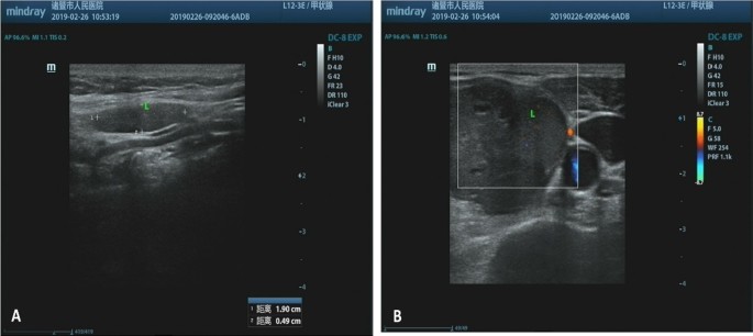
Neck ultrasonography showing an elliptical tumor with soft tissue density and ill-defined boundary adhered to the left lobe of the thyroid ( A and B )
Given the suspicion for thyroid carcinoma, she underwent left unilateral thyroidectomy. Intraoperatively, the tumor was located on the dorsal side of the left central pole of the thyroid gland, and clearly originated inside the thyroid. No obvious adhesion of the trachea and cricothyroid muscle was observed. Macroscopically, the mass measured approximately 35 mm × 20 mm × 18 mm in size and was obtained from the thyroid lobe. The cutting surface was solid and revealed a grayish brown, heterogeneous texture. Areas of necrosis and hemorrhage were not obvious. Histopathological examination with hematoxylin and eosin staining showed the tumor as multinodular and well circumscribed that in areas interlaced with the surrounding thyroid gland. The tumor was composed of mononuclear cells and multinucleated osteoclast-like giant cells. The nuclear morphology of mononuclear cells was similar to those of osteoclast multinucleated giant cells, and osteoclast giant cells were evenly distributed in the nodules (Fig. 2 ). Monocytes exhibited minimal pleomorphism in tumors, and mitosis was rare in our case, ranging from 1 to 2 per 10 high-power fields (HPFs). The osteoclastic cells were mainly found in oval-to-round monocytes. The number of nuclei in the multinucleated cells ranged from 3 to more than 50. In addition, these cells tended to cluster at the site of erythrocyte extravasation.
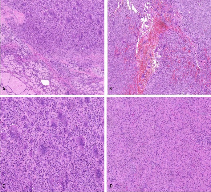
Histological features of thyroid giant cell tumor of soft tissue. A The tumor was composed of mononuclear stromal cells and a large number of multinucleated cells. B Hemorrhagic cystic formation is seen in the nodules, with marked interstitial fibrosis. C Osteoclast-like giant cells can be very large, with more than 50 nuclei. There are recognizable mitotic forms, but the cells exhibit minimal nuclear pleomorphism. D Mononuclear cells are histological and locally spindle shaped, with no atypia nor abnormal mitotic activity (hematoxylin and eosin, with original magnifications of 100×, 100×, 200×, and 200×, respectively)
On immunohistochemical analysis, these tumor cells were positive for CD68 and vimentin. The multicellular cells showed a higher expression of CD68 than that of monocytes (Fig. 3 ). The mononuclear cells were positive for synaptophysin (SYN), while negative for smooth muscle actin (SMA), desmin, and calcitonin. Cytokeratin AE1/AE3, thyroid transcription factor-1 (TTF-1), epithelial membrane antigen (EMA), S-100, and HMB45 were negative in the tumor cells. The Ki-67 labeling index ranged from 10% to 30%. Since there was no evidence of skeletal disease, we made a diagnosis of primary thyroid GCT-ST on the basis of the histological and immunohistochemical features.
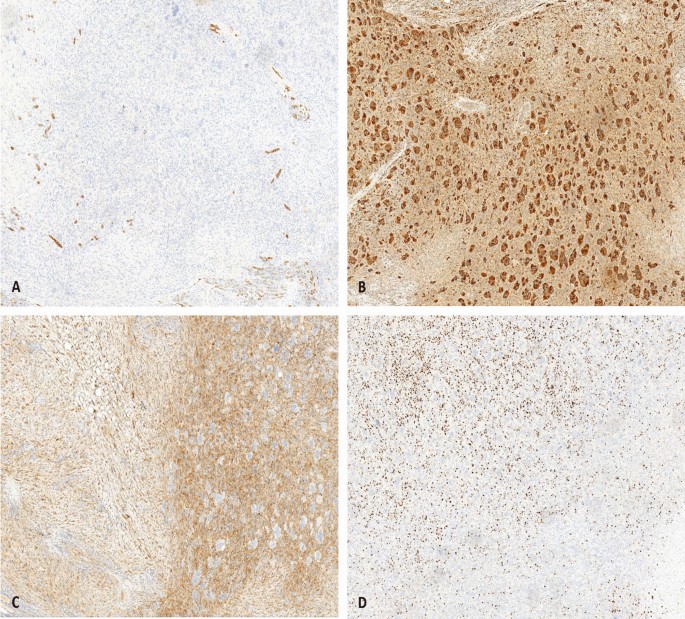
Immunohistochemical staining characteristics of thyroid giant cell tumor of soft tissue. A The tumor cells are negative for AE1/AE3 by immunohistochemical staining. B Immunostaining for CD68 is diffusely strong positive in multicellular cells. The mononuclear cells demonstrate reactivity for SYN C and the proliferation marker Ki-67 D mainly existed in the mononuclear cells (IHC staining, 100×)
The patient was admitted to Fudan University Shanghai Cancer Center with recurrence of left thyroid tumor 3 month later, and total thyroidectomy was performed. The tumor was diagnosed as primary GCT-ST of the thyroid and p-T1N0M0GX Stage IA. The postoperative process was satisfactory, and the patient was completely asymptomatic, without hypocalcemia or nerve damage. The patient did not receive adjuvant chemotherapy, and remained well without local recurrence or metastasis during 12 months of follow-up.
Giant cell tumor of soft tissue (GCT-ST) is a rare neoplasm commonly found in the superficial and deep soft tissue of the extremities [ 7 , 8 , 9 ]. GCT-ST affects both sexes equally and tends to occur in middle-aged adults. Since its morphologic characteristics and immunophenotype are similar to giant cell tumor of bone (GCTB), GCT-ST is regarded as its bony counterpart [ 10 , 11 ]. Folpe et al . proposed the term “low-malignant potential giant cell tumor” for patients with biological behavior of frequent local recurrence and uncertain metastatic potential [ 4 ]. At present, these tumors are classified into low malignant potential GCT-ST and malignant GCT-ST according to the 5th edition of World Health Organization Classification of Soft Tissue and Bone [ 12 ].
It has been reported that GCT-ST occurs in various sites, such as salivary glands, pancreas, lung, and breast [ 13 , 14 , 15 , 16 ]. However, GCT-ST arising in the thyroid is particularly rare. In the English literature, two cases of giant cell tumor of bone involving the thyroid have been described: one in a 38-year-old male patient with a 35 mm diameter nodule located within the right thyroid lobe [ 17 ], and another in a 40-year-old male patient with a nodule in left thyroid lobe [ 18 ]. Both cases of thyroid GCTB were identified in the thyroid cartilage. Our patient also showed a nodular in the thyroid lobe, and underwent surgery to remove the thyroid along with the tumor. Clinical and radiological examinations revealed no evidence of bone involvement, and the tumor was found in the thyroid gland, not adjacent to the thyroid cartilage. Finally, the possibility of giant cell tumor of bone was ruled out in our case. Shahd et al . reviewed 12 cases of GCT-ST in the head and neck, and 5 were located in different neck triangles [ 19 ]. These tumors were all described as well circumscribed, and not related to the bone or cartilage.
Microscopically, GCT-ST consists of neoplastic mononuclear cells mixed with osteoclast-like multinucleated giant cells, both of cell types immersed in a vascularized matrix [ 11 ]. The mononuclear cells were short fusiform, with red stained cytoplasm and round-to-ovoid nuclei. Multinuclear giant cells, similar to osteoclasts, are larger in size, cytoplasmic eosinophilic, and had a central accumulation of dozens of pale or solid nuclei. Lau et al . proposed that the monocyte cell components fused to form multinuclear giant cells [ 10 ]. Mitotic activity was generally observed in GCT-ST, usually ranging from 1 to more than 30 per 10 HPFs. The tumor cells of malignant GCT-ST showed nuclear atypia, pleomorphism, and frequent abnormal mitotic activity. Immunohistochemical staining exhibited strong diffusely positive for CD68 within multinucleated osteoclast-like cells, whereas mononuclear cells were focal positive. All tumors were negative for a group of cytokeratin, epithelial membrane antigen (EMA), and leukocyte common antigen (LCA), suggesting mesenchymal lineage. Irene et al . analyzed the immunophenotype and molecular characteristics between GCT-ST and GCTB, and found that osteoclastogenesis was considered to be the result of fusion of preosteoclasts with CD14+ mononuclear cells [ 20 ]. However, GCT-ST might be genetically different from GCTB, and might be considered as two distinct entities in the study performed by Lee et al . [ 21 ].
The differential diagnosis of this disease includes benign lesion (subacute thyroiditis, true granulomatous such as tuberculosis or fungal infection, and sarcoidosis) and malignant lesions (papillary carcinoma, anaplastic carcinomas, and extraskeletal osteosarcoma). The two-cell pattern and immunohistochemical features of GCT-ST contribute to resolving this diagnostic difficulty. In our case, thyroid carcinoma was ruled out because of the absence of staining for epithelial markers. No significant nuclear atypia, pleomorphism, mitotic activity, and necrosis were observed, which ruled out the possibility of giant cell malignant fibrous histiocytoma. GCT-ST differs from plexiform fibrohistiocytic tumor (PFT) by lack of plexiform pattern of growth, larger nodules, metaplastic bone formation, and uniform distribution of multicellular osteoclast-like giant cell, which were absent in PFT. Previous studies have reported that small amounts of osteoid were found in some of GCT-ST, raising the differential diagnostic considerations for extra-skeletal osteosarcoma (ES-OGS). The giant cell-rich ES-OGS is a distinctly malformed tumor, with the morphologic features of conspicuous atypia and pleomorphism, as well as numerous bizarre, mitotic figures, which were not characteristic of GCT-ST. In ES-OGS, bone and osteoid formation is generated by the sarcomatoid, spindle cell stroma. In contrast, the osteoblast-lined, metaplastic bone trabeculae were seen in the cases of GCT-ST. Mana et al . described a case of extraskeletal giant cell tumor of the larynx, which definitely originated outside the laryngeal cartilage and was also diagnosed as a GCT-ST [ 22 ].
Since the majority of GCT-ST have a benign clinical course, the first-line treatment is complete surgical resection. There is no definitive evidence to support postoperative radiotherapy, but it should be considered in the case of incomplete surgical margin resection [ 5 , 23 ]. Righi et al . found that the aggregation of osteoclast-like giant cells in GCTB and GCT-ST occurs through the RANKL-dependent mechanism, and drug inhibitors of osteoclast-like activity may control their absorption [ 20 ]. Several clinical studies and a retrospective case–control study have shown that denosumab (RANKL inhibitor) may reduce local recurrence after surgery [ 24 ], but there is no conclusive evidence for the use of denosumab in the recurrence of GCT-ST. In general, tumors lacking significant atypia or nuclear pleomorphism have benign behavior and rarely have local recurrences or lung metastasis. Guccion et al . found that tumors beneath a fascial plane were more aggressive [ 7 ]. On the contrary, the depth of involvement as having no significant effect on clinical course was reported by Folpe et al . The prognosis factors of GCT-ST are still unclear, and close follow-up is necessary due to the inability to predict local recurrence and metastasis.
In summary, primary thyroid GCT-ST is a rare tumor, composed of mononuclear and multinucleated cells. The clinical behavior of GCT-ST may vary widely, with some patients presenting with relatively indolent disease and others with rapidly distant metastasis. Familiarity with this rare disease and its characteristic histopathology features ensures establishing a correct diagnosis and distinguishing between other benign and malignant tumors. Clinicians and pathologists need to be aware of the rare diagnosis of GCT-ST in thyroid and should carefully individualize management and define its clinical prognosis.
Giant cell tumor of soft tissue is a tumor with low malignant potential, which should be distinguished from other giant cell-rich soft tissue neoplasms. The significance of this case lies in the rarity of this entity, the challenge in preoperative diagnosis, and the differential diagnosis with other malignancies. More cases should be collected to improve the understanding of clinical characteristics, histopathology, pathogenesis, and relevant treatment.
Availability of data and materials
The datasets used and analyzed during the current study are available from the corresponding author upon request.
Abbreviations
Thyroid stimulating hormone
Free triiodothyronine
Free thyroxine
Synaptophysin
Smooth muscle actin
Thyroid transcription factor-1
Epithelial membrane antigen
O’Connell JX, Wehrli BM, Nielsen GP, Rosenberg AE. Giant cell tumors of soft tissue: a clinicopathologic study of 18 benign and malignant tumors. Am J Surg Pathol. 2000;24(3):386–95.
Article CAS PubMed Google Scholar
Doussis IA, Puddle B, Athanasou NA. Immunophenotype of multinucleated and mononuclear cells in giant cell lesions of bone and soft tissue. J Clin Pathol. 1992;45(5):398–404.
Article CAS PubMed PubMed Central Google Scholar
Salm R, Sissons HA. Giant-cell tumours of soft tissues. J Pathol. 1972;107(1):27.
Folpe AL, Morris RJ, Weiss SW. Soft tissue giant cell tumor of low malignant potential: a proposal for the reclassification of malignant giant cell tumor of soft parts. Mod Pathol. 1999;12(9):894–902.
CAS PubMed Google Scholar
Icihikawa K, Tanino R. Soft tissue giant cell tumor of low malignant potential. Tokai J Exp Clin Med. 2004;29(3):91–5.
PubMed Google Scholar
Grabellus F, Winterfeld FV, Sheu SY, et al . Unusual aggressive course of a giant cell tumor of soft tissue during immunosuppressive therapy. Virchows Arch. 2006;448(6):847–51.
Article PubMed Google Scholar
Guccion JG, Enzinger FM. Malignant giant cell tumor of soft parts. An analysis of 32 cases. Cancer. 1972;29(6):1518–29.
Rodriguez-Peralto JL, Lopez-Barea F, Fernandez-Delgado J. Primary giant cell tumor of soft tissues similar to bone giant cell tumor: a case report and literature review. Pathol Int. 2001;51(1):60–3.
Holst VA, Elenitsas R. Primary giant cell tumor of soft tissue. J Cutan Pathol. 2001;28(9):492–5.
Lau YS , Sabokbar A, Gibbons CL, et al . Phenotypic and molecular studies of giant-cell tumors of bone and soft tissue. Hum Pathol 2005, 36(9):0–954.
Wylie J . Pathology and genetics of tumours of soft tissue and bone. Published 2002, 1st edition, ISBN 92 832 24132. Surg Oncol 2004, 13(1):43–0.
Rosenberg AE. WHO classification of soft tissue and bone, fourth edition: summary and commentary. Curr Opin Oncol. 2013;25(5):571–3.
Fukunaga M. Giant cell tumor of the breast. Virchows Archiv Int J Pathol. 2002;441(1):93–5.
Article Google Scholar
Itoh Y, Taniguti Y, Arai K. A case of giant cell tumor of the parotid gland. Ann Plast Surg. 1992;28(2):183–6.
Shiozawa M, Imada T, Ishiwa N, et al . Osteoclast-like giant cell tumor of the pancreas[J]. Int J Clin Oncol. 2002;7(6):0376–80.
Righi S, Boffano P, et al . Soft tissue giant cell tumor of low malignant potential with 3 localizations: report of a case. Oral Surg Oral Med Oral Pathol Oral Radiol. 2014;118(5):e135–8.
Derbel O, Zrounba P, Chassagne-Clément C, et al . An unusual giant cell tumor of the thyroid: case report and review of the literature. J Clin Endocrinol Metab. 2013;98(1):1–6.
Zhang X, Zhu X, Li J, et al . Fine needle aspiration of giant cell tumor involving thyroid gland: a case report of an unprecedented entity. Diagn Cytopathol. 2018;46(10):879–82.
Hafiz SM, Bablghaith ES, Alsaedi AJ, et al . Giant-cell tumors of soft tissue in the head and neck: a review article. Int J Health Sci. 2018;12(4):88.
Google Scholar
Mancini I, Righi A, Gambarotti M, et al . Phenotypic and molecular differences between giant-cell tumour of soft tissue and its bone counterpart. Histopathology. 2017;71(3):453–60.
Lee JC, Liang CW, Fletcher CD. Giant cell tumor of soft tissue is genetically distinct from its bone counterpart. ModPathol. 2017;30(5):728–33.
CAS Google Scholar
Rochanawutanon M, Praneetvatakul P, Laothamatas J, et al . Extraskeletal giant cell tumor of the larynx: case report and review of the literature. Ear Nose Throat J. 2011;90(5):226–30.
Pepper T, Falla L, Brennan PA. Soft tissue giant cell tumour of low malignant potential arising in the masseter—a rare entity in the head and neck. Br J Oral Maxillofac Surg 2010:48(2):0–151.
Balke M, Hardes J. Denosumab: a breakthrough in treatment of giant-cell tumour of bone. Lancet Oncol. 2010;11(3):218–9.
Download references
Acknowledgements
We greatly appreciate the assistance of the staff of the Department of Pathology, Zhuji People’s Hospital, Zhuji Affiliated Hosptal of Wenzhou Medical University, and thank them for their efforts.
No funding was received.
Author information
Authors and affiliations.
Department of Pathology, Zhuji People’s Hospital of Zhejiang Province, Jianming Road, Taozhu Street, Zhuji, Zhejiang, China
Jianrong Chen, Haiyong Zhang, Xiufang Li & Mengjun Hu
Department of Pathology, Zhejiang Hospital, 1229 Gudun Road, Xihu District, Hangzhou, Zhejiang, China
Xiaomin Dai
You can also search for this author in PubMed Google Scholar
Contributions
JC and HZ were responsible for the conception and design of the experiments; XL and MH contributed to the acquisition, analysis, and interpretation of the data; JC drafted the manuscript; and XD revised the manuscript. All authors have read and approved the final manuscript.
Corresponding author
Correspondence to Xiaomin Dai .
Ethics declarations
Ethics approval and consent to participate.
The Ethics Review Committee of Zhuji People’s Hospital of Zhejiang Province. A copy of the written consent is available for review by the author of this paper.
Consent for publication
Written informed consent was obtained from the patient for publication of this case report and any accompanying images. A copy of the written consent is available for review by the Editor-in-Chief of this journal.
Competing interests
The authors declare that they have no competing interest in this work.
Additional information
Publisher’s note.
Springer Nature remains neutral with regard to jurisdictional claims in published maps and institutional affiliations.
Rights and permissions
Open Access This article is licensed under a Creative Commons Attribution 4.0 International License, which permits use, sharing, adaptation, distribution and reproduction in any medium or format, as long as you give appropriate credit to the original author(s) and the source, provide a link to the Creative Commons licence, and indicate if changes were made. The images or other third party material in this article are included in the article's Creative Commons licence, unless indicated otherwise in a credit line to the material. If material is not included in the article's Creative Commons licence and your intended use is not permitted by statutory regulation or exceeds the permitted use, you will need to obtain permission directly from the copyright holder. To view a copy of this licence, visit http://creativecommons.org/licenses/by/4.0/ . The Creative Commons Public Domain Dedication waiver ( http://creativecommons.org/publicdomain/zero/1.0/ ) applies to the data made available in this article, unless otherwise stated in a credit line to the data.
Reprints and permissions
About this article
Cite this article.
Chen, J., Zhang, H., Li, X. et al. Giant cell tumor of soft tissue involving thyroid gland: a case report and review of the literature. J Med Case Reports 18 , 123 (2024). https://doi.org/10.1186/s13256-024-04450-1
Download citation
Received : 11 January 2024
Accepted : 09 February 2024
Published : 22 March 2024
DOI : https://doi.org/10.1186/s13256-024-04450-1
Share this article
Anyone you share the following link with will be able to read this content:
Sorry, a shareable link is not currently available for this article.
Provided by the Springer Nature SharedIt content-sharing initiative
- Thyroid gland
- Tumor of soft tissue
- Giant cell tumor of soft tissue
- Immunohistochemistry
- Differential diagnosis
Journal of Medical Case Reports
ISSN: 1752-1947
- Submission enquiries: Access here and click Contact Us
- General enquiries: [email protected]
Seizures associated with levofloxacin: case presentation and literature review
Affiliation.
- 1 Centre de Psychiatrie et Neurosciences, INSERM U894 Laboratoire de Physiopathologie des Maladies Psychiatriques, Paris, France. [email protected]
- PMID: 19707748
- DOI: 10.1007/s00228-009-0717-5
Purpose: We present a case of a patient who developed seizures shortly after initiating treatment with levofloxacin and to discuss the potential drug-drug interactions related to the inhibition of cytochrome P450 (CYP) 1A2 in this case, as well as in other cases, of levofloxacin-induced seizures.
Methods: Several biomedical databases were searched including MEDLINE, Cochrane and Ovid. The main search terms utilized were case report and levofloxacin. The search was limited to studies published in English.
Results: Six cases of levofloxacin-induced seizures have been reported in the literature. Drug-drug interactions related to the inhibition of CYP1A2 by levofloxacin are likely involved in the clinical outcome of these cases.
Conclusions: Clinicians are exhorted to pay close attention when initiating levofloxacin therapy in patients taking medications with epileptogenic properties that are CYP1A2 substrates.
Publication types
- Case Reports
- Research Support, Non-U.S. Gov't
- Adrenergic alpha-Antagonists / adverse effects
- Aged, 80 and over
- Anti-Bacterial Agents / administration & dosage
- Anti-Bacterial Agents / adverse effects*
- Bacteremia / diagnosis
- Bacteremia / microbiology*
- Cytochrome P-450 CYP1A2 / drug effects
- Cytochrome P-450 CYP1A2 / metabolism
- Dopamine Antagonists / adverse effects
- Drug Administration Schedule
- Drug Synergism
- Klebsiella Infections / diagnosis*
- Klebsiella Infections / drug therapy
- Levofloxacin*
- Metoclopramide / adverse effects
- Mianserin / adverse effects
- Mianserin / analogs & derivatives
- Middle Aged
- Mirtazapine
- Ofloxacin / administration & dosage
- Ofloxacin / adverse effects*
- Phosphodiesterase Inhibitors / adverse effects
- Seizures / chemically induced*
- Theophylline / adverse effects
- Adrenergic alpha-Antagonists
- Anti-Bacterial Agents
- Dopamine Antagonists
- Phosphodiesterase Inhibitors
- Levofloxacin
- Theophylline
- Cytochrome P-450 CYP1A2
- Metoclopramide

IMAGES
VIDEO
COMMENTS
Examples of literature reviews. Step 1 - Search for relevant literature. Step 2 - Evaluate and select sources. Step 3 - Identify themes, debates, and gaps. Step 4 - Outline your literature review's structure. Step 5 - Write your literature review.
A case report describes several aspects of an individual patient's presentation, investigations, management decisions, and/or outcomes. This is a type of observational study and has been described as the smallest publishable unit in medical literature. A case series involves a group of patients with similar presentations or treatments.
The oldest example of a preserved clinical case in medical literature is a text from an ancient Egyptian papyrus dating from the 16 th to the ... but not mandatory, accompanied by a review of the literature of other reported cases. Although case reports are considered the lowest in the hierarchy of evidence-based practice in ... Case presentation.
Presenting patient cases is a key part of everyday clinical practice. A well delivered presentation has the potential to facilitate patient care and improve efficiency on ward rounds, as well as a means of teaching and assessing clinical competence.1 The purpose of a case presentation is to communicate your diagnostic reasoning to the listener, so that he or she has a clear picture of the ...
a description of the publication. a summary of the publication's main points. an evaluation of the publication's contribution to the topic. identification of critical gaps, points of disagreement, or potentially flawed methodology or theoretical approaches. indicates potential directions for future research.
Writing up. Write up the case emphasising the interesting points of the presentation, investigations leading to diagnosis, and management of the disease/pathology. Get input on the case from all members of the team, highlighting their involvement. Also include the prognosis of the patient, if known, as the reader will want to know the outcome.
Case reports are a time-honored, important, integral, and accepted part of the medical literature. Both the Journal of Medical Case Reports and the Case Report section of BioMed Central Research Notes are committed to case report publication, and each have different criteria.Journal of Medical Case Reports was the world's first international, PubMed-listed medical journal devoted to ...
(How to write a Literature Review is the subject of a future paper). Significant value in writing a Case report is that it provides medical students and junior doctors with an excellent opportunity to develop their writing skills. ... Case presentation. First, tackle the details of the case. The case presentation is a fundamental part of the ...
The reviewers need to serve as content experts regarding the drugs and other technologies used in the case. A literature search by the reviewer provides the data to comment on this aspect. Competing interests (or conflicts of interest) are concerns that interfere or potentially interfere with presentation, review, or publication.
Introduction: Provide a brief literature review that presents the background, significance, and aims of the case study presentation. As we particularly are interested in case presentations that make substantive clinical or theoretical contributions to the literature, this section should ground the presentation in the relevant scholarly literature.
The literature review for a case study research paper is generally structured the same as it is for any college-level research paper. The difference, however, is that the literature review is focused on providing background information and enabling historical interpretation of the subject of analysis in relation to the research problem the case ...
Literature review and case discussion Limb body wall complex (LBWC) is a rare, polimalformative fetal syndrome, appearing in .21-.31/10.000 deliveries, with onl y about 245 cases described in ...
A case study provides a meaningful learning tool for critical care nurses, 1 particularly those beginning their practice, by building knowledge through application of literature to practice in complex clinical presentations. In Part I of this case study, Peter, 32, was admitted to hospital with multiple injuries including a traumatic Sub ...
Posttraumatic subarachnoid fat embolism: Case presentation and literature review Clin Imaging. 2020 Dec;68:121-123. doi: 10.1016/j.clinimag.2020.06.035. Epub 2020 Jun 20. Authors Rahul Chaturvedi 1 ... Herein, we report the only case in the United States, to our knowledge, of a patient diagnosed with subarachnoid fat emboli secondary to sacral ...
Therefore, little is known about its epidemiology, clinical presentation, diagnostic methods, treatment, and outcomes. Methods: In this paper, we report a case of an adult patient with C. jejuni meningitis. In addition, we reviewed 16 cases of C. jejuni published since 1980. Results: We described a 62-year-old immunocompromised patient with ...
Literature review. Pediatric onset multiple sclerosis (POMS), defined as multiple sclerosis (MS) with onset in patients younger than 18 years, occurs in 3-10% of all MS patients ( 1 ). The incidence varies by country and is estimated to range from 0.57 to 2.85/100000 children ( 2 - 5 ). Onset before the age of 10 is extremely rare ( 6 - 9 ...
Reactive arthritis (ReA) is a clinical condition typically triggered by extra-articular bacterial infections and often associated with the presence of HLA-B27. While ReA has traditionally been associated with gastrointestinal and genitourinary infections, its pathogenesis involves immune and inflammatory responses that lead to joint affections. The emergence of COVID-19, caused by SARS-CoV-2 ...
Background Primary brain rhabdomyosarcoma is a rare primary brain malignancy with few case reports. The vast majority of cases of primary brain rhabdomyosarcoma occur in pediatric patients, and immunohistochemistry can distinguish it from embryonal subtypes; however, few cases of primary brain rhabdomyosarcoma in adults have been reported in the literature. Case presentation We report the case ...
Template 1: Literature Review PPT Template. This literature review design is a perfect tool for any student looking to present a summary and critique of knowledge on their research statement. Using this layout, you can discuss theoretical and methodological contributions in the related field.
A case report is a detailed report of the symptoms, signs, diagnosis, treatment, and follow-up of an individual patient. Case reports usually describe an unusual or novel occurrence and as such, remain one of the cornerstones of medical progress and provide many new ideas in medicine. Some reports contain an extensive review of the relevant ...
Abstract. Bilateral facial paralysis is a diagnostic challenge, which may manifest itself as either a simultaneous or alternating form, occurring in 0.3-2.0% of patients that present with facial paralysis. The differential diagnosis of facial paralysis includes congenital, traumatic, neurologic, infectious, metabolic, neoplastic, toxic ...
Central hyperthyroidism due to an ectopic TSH-secreting pituitary tumor: a case report and literature review. ... Case presentation. A 60-year-old woman with a complaint of weight loss, palpitation, tremor, and heat intolerance was admitted to Endocrinology and Metabolism Department of the Tianjin Medical University General Hospital due to ...
At present, English literature reports on the close growth of LOTs and AML are rare. In this case, the patient was only 29 years old but had multiple large tumors, which is relatively rare among reported cases. The patient refused to undergo genetic testing; genetic detection may have revealed a relationship between the LOTs and AML.
A case of ReA is presented in a patient who survived COVID-19 and presented with joint affections and this article encompasses a study of similar clinical cases of reactive arthritis following CO VID-19 reported worldwide. Reactive arthritis (ReA) is a clinical condition typically triggered by extra-articular bacterial infections and often associated with the presence of HLA-B27. While ReA has ...
The aim of the present manuscript is: 1) to report a case series of 16 patients diagnosed with PPCM following arrhythmic clinical presentation, and: 2) to present an updated review of the literature about the arrhythmic manifestations of PPCM.
Clinical case presentation is part of daily routine for doctors to communicate with each other to facilitate learning, and ultimately patient management. ... Therefore, the review of literature needs to be brief and succinct. Its main purpose is to articulate the lessons learnt from this case and should illustrate how a similar case should be ...
Background Giant cell tumor of soft tissue is a low malignant uncommon neoplasm, with histologic features and immunophenotype similar to its bone counterpart. Primary giant cell tumor of soft tissue in the thyroid gland is considered an exceedingly rare entity. Case presentation We describe a case of primary thyroid giant cell tumor of soft tissue in a 69-year-old Chinese female patient. Neck ...
Charles Bonnet syndrome: case presentation and literature review Optometry. 2009 Jul;80(7):360-6. doi: 10.1016/j.optm.2008.10.017. Author Elizabeth M ... Case 2 is an 86-year-old man who presented with a 2-month history of hallucinations. Both patients are legally blind from age-related macular degeneration. In both cases, the ocular ...
Herein, we present two cases aged 14 days and 35 days old with severe symptoms of deviated nasal septum, balloon dilatation septoplasty, a minimally invasive approach, was employed, with low-risk complications and good outcomes. Up to this date, this approach has not been reported in this age group.
The main search terms utilized were case report and levofloxacin. The search was limited to studies published in English. Results: Six cases of levofloxacin-induced seizures have been reported in the literature. Drug-drug interactions related to the inhibition of CYP1A2 by levofloxacin are likely involved in the clinical outcome of these cases.