59 Ultrasound Essay Topic Ideas & Examples
🏆 best ultrasound topic ideas & essay examples, ✅ good essay topics on ultrasound, 📑 interesting topics to write about ultrasound.
- Use of Ultrasound-Guidance for Arterial Puncture All the anthropometric and demographic variables were recorded, as well as the main diagnosis of admission, comorbidities, the placement of the central venous catheter, and the course of the procedure.
- MRI and Ultrasound for Determining Abnormalities in Preterm Infants Neonatal cranial ultrasound is used in detecting brain injury in preterm infants and can be used repetitively without harming the infant. We will write a custom essay specifically for you by our professional experts 808 writers online Learn More
- Benefits of 3D Ultrasound to Pregnant Mothers This is coherent to the 3D planar imaging are improved technology previously applied in the 2D ultrasound technology. As an extrapolation from 3D technology, 3D ultrasound is applied as a medical diagnostic technique that utilizes […]
- The Biological Effects of Ultrasound The paper also evaluates the physical mechanisms for the biological effects of ultrasound and the effects of ultrasound on living tissues in vivo and vitriol.
- The Recent Advances in Real Time Imaging in Ultrasound In point of fact, Medical imaging provides the most perfect task of diagnostic to Ultrasound, whereas, the main usage of therapeutic Ultrasound is to treat the numerous types of diseases and disorders in human beings.
- Comparison of MRI and Ultrasound for Determining Abnormalities in Preterm Infants Medical Imaging helps in detecting and diagnosing diseases at its earliest and treatable stage and helps in determining most appropriate and effective care for the patient.”Medical imaging provides a picture of the inside of the […]
- Ultrasound Techniques Applied to Body Fat Measurement in Male and Female The main objective of this paper is to evaluate the accuracy of body fat by using portable ultra sound device which results are reliable and authentic. The ultra sound technique is widely used to measure […]
- Benefits of 3D/4D Ultrasound in Prenatal Care The information that is obtained from this exam assists the health care providers in counseling parents on the development of the fetus especially in the nature of anomalies, prognosis, and the postnatal consideration of the […]
- Biologic Effects of Ultrasound in Healthcare Setting The instrument performing the emission of the sound waves and the recording of their bouncing back is referred to as the transducer and the medical practitioner generally gently presses the transducer against the skin of […]
- Ultrasound Scanning: Diagnosing Health Conditions The calculation is based on the time taken by the wave to return by measuring the distance, mass, nature, and stability of the object hit.
- Mammography vs. Ultrasound for Breast Tissue Analysis Mammography screening is one of the most recognized options for analyzing breast tissue in adult women. In contrast, the accuracy of this procedure allows it to be an alternative for women who cannot undergo mammography […]
- Ultrasound Physics and Instrumentation The camera is often not in harmony with the perception of the depth of a human vision. The level of such an acoustic signal distortion within a tissue is dependent on the emitted pulse’s amplitude […]
- Low-Back Pain and Ultrasound Therapy In the meantime, their opponents highlight that the beneficial aspects of the treatment course outweigh the risks related to the use of ultrasound equipment.
- Ultrasound in Treatment and Side-Effect Reduction Within the framework of the research project conducted by Ebadi et al, the research problem consisted in the fact that the effects of continuous ultrasound were underresearched.
- Ultrasound in Achilles Tendinitis Diagnosis In this research, the case study approach is applicable due to the fact that various patients suffering from tendon Achilles problem will be used as a basis for gauging the effectiveness of the method of […]
- Ultrasound Technology in Podiatry Surgery First, it is important to briefly outline the peculiarities of the RCT to understand the researchers’ point. They will be able to use the technology in numerous settings.
- Abdominal Ultrasound and Diagnoses The examiner explains to the patient how the procedure will be performed and how much time is necessary to finish the examination.
- Ultrasound and Color Doppler-Guided Surgery The purpose of the study is to examine the opinions of the trainees attending a training course concerning the use of technology.
- Contrast-Enhanced Ultrasound in Focal Liver Lesions In addition, inaccessibility to the eighth of the liver is a major setback in detecting lesions in the segment. With the advent of Doppler ultrasound, more insight in the diagnosis of liver lesions has been […]
- Ultrasound in Chemistry: Sonochemistry
- Intravascular Ultrasound: Current Role and Future Perspectives
- The Difference Between an Echocardiogram and an Ultrasound of the Heart
- Cooperative Control With Ultrasound Guidance for Radiation Therapy
- Optically Generated Ultrasound: A New Paradigm for Intracoronary Imaging
- Nondestructive Testing: Principle of Flaw Detection With Ultrasound
- Closed-Loop Transcranial Ultrasound Stimulation for Real-Time Non-invasive Neuromodulation
- Consumer Application of Ultrasound: A Television Remote
- Roles of Low-Intensity Ultrasound in Differentiating Cell Death
- High-Intensity Focused Ultrasound Development: Destroying the Target Tissue
- Using Ultrasound to Enhance the Mechanical and Physical Properties of Metals
- The Security Implications of the Machine-Learning Supply Chain: Professionalism in the Ultrasound Department
- Ultrasound Neuromodulation: Mechanisms and the Potential of Multimodal Stimulation for Neuronal Function Assessment
- How Ultrasound Can Produce Sonoluminescence
- Preparation for an Ultrasound of a Gallbladder and a Pelvic
- Endorectal and Endoanal Ultrasound Technique
- Transvaginal Ultrasound: Is It Painful, Purpose, and Results
- Endoscopic Ultrasound in Diagnosing Cancer: One of the Most Common Imaging Procedures
- Ultrasound Skin Imaging in Dermatology, Aesthetic Medicine, and Cosmetology
- Ultrasound Physical Medical Treatment in Healing Following an Acute Injury or a Chronic Condition
- How Do Ultrasound Scans Work
- Americas Ultrasound Systems Market: From USD 7.9 Billion to USD 10.23 Billion
- How Ultrasound Imaging Helps Us Understand Speech and Accent Variation
- Cerebral Ultrasound Time-Harmonic Elastography: Softening of the Human Brain Due to Dehydration
- Wireless Communication in “Audio Beacons” Using Ultrasound
- Low-Intensity Focused Ultrasound for Posttraumatic Stress Disorder
- Thyroid Ultrasound: Purpose, Procedure, Benefits
- Blood Pressure Modulation With Low-Intensity Focused Ultrasound
- Accuracy of Ultrasounds in Diagnosing Birth Defects
- Functional Ultrasound During Awake Brain Surgery
- Live Animal Ultrasound Explained by Dr. Allen Williams
- Ultrasound Waves in Acoustic Microscopy
- Chemical and Physical Effects of Ultrasound: Sonoluminescence and Materials
- Focused Ultrasound for Noninvasive, Focal Pharmacologic Neurointervention
- Lung Ultrasound Findings in COVID-19 Pneumonia
- High-Power Ultrasound in Dry Corn Milling Plants
- History of Ultrasound of Physics and the Properties of the Transducer
- Relationship Between Ultrasound Viewing and Proceeding to Abortion
- Transrectal Ultrasound of the Prostate With a Biopsy
- Iron-Based Catalysts Used in Water Treatment Assisted by Ultrasound
- Chicago (A-D)
- Chicago (N-B)
IvyPanda. (2023, January 24). 59 Ultrasound Essay Topic Ideas & Examples. https://ivypanda.com/essays/topic/ultrasound-essay-topics/
"59 Ultrasound Essay Topic Ideas & Examples." IvyPanda , 24 Jan. 2023, ivypanda.com/essays/topic/ultrasound-essay-topics/.
IvyPanda . (2023) '59 Ultrasound Essay Topic Ideas & Examples'. 24 January.
IvyPanda . 2023. "59 Ultrasound Essay Topic Ideas & Examples." January 24, 2023. https://ivypanda.com/essays/topic/ultrasound-essay-topics/.
1. IvyPanda . "59 Ultrasound Essay Topic Ideas & Examples." January 24, 2023. https://ivypanda.com/essays/topic/ultrasound-essay-topics/.
Bibliography
IvyPanda . "59 Ultrasound Essay Topic Ideas & Examples." January 24, 2023. https://ivypanda.com/essays/topic/ultrasound-essay-topics/.
- X-Ray Questions
- Health Promotion Research Topics
- Gynecology Research Ideas
- Osteoarthritis Ideas
- Pneumonia Questions
- Cardiomyopathy Titles
- Heart Disease Titles
- Epigenetics Essay Titles
- Hypertension Topics
- Biomedicine Essay Topics
- Deontology Questions
- Breast Cancer Ideas
- Health Insurance Research Topics
- Evidence-Based Practice Titles
- Healthcare Questions


Top 100 Ultrasound
Litfl 100+ ultrasound quiz.
Clinical cases and self assessment problems from the Ultrasound library to enhance interpretation skills through Ultrasound problems. Preparation for examinations.
Each case presents a clinical scenario; a series of questions; clinical images and finally some pearls to highlight the key learning points. We trust that you will find a few clinical pearls or reminders that you could apply to your patients that you care for in your emergency department or other health setting
Search by keywords; disease process; condition; eponym or clinical features…
LITFL Top 100 Self Assessment Quizzes
ULTRASOUND LIBRARY
POCUS, eFAST and basic principles
Privacy Overview
- Join or Renew
- Member Benefits
- Journal (JDMS)
- Collaborate (Communities)
SDMS's core purpose is to enhance the art and science of medicine by advancing medical sonography. As an SDMS member you will join an exclusive network of over 28,000 sonographers and sonography students.

- My SDMS CME Tracker
- Earn SDMS CME Credit
- Offer SDMS CME Credit
- SDMS Calendar of Events
- SDMS Annual Conference
The SDMS provides opportunities to earn and offer continuing medical education (CME). We also have educational resources for the sonography community.

- FAQs
- What is Sonography?
- Career Resources
- SDMS Career Center
- Salary Resources
- White Papers
The SDMS offers resources for the sonography industry.

- Certification
- State Licensure
- Coding & Reimbursement
The SDMS supports credentialing for sonographers and provides representation on legislative and regulatory issues that affect the sonography profession.

SDMS Foundation
Formed in 2009, the SDMS Foundation is a nonprofit charitable organization affiliated with the SDMS. The SDMS Foundation fosters professional learning and excellence by working to improve the field of diagnostic medical sonography.
- Shopping Cart
- Search Products
The SDMS provides various products for the sonography community.

- Advertising & Partnership Opportunities
About the SDMS
The Society of Diagnostic Medical Sonography (SDMS) is a professional membership organization founded in 1970 to promote, advance, and educate its members and the medical community in the science of diagnostic medical sonography. The SDMS is the largest association of sonographers and sonography students in the world. Learn More

Society of Diagnostic Medical Sonography

- Resources /
- What is Sonography? /
Understanding Sonography
Images of health.
Doctors use ultrasound in women, men, and children to gain advanced insights into the inner workings of the body. In fact, after x-ray exams, ultrasound is the most utilized form of diagnostic imaging available today. Using ultrasound, doctors can monitor a variety of women’s health conditions from heart disease to breast abnormalities to several gynecological problems-accurately while limiting invasive procedures. Ultrasound can help diagnose a wide variety of conditions in men, ranging from heart disease to abnormalities in the prostate gland or testicles. With children, doctors commonly use ultrasound to detect a variety of illnesses and disorders. A physician may use ultrasound to examine a child's gastrointestinal tract for signs of appendicitis or a baby's bone structure for alignment problems like congenital hip dislocation or spina bifida. An ultrasound exam of the head can detect hydrocephaly (water on the brain), intracranial hemorrahage (bleeding in the skull), and other conditions of the head. Despite today's sophisticated, high-tech systems, ultrasound remains a science built upon the simple sound wave. By beaming high-frequency sound waves into the body, physicians translate the echoes that bounce off body tissues and organs into colorful, visual images that provide valuable medical information. Heart disease, stroke, abnormalities in the abdomen or reproductive system, gallstones, liver damage, and kidney dysfunction all exhibit telltale signs that ultrasound can help to detect. Safe, affordable, and non-invasive, ultrasound is also portable. Very sick or fragile patients, who might not be able to travel to a radiology lab without risking further injury, can have the lab wheeled to them. Ultrasound helps doctors make a diagnosis and determine the best and most effective means possible to achieve health.
Ultrasound Makes History
1998 marked the 50th anniversary of diagnostic ultrasound. From the late 1940s, when scans were done on patients seated in water-filled gun turrets to the late 1990s, when Color Doppler (as seen in TV weather reports) introduced color to black and white images, ultrasound has always been at the forefront of modern medicine. Beginning as a science of navigation, the first form of ultrasound, called SONAR, was used on war ships during WWII for navigating the seas by bouncing sound waves off the ocean floor and interpreting the echoes. In the late 1940s, scientists began experimenting with these sound waves as a new way to image the human body similar to X-ray. In the 1950s and '60s, doctors used ultrasound with heart patients, while also branching out into obstetrics, gynecology, and abdominal uses. These early images looked like a seismograph output (a record of an earthquake) with spikes and lines, depicting returning sound waves. In the 1970s, grayscale imaging was introduced, providing physicians with the first opportunity to see a cross-section of a person's anatomy. In the early 1980s, computer software was joined to ultrasound technology to usher in the age of modern ultrasound. Today, ultrasound is a sophisticated, computer-integrated diagnostic tool that provides dynamic and crisp visual insights into the human body.
A physician can use ultrasound to detect a wide array of gynecological conditions. For people experiencing pelvic pain, ultrasound may be used as part of a standard pelvic exam to find or rule out conditions such as internal bleeding, pelvic inflammatory disease, abscesses, pelvic masses, and endometriosis. If doctors suspect any of these problems, an ultrasound examination can confirm or identify these concerns. Ultrasound can also help address infertility issues. New higher resolution ultrasound systems allow physicians to safely monitor and examine the reproductive system in the early stages of embryo development in the fertility process. Pregnancy
Ultrasound can be useful in high-risk pregnancy. Doctors can predict premature delivery by examining the size of the cervix. They can also screen for fallopian tube patency and detect ectopic pregnancies (when the fertilized egg grows outside the uterus). Fetal Imaging
Because ultrasound uses sound waves to create its images, it has been shown to be safe for both mother and child. A complete fetal examination using ultrasound involves imaging the baby's head, heart, kidneys, spine, stomach, umbilical cord, bladder and placenta to determine if any abnormalities exist. The same fetal exam can also be used to check for the possibility of multiple births, unusual orientation, and if the baby is positioned correctly, the sex can also be determined. By taking measurements of the fetus, a doctor can also determine the gestational age of the baby to help date the pregnancy. In some cases, early in the first trimester, a special examination using an endovaginal (inside the vagina) transducer may be conducted to check for conditions not easily recognized with the standard, more widely used trans-abdominal (outside the abdomen) examination.
Breast Imaging
After skin cancer, breast cancer is the most common type of cancer among women. According to the Susan G. Komen Breast Cancer Foundation , each year in the United States, more than 200,000 women are diagnosed with invasive breast cancer and 64,000 are diagnosed with non-invasive breast cancer. More than 2,000 cases in men will also be diagnosed with breast cancer each year in the United States. Data shows that 95 percent of patients whose cancer is detected early have a greater survival rate than those whose cancer is detected in later stages.
As an adjunct to mammography, ultrasound can be a primary breast imaging application for women under the age of 40 who have dense breasts, or breast implants, or breastfeed. Ultrasound can also help doctors assess masses that are detected in mammograms, aid in the detection of cysts and guide breast biopsies. If you are 35 or over, physicians suggest a breast exam each year as a part of your routine health regimen.
Heart Disease
According to the American Heart Association , cardiovascular disease is the leading cause of death around the world, accounting for 17.3 million deaths per year. Event when all forms of cancer are combined, heart disease is the No. 1 killer of women.
By using ultrasound, doctors can pinpoint trouble spots and patients avoid life-threatening heart conditions as well as stroke and hypertension. Using ultrasound, a doctor can image the heart muscle to detect damage, congenital defects or hereditary abnormalities. By imaging the carotid artery using Color Doppler ultrasound, doctors can check for the development of plaque which is the precursor to potentially, deadly coronary artery disease. In some cases, this can help to predict the chances of developing coronary artery disease, allowing doctors the opportunity to prescribe early treatment options.
Prostate Cancer
According to the Prostate Cancer Foundation , cancer of the prostate is the most common non-skin cancer among American men and affects one in seven men during the course of a lifetime. Advanced age, African American race, and a family history of prostrate cancer can all increase the chances of being diagnosed with the disease.
As with other cancers for which a cause is unknown, early detection is the most valuable weapon in this battle. If a problem in the prostate is suspected, ultrasound can allow a physician or radiologist to accurately acquire a tissue sample or biopsy from areas of the prostate. The exam is conducted through the use of a small probe and may be carried out in conjunction with other exams, including the important prostate-specific antigen (PSA) blood test. Most men with prostate cancer show elevated levels of prostate-specific antigen, a protein that is produced by the prostate gland.
Musculoskeletal
Ultrasound can be used to determine soft-tissue injury such as nerve damage, ganglions (tumors growing on a tendon), or other superficial injuries. Ultrasound has allowed sports medicine to exam sports-related injuries such stressed ligaments in the knee or an injured rotator cuff. The ability to look closely at moving tendons in the hand is of significant clinical benefit to orthopedic and hand surgeons. Using ultrasound, doctors can see internal structures of nerves in the wrist to determine if they're trapped or swollen, potentially causing carpal tunnel syndrome. Muscle injuries to the ankle, hip, knee or elbow can also be addressed using this portable and safe diagnostic modality.
Take An Active Role in Your Ultrasound Exam
As a patient, you should take an active role in ensuring your ultrasound provides the most diagnostic information possible. This starts with asking questions about what is involved and following your doctor's instructions about pre-exam procedures, such as drinking water before an obstetric exam.
This also includes requesting that a certified sonographer perform your exam. The American Registry for Diagnostic Medical Sonography®, which tests the competency of sonographers, has certified more than 90,000 sonographers to perform ultrasound exam. Once certified, these individuals use credentials, much like nurses use the RN credential. The four credentials are RDMS - Registered Diagnostic Medical Sonographer; RDCS - Registered Diagnostic Cardiac Sonographer; RVT - Registered Vascular Technologist; and RPVI – Registered Physician in Vascular Interpretation.
Learn more about who is qualified to perform your exam.
Please note that all links provided are for informational purposes only. The SDMS does not endorse the websites or their content.

2745 Dallas Pkwy Ste 350, Plano, TX 75093 USA Tel: +1 214.473.8057 or 1.800.229.9506 Fax: +1 214.473.8563
© 2024 Society of Diagnostic Medical Sonography Privacy Terms of Use Contact Us System Requirements Support Refund Policy
Open Access is an initiative that aims to make scientific research freely available to all. To date our community has made over 100 million downloads. It’s based on principles of collaboration, unobstructed discovery, and, most importantly, scientific progression. As PhD students, we found it difficult to access the research we needed, so we decided to create a new Open Access publisher that levels the playing field for scientists across the world. How? By making research easy to access, and puts the academic needs of the researchers before the business interests of publishers.
We are a community of more than 103,000 authors and editors from 3,291 institutions spanning 160 countries, including Nobel Prize winners and some of the world’s most-cited researchers. Publishing on IntechOpen allows authors to earn citations and find new collaborators, meaning more people see your work not only from your own field of study, but from other related fields too.
Brief introduction to this section that descibes Open Access especially from an IntechOpen perspective
Want to get in touch? Contact our London head office or media team here
Our team is growing all the time, so we’re always on the lookout for smart people who want to help us reshape the world of scientific publishing.
Home > Books > Medical Imaging
Ultrasound Imaging - Current Topics

Book metrics overview
3,903 Chapter Downloads
Impact of this book and its chapters
Total Chapter Downloads on intechopen.com
Total Chapter Citations
Academic Editor
Gregory University , Nigeria
Published 11 May 2022
Doi 10.5772/intechopen.95178
ISBN 978-1-78985-186-1
Print ISBN 978-1-78984-877-9
eBook (PDF) ISBN 978-1-78985-331-5
Copyright year 2022
Number of pages 154
Ultrasound Imaging - Current Topics presents complex and current topics in ultrasound imaging in a simplified format. It is easy to read and exemplifies the range of experiences of each contributing author. Chapters address such topics as anatomy and dimensional variations, pediatric gastrointestinal emergencies, musculoskeletal and nerve imaging as well as molecular sonography. The book is...
Ultrasound Imaging - Current Topics presents complex and current topics in ultrasound imaging in a simplified format. It is easy to read and exemplifies the range of experiences of each contributing author. Chapters address such topics as anatomy and dimensional variations, pediatric gastrointestinal emergencies, musculoskeletal and nerve imaging as well as molecular sonography. The book is a useful resource for researchers, students, clinicians, and sonographers looking for additional information on ultrasound imaging beyond the basics.
By submitting the form you agree to IntechOpen using your personal information in order to fulfil your library recommendation. In line with our privacy policy we won’t share your details with any third parties and will discard any personal information provided immediately after the recommended institution details are received. For further information on how we protect and process your personal information, please refer to our privacy policy .
Cite this book
There are two ways to cite this book:
Edited Volume and chapters are indexed in
Table of contents.
By Solomon Demissie, Mulatie Atalay and Yonas Derso
By Ercan Ayaz
By Haithem Zaafouri, Meryam Mesbahi, Nizar Khedhiri, Wassim Riahi, Mouna Cherif, Dhafer Haddad and Anis Ben Maamer
By Felix Okechukwu Erondu
By Stefan Cristian Dinescu, Razvan Adrian Ionescu, Horatiu Valeriu Popoviciu, Claudiu Avram and Florentin Ananu Vreju
By María Eugenia Aponte-Rueda and María Isabel de Abreu
By Jong Hwa Lee, Jae Uk Lee and Seung Wan Yoo
By J.M. López Álvarez, O. Pérez Quevedo, S. Alonso-Graña López-Manteola, J. Naya Esteban, J.F. Loro Ferrer and D.L. Lorenzo Villegas
By Arthur Fleischer and Sai Chennupati
IMPACT OF THIS BOOK AND ITS CHAPTERS
3,903 Total Chapter Downloads
1 Crossref Citations
2 Dimensions Citations
Order a print copy of this book
Available on

Delivered by
£119 (ex. VAT)*
Hardcover | Printed Full Colour
FREE SHIPPING WORLDWIDE
* Residents of European Union countries need to add a Book Value-Added Tax Rate based on their country of residence. Institutions and companies, registered as VAT taxable entities in their own EU member state, will not pay VAT by providing IntechOpen with their VAT registration number. This is made possible by the EU reverse charge method.
As an IntechOpen contributor, you can buy this book for an Exclusive Author price with discounts from 30% to 50% on retail price.
Log in to your Author Panel to purchase a book at the discounted price.
For any assistance during ordering process, contact us at [email protected]
Related books
Medical imaging.
Edited by Felix Okechukwu Erondu
Medical Imaging in Clinical Practice
Medical and biological image analysis.
Edited by Robert Koprowski
Edited by Yongxia Zhou
Ultrasound Elastography
Edited by Monica Lupsor Platon
New Advances in Magnetic Resonance Imaging
Edited by Denis Larrivee
Frontiers in Neuroimaging
Edited by Xianli Lv
Optical Coherence Tomography
Edited by Giuseppe Lo Giudice
Updates in Endoscopy
Edited by Somchai Amornyotin
Elastography
Edited by Dana Stoian
Call for authors
Submit your work to intechopen.


- New To ASRT?
- Member ID
- Earn CE Credits
- Track CE Credits
- Request for CE Approval
- Publications
- Professional Practice
- Legislation, Regulations & Advocacy
- Careers in Radiologic Technology
- Plan Your Career
- JobBank®
- Salary Estimator
- Governance, Chapters & Affiliates
- Museum & Archives
- Careers at ASRT
- Advertising & Sponsorship
- ASRT Foundation
- Join or Renew
- Member Benefits
- Membership Categories
- ASRT Home /
- Student Center /
- Study Guides

Study Guides Sonography Registry Review
The following study materials and resources are suggested by experienced R.T.s to help you prepare for the sonography certification exam offered by the American Registry of Radiologic Technologists or the American Registry for Diagnostic Medical Sonography.

Educational webcasts including sonography topics.
Additional Resources
- ASRT Scope of Practice - Review the ASRT position statements and the ASRT Practice Standards for sonography, including the scope of practice and advisory opinion statements.
- Joint Review Committee on Education in Diagnostic Medical Sonography National Education Curriculum (JRC-DMS) - The objective of this curriculum is to assist in the achievement of educational objectives to become an entry-level sonographer.
- American Registry of Radiologic Technologists (ARRT) - Review the requirements and understand the application process to earn ARRT certification and registration in sonography.
- American Registry for Diagnostic Medical Sonography (ARDMS) - Review the requirements and understand the application process to become an ARDMS-certified registered diagnostic medical sonographer.

Choose From More Than 575 CE Courses
Become an ASRT member and receive access to 17 CE credits each membership year. Choose from more than 575 CE courses in a variety of topics. Earn CE to fulfill biennium, CQR prescription, state and regulatory requirements.
Advertising

The mission of the American Society of Radiologic Technologists is to advance and elevate the medical imaging and radiation therapy profession and to enhance the quality and safety of patient care.
ASRT strives to be the premier professional association for the medical imaging and radiation therapy community through education, advocacy, research and innovation.
15000 Central Ave. SE Albuquerque, NM 87123-3909
- 800-444-2778
505-298-4500
- 505-298-5063 fax
- [email protected]
Member Services Hours: 7:30 a.m. to 4:30 p.m. Mountain time, Monday-Friday
Earn and Track CE
- ASRT Directed Reading Quizzes
- Events and Conferences
Featured CE Courses
- My Learning
News, Research and Publications
Standards and regulations.
- Regulatory and Legislative News
- ASRT Advocacy
Career Center
- ASRT JobBank®
- Who ASRT Represents
- Why Join ASRT
- ASRT Affiliates
- Doing Business With ASRT
- Computed Tomography
- Mammography Toolkit
- Clinical Instructor Academy
- Fluoroscopy
- Radiography
- California Fluoroscopy Permit
Have a Question?
Member Services Hours: 7:30 a.m. to 4:30 p.m. Mountain Time, Monday-Friday
© 2024 American Society of Radiologic Technologists (ASRT) | Privacy | Refund Policy | Terms | Help | Sitemap | Feedback | Contact
Spring Open House, May 15th See Details
Career Insights
The Ultimate Guide to a Career in Sonography: Here’s What To Know
Concorde Staff
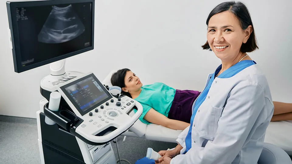
Sonography is a diagnostic test that uses high-frequency sound waves to create detailed images of different parts of the body to detect anomalies and make diagnoses. Without sonography, it would be much harder to treat a range of conditions. The sonography field has a lot to offer students who have the desire to make a difference in the medical field and grow their technical skills. By learning more about this field, prospective students can ensure that they make the right career decision.
What Is Sonography?
Introducing Concorde's Diagnostic Medical Sonography Program
Sonography or diagnostic ultrasound is an imaging method that uses sound waves to produce images of structures inside the body, including tissues, organs, and blood vessels. Medical professionals use it to detect everything from heart disease to gland abnormalities. Ultrasounds are one of the most common forms of diagnostic imaging, and they can be especially useful during pregnancy. Women typically receive a few ultrasounds during pregnancy to confirm the due date and check to see that the baby is developing properly.
Benefits of Sonography
Sonography offers several advantages in comparison to more traditional diagnostic approaches. Since it's noninvasive, it's much easier to get images of the inside of the body and pinpoint the source of a patient's condition. Little prep is necessary beforehand and there are no incisions required, so patients can get it over and done fairly quickly. Unlike an X-ray, sonography uses sound waves rather than radiation, so no ionizing radiation exposure occurs. This makes it the safest and most preferable way to image a pregnant woman or a young child.
Types of Sonography Careers
There are many different types of sonography , and each serves a distinct purpose and offers a unique career path. Here are several concentrations within the field:
Diagnostic Medical Sonography
Diagnostic medical sonography uses imaging equipment to create images of internal organs. A diagnostic medical sonographer would then analyze those images to diagnose and treat various medical conditions. There are many smaller concentrations within medical sonography. For example, a medical sonographer might specialize in imaging the musculoskeletal system or the body's blood vessels. The use of specialized equipment may be common in this concentration, and professionals typically need a high degree of technical expertise.
Cardiovascular Sonography
Cardiovascular sonography involves the use of imaging equipment to detect heart problems in patients. It captures images of the heart, including its chambers, valves, and blood vessels, allowing cardiologists to recommend a treatment plan quickly. Two- and three-dimensional equipment helps physicians observe the structure of the heart and the blood flow to determine if there are any blockages or significant issues that need addressing.
Abdominal Sonography
Abdominal sonography involves examining the internal organs that make up the abdomen, including the liver, pancreas, gallbladder, spleen, and kidneys. Abdominal sonography helps treat a wide range of diseases, including kidney stones and cancers. Sonograms and ultrasounds can help guide procedures like biopsies and help physicians uncover the source of strange abdominal pains.
Obstetric Sonography
Obstetric sonography looks at the fetus while it's still inside the womb. It assesses the condition of the fetus and the pregnant woman to diagnose any congenital abnormalities, observe the position of the fetus in the womb, and estimate the expected date of birth. Doctors usually only recommend obstetric sonography when there's an important medical need for it, but some women might choose an elective ultrasound to see the fetus for themselves. Most women get at least two ultrasounds during pregnancy (1).
Neurosonology
Neurosonology takes diagnostic images of the brain and nervous system using methods other than traditional ultrasounds and sonograms, such as a transcranial Doppler machine. This machine uses sound waves to explore the blood flow to the patient's brain and detect aneurysms and abnormalities. The internal images the machine takes can also help doctors diagnose medical conditions like cerebral palsy and encephalitis.
Musculoskeletal sonography
Musculoskeletal sonography involves taking pictures of the muscles and parts of the skeletal system to allow doctors to see the muscles, joints, and tendons inside the body. This type of sonography is useful for locating muscle tears and strains before more damage can occur. It can also help doctors look for signs of arthritis and osteoporosis. Sonographers in this concentration may specialize in sports medicine and help athletes recover from an injury on the field.
What Do Sonographers Do?
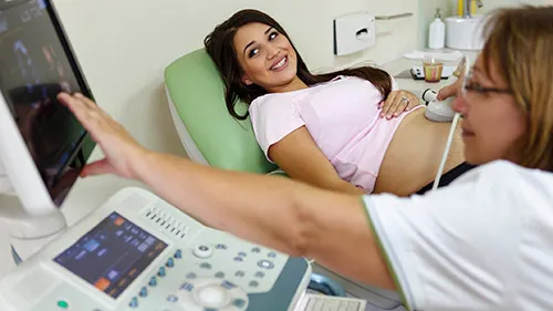
A Day in the Life of Two Sonographers
Sonographers have a wide range of duties that can vary slightly depending on their specialty. Their first responsibility usually involves collecting their patient's medical history, asking them about the concerns they might have, and answering any questions about the procedure. They then conduct the ultrasound scan with the observing physician and create the sonograms that they'll then use to make a formal diagnosis.
It's necessary that sonographers understand how to use the equipment to obtain sonographic images properly. Diagnostic medical sonographers typically use a transabdominal machine to create two-dimensional images to examine the abdominal organs. They might also use two-dimensional and four-dimensional ultrasound equipment and transvaginal or transrectal scanners. Many advancements in medical technology directly affect sonographers and help them perform their duties more effectively. It's often important for sonographers to keep up to date with the latest advancements in the field.
Do Sonographers Differ From Ultrasound Technicians?
Sonographers and ultrasound technicians often have the same job. They both might perform an ultrasound and produce a sonogram to help treat patients. In some cases, sonographers might have more responsibilities and more specialized knowledge related to sonograms and equipment. They might also take on a more active role in helping diagnose patients and constructing treatment plans. Due to their additional expertise, it may be necessary that these professionals complete additional training before they can begin working in a medical facility.
Is Sonography a Good Career Field?
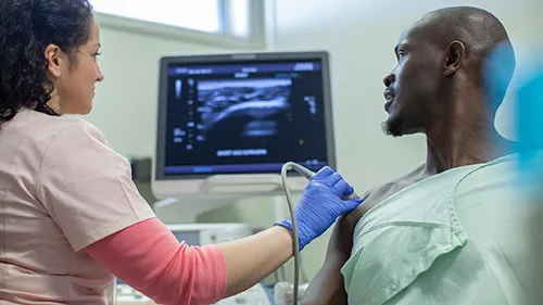
Top Reasons To Train for a Career in Cardiovascular Sonography
Sonography is one of the fastest-growing medical careers that require an associate degree and specialization. With a projected growth rate of 10% through 2031 (2), there's a demand for the position in a wide range of health care settings, including hospitals, walk-in clinics, and doctor's offices. Although most sonographers work in full-time roles, part-time work can also be common. The job can offer a higher degree of flexibility than many jobs in the medical field, allowing professionals to have a good work-life balance and establish more positive working relationships.
Sonographers also play an important role in the early detection and treatment of disease and injury, making it an especially rewarding career. These medical technicians have the opportunity to work closely with both doctors and patients and make a real difference in the lives of the patients they treat. They're often among the first to access all the cutting-edge new technologies available to mitigate and prevent disease. They're also not usually tied down to a single facility or concentration.
What Are the Licensing or Certification Requirements?
Licensing and certifications aren't typically necessary to work as a sonographer , but individual employers may have varying requirements. Individuals interested in pursuing a full-time career in sonography should consider enrolling in an educational program to help them acquire the skills needed to perform ultrasounds and diagnose patients.
What Education Level Is Required To Become a Sonographer?
Like most medical careers, employers expect sonographers to have a certain level of education. To work as a sonographer, it's necessary to first complete a one-year certificate program or a two-year associate degree program. Completing a program allows aspiring sonographers to develop the skills they need to perform a variety of diagnostic procedures, obtain clear images, and summarize their findings to doctors and other medical professionals.
Compared to other health care professionals, sonographers don't need to spend years poring over books and preparing for exams. Concorde offers two associate degree programs in sonography that take as little as 20 months to complete. Both programs include lab work designed to equip students with the technical expertise needed to perform examinations, use an ultrasound machine to conduct ultrasound images of the body's internal organs, and become skilled professionals.
The program also integrates traditional classroom instruction with clinical instruction to provide students with the opportunity to learn how to record a variety of medical data and apply critical thinking and problem-solving to a range of different situations. Concorde's education programs offer hands-on training and clinical experiences to provide students with the opportunity to gain the experience necessary to work with the human body and obtain entry-level employment. In addition, financial aid and a variety of scholarships are available to students concerned with the expenses related to continuing their education.
Interested candidates can contact us online for more information about our sonography programs and the curriculum we offer. Simply complete the online form and instantly get a helpful resource to use as guidance when exploring various career training options. We also offer several other in-demand health care degree and diploma programs with educational and vocational training. Our programs take less than two years and can prepare students for a rewarding career in a range of health care positions.
Footnotes
1. "Ultrasounds During Pregnancy: How Many and How Often?", Beth Israel Deaconess Medical Center, https://www.bidmc.org/about-bidmc/wellness-insights/pregnancy/2018/09/ultrasounds-during-pregnancy-how-many-and-how-often
2. "Diagnostic Medical Sonographers and Cardiovascular Technologists and Technicians," U.S. Bureau of Labor Statistics, https://www.bls.gov/ooh/healthcare/diagnostic-medical-sonographers.htm

Take The Next Step Towards a Brighter Future
Interested in learning more about our Diagnostic Medical Sonography program ? We have a Concorde representative ready to talk about what matters most to you. Get answers about start dates, curriculum, financial aid, scholarships and more!
Request Info
Related Articles
September 15
What Is The Difference Between CVS & DMS
How To Become a Diagnostic Medical Sonographer
How to Become a Sonographer

December 15
How To Prepare For An Admissions Appointment At Concorde
Latest Articles

National Nurses Month: Celebrating Courage and Compassion

Amy Marks: From Medical Assistant to OTA Success
Shining a Light on Occupational Therapy
Program length may be subject to change dependent on transfer credits and course load. Please refer to current course catalog for more information. Concorde does not guarantee admittance, graduation, subsequent employment or salary amount.
Professional certification is not a requirement for graduation, may not be a requirement for employment nor does it guarantee employment.
Financial aid is available to those who qualify but may not be available for all programs. Concorde does not guarantee financial aid or scholarship awards or amounts.
Clinical hour requirements and delivery may vary by campus location and may be subject to change. Concorde does not guarantee clinical site assignments based upon student preference or geographic convenience; nor do clinical experiences guarantee graduation, post-clinical employment or salary outcomes.
Registration and certification requirements for taking and passing these examinations are not controlled by Concorde, but by outside agencies, and are subject to change by the agency without notice. Therefore, Concorde cannot guarantee that graduates will be eligible to take these exams, at all or at any specific time, regardless of their eligibility status upon enrollment.
Request Information
Head Start Your Radiology Residency [Online] ↗️
- Radiology Thesis – More than 400 Research Topics (2022)!
Please login to bookmark

Introduction
A thesis or dissertation, as some people would like to call it, is an integral part of the Radiology curriculum, be it MD, DNB, or DMRD. We have tried to aggregate radiology thesis topics from various sources for reference.
Not everyone is interested in research, and writing a Radiology thesis can be daunting. But there is no escape from preparing, so it is better that you accept this bitter truth and start working on it instead of cribbing about it (like other things in life. #PhilosophyGyan!)
Start working on your thesis as early as possible and finish your thesis well before your exams, so you do not have that stress at the back of your mind. Also, your thesis may need multiple revisions, so be prepared and allocate time accordingly.
Tips for Choosing Radiology Thesis and Research Topics
Keep it simple silly (kiss).
Retrospective > Prospective
Retrospective studies are better than prospective ones, as you already have the data you need when choosing to do a retrospective study. Prospective studies are better quality, but as a resident, you may not have time (, energy and enthusiasm) to complete these.
Choose a simple topic that answers a single/few questions
Original research is challenging, especially if you do not have prior experience. I would suggest you choose a topic that answers a single or few questions. Most topics that I have listed are along those lines. Alternatively, you can choose a broad topic such as “Role of MRI in evaluation of perianal fistulas.”
You can choose a novel topic if you are genuinely interested in research AND have a good mentor who will guide you. Once you have done that, make sure that you publish your study once you are done with it.
Get it done ASAP.
In most cases, it makes sense to stick to a thesis topic that will not take much time. That does not mean you should ignore your thesis and ‘Ctrl C + Ctrl V’ from a friend from another university. Thesis writing is your first step toward research methodology so do it as sincerely as possible. Do not procrastinate in preparing the thesis. As soon as you have been allotted a guide, start researching topics and writing a review of the literature.
At the same time, do not invest a lot of time in writing/collecting data for your thesis. You should not be busy finishing your thesis a few months before the exam. Some people could not appear for the exam because they could not submit their thesis in time. So DO NOT TAKE thesis lightly.
Do NOT Copy-Paste
Reiterating once again, do not simply choose someone else’s thesis topic. Find out what are kind of cases that your Hospital caters to. It is better to do a good thesis on a common topic than a crappy one on a rare one.
Books to help you write a Radiology Thesis
Event country/university has a different format for thesis; hence these book recommendations may not work for everyone.

- Amazon Kindle Edition
- Gupta, Piyush (Author)
- English (Publication Language)
- 206 Pages - 10/12/2020 (Publication Date) - Jaypee Brothers Medical Publishers (P) Ltd. (Publisher)
In A Hurry? Download a PDF list of Radiology Research Topics!
Sign up below to get this PDF directly to your email address.
100% Privacy Guaranteed. Your information will not be shared. Unsubscribe anytime with a single click.
List of Radiology Research /Thesis / Dissertation Topics
- State of the art of MRI in the diagnosis of hepatic focal lesions
- Multimodality imaging evaluation of sacroiliitis in newly diagnosed patients of spondyloarthropathy
- Multidetector computed tomography in oesophageal varices
- Role of positron emission tomography with computed tomography in the diagnosis of cancer Thyroid
- Evaluation of focal breast lesions using ultrasound elastography
- Role of MRI diffusion tensor imaging in the assessment of traumatic spinal cord injuries
- Sonographic imaging in male infertility
- Comparison of color Doppler and digital subtraction angiography in occlusive arterial disease in patients with lower limb ischemia
- The role of CT urography in Haematuria
- Role of functional magnetic resonance imaging in making brain tumor surgery safer
- Prediction of pre-eclampsia and fetal growth restriction by uterine artery Doppler
- Role of grayscale and color Doppler ultrasonography in the evaluation of neonatal cholestasis
- Validity of MRI in the diagnosis of congenital anorectal anomalies
- Role of sonography in assessment of clubfoot
- Role of diffusion MRI in preoperative evaluation of brain neoplasms
- Imaging of upper airways for pre-anaesthetic evaluation purposes and for laryngeal afflictions.
- A study of multivessel (arterial and venous) Doppler velocimetry in intrauterine growth restriction
- Multiparametric 3tesla MRI of suspected prostatic malignancy.
- Role of Sonography in Characterization of Thyroid Nodules for differentiating benign from
- Role of advances magnetic resonance imaging sequences in multiple sclerosis
- Role of multidetector computed tomography in evaluation of jaw lesions
- Role of Ultrasound and MR Imaging in the Evaluation of Musculotendinous Pathologies of Shoulder Joint
- Role of perfusion computed tomography in the evaluation of cerebral blood flow, blood volume and vascular permeability of cerebral neoplasms
- MRI flow quantification in the assessment of the commonest csf flow abnormalities
- Role of diffusion-weighted MRI in evaluation of prostate lesions and its histopathological correlation
- CT enterography in evaluation of small bowel disorders
- Comparison of perfusion magnetic resonance imaging (PMRI), magnetic resonance spectroscopy (MRS) in and positron emission tomography-computed tomography (PET/CT) in post radiotherapy treated gliomas to detect recurrence
- Role of multidetector computed tomography in evaluation of paediatric retroperitoneal masses
- Role of Multidetector computed tomography in neck lesions
- Estimation of standard liver volume in Indian population
- Role of MRI in evaluation of spinal trauma
- Role of modified sonohysterography in female factor infertility: a pilot study.
- The role of pet-CT in the evaluation of hepatic tumors
- Role of 3D magnetic resonance imaging tractography in assessment of white matter tracts compromise in supratentorial tumors
- Role of dual phase multidetector computed tomography in gallbladder lesions
- Role of multidetector computed tomography in assessing anatomical variants of nasal cavity and paranasal sinuses in patients of chronic rhinosinusitis.
- magnetic resonance spectroscopy in multiple sclerosis
- Evaluation of thyroid nodules by ultrasound elastography using acoustic radiation force impulse (ARFI) imaging
- Role of Magnetic Resonance Imaging in Intractable Epilepsy
- Evaluation of suspected and known coronary artery disease by 128 slice multidetector CT.
- Role of regional diffusion tensor imaging in the evaluation of intracranial gliomas and its histopathological correlation
- Role of chest sonography in diagnosing pneumothorax
- Role of CT virtual cystoscopy in diagnosis of urinary bladder neoplasia
- Role of MRI in assessment of valvular heart diseases
- High resolution computed tomography of temporal bone in unsafe chronic suppurative otitis media
- Multidetector CT urography in the evaluation of hematuria
- Contrast-induced nephropathy in diagnostic imaging investigations with intravenous iodinated contrast media
- Comparison of dynamic susceptibility contrast-enhanced perfusion magnetic resonance imaging and single photon emission computed tomography in patients with little’s disease
- Role of Multidetector Computed Tomography in Bowel Lesions.
- Role of diagnostic imaging modalities in evaluation of post liver transplantation recipient complications.
- Role of multislice CT scan and barium swallow in the estimation of oesophageal tumour length
- Malignant Lesions-A Prospective Study.
- Value of ultrasonography in assessment of acute abdominal diseases in pediatric age group
- Role of three dimensional multidetector CT hysterosalpingography in female factor infertility
- Comparative evaluation of multi-detector computed tomography (MDCT) virtual tracheo-bronchoscopy and fiberoptic tracheo-bronchoscopy in airway diseases
- Role of Multidetector CT in the evaluation of small bowel obstruction
- Sonographic evaluation in adhesive capsulitis of shoulder
- Utility of MR Urography Versus Conventional Techniques in Obstructive Uropathy
- MRI of the postoperative knee
- Role of 64 slice-multi detector computed tomography in diagnosis of bowel and mesenteric injury in blunt abdominal trauma.
- Sonoelastography and triphasic computed tomography in the evaluation of focal liver lesions
- Evaluation of Role of Transperineal Ultrasound and Magnetic Resonance Imaging in Urinary Stress incontinence in Women
- Multidetector computed tomographic features of abdominal hernias
- Evaluation of lesions of major salivary glands using ultrasound elastography
- Transvaginal ultrasound and magnetic resonance imaging in female urinary incontinence
- MDCT colonography and double-contrast barium enema in evaluation of colonic lesions
- Role of MRI in diagnosis and staging of urinary bladder carcinoma
- Spectrum of imaging findings in children with febrile neutropenia.
- Spectrum of radiographic appearances in children with chest tuberculosis.
- Role of computerized tomography in evaluation of mediastinal masses in pediatric
- Diagnosing renal artery stenosis: Comparison of multimodality imaging in diabetic patients
- Role of multidetector CT virtual hysteroscopy in the detection of the uterine & tubal causes of female infertility
- Role of multislice computed tomography in evaluation of crohn’s disease
- CT quantification of parenchymal and airway parameters on 64 slice MDCT in patients of chronic obstructive pulmonary disease
- Comparative evaluation of MDCT and 3t MRI in radiographically detected jaw lesions.
- Evaluation of diagnostic accuracy of ultrasonography, colour Doppler sonography and low dose computed tomography in acute appendicitis
- Ultrasonography , magnetic resonance cholangio-pancreatography (MRCP) in assessment of pediatric biliary lesions
- Multidetector computed tomography in hepatobiliary lesions.
- Evaluation of peripheral nerve lesions with high resolution ultrasonography and colour Doppler
- Multidetector computed tomography in pancreatic lesions
- Multidetector Computed Tomography in Paediatric abdominal masses.
- Evaluation of focal liver lesions by colour Doppler and MDCT perfusion imaging
- Sonographic evaluation of clubfoot correction during Ponseti treatment
- Role of multidetector CT in characterization of renal masses
- Study to assess the role of Doppler ultrasound in evaluation of arteriovenous (av) hemodialysis fistula and the complications of hemodialysis vasular access
- Comparative study of multiphasic contrast-enhanced CT and contrast-enhanced MRI in the evaluation of hepatic mass lesions
- Sonographic spectrum of rheumatoid arthritis
- Diagnosis & staging of liver fibrosis by ultrasound elastography in patients with chronic liver diseases
- Role of multidetector computed tomography in assessment of jaw lesions.
- Role of high-resolution ultrasonography in the differentiation of benign and malignant thyroid lesions
- Radiological evaluation of aortic aneurysms in patients selected for endovascular repair
- Role of conventional MRI, and diffusion tensor imaging tractography in evaluation of congenital brain malformations
- To evaluate the status of coronary arteries in patients with non-valvular atrial fibrillation using 256 multirow detector CT scan
- A comparative study of ultrasonography and CT – arthrography in diagnosis of chronic ligamentous and meniscal injuries of knee
- Multi detector computed tomography evaluation in chronic obstructive pulmonary disease and correlation with severity of disease
- Diffusion weighted and dynamic contrast enhanced magnetic resonance imaging in chemoradiotherapeutic response evaluation in cervical cancer.
- High resolution sonography in the evaluation of non-traumatic painful wrist
- The role of trans-vaginal ultrasound versus magnetic resonance imaging in diagnosis & evaluation of cancer cervix
- Role of multidetector row computed tomography in assessment of maxillofacial trauma
- Imaging of vascular complication after liver transplantation.
- Role of magnetic resonance perfusion weighted imaging & spectroscopy for grading of glioma by correlating perfusion parameter of the lesion with the final histopathological grade
- Magnetic resonance evaluation of abdominal tuberculosis.
- Diagnostic usefulness of low dose spiral HRCT in diffuse lung diseases
- Role of dynamic contrast enhanced and diffusion weighted magnetic resonance imaging in evaluation of endometrial lesions
- Contrast enhanced digital mammography anddigital breast tomosynthesis in early diagnosis of breast lesion
- Evaluation of Portal Hypertension with Colour Doppler flow imaging and magnetic resonance imaging
- Evaluation of musculoskeletal lesions by magnetic resonance imaging
- Role of diffusion magnetic resonance imaging in assessment of neoplastic and inflammatory brain lesions
- Radiological spectrum of chest diseases in HIV infected children High resolution ultrasonography in neck masses in children
- with surgical findings
- Sonographic evaluation of peripheral nerves in type 2 diabetes mellitus.
- Role of perfusion computed tomography in the evaluation of neck masses and correlation
- Role of ultrasonography in the diagnosis of knee joint lesions
- Role of ultrasonography in evaluation of various causes of pelvic pain in first trimester of pregnancy.
- Role of Magnetic Resonance Angiography in the Evaluation of Diseases of Aorta and its Branches
- MDCT fistulography in evaluation of fistula in Ano
- Role of multislice CT in diagnosis of small intestine tumors
- Role of high resolution CT in differentiation between benign and malignant pulmonary nodules in children
- A study of multidetector computed tomography urography in urinary tract abnormalities
- Role of high resolution sonography in assessment of ulnar nerve in patients with leprosy.
- Pre-operative radiological evaluation of locally aggressive and malignant musculoskeletal tumours by computed tomography and magnetic resonance imaging.
- The role of ultrasound & MRI in acute pelvic inflammatory disease
- Ultrasonography compared to computed tomographic arthrography in the evaluation of shoulder pain
- Role of Multidetector Computed Tomography in patients with blunt abdominal trauma.
- The Role of Extended field-of-view Sonography and compound imaging in Evaluation of Breast Lesions
- Evaluation of focal pancreatic lesions by Multidetector CT and perfusion CT
- Evaluation of breast masses on sono-mammography and colour Doppler imaging
- Role of CT virtual laryngoscopy in evaluation of laryngeal masses
- Triple phase multi detector computed tomography in hepatic masses
- Role of transvaginal ultrasound in diagnosis and treatment of female infertility
- Role of ultrasound and color Doppler imaging in assessment of acute abdomen due to female genetal causes
- High resolution ultrasonography and color Doppler ultrasonography in scrotal lesion
- Evaluation of diagnostic accuracy of ultrasonography with colour Doppler vs low dose computed tomography in salivary gland disease
- Role of multidetector CT in diagnosis of salivary gland lesions
- Comparison of diagnostic efficacy of ultrasonography and magnetic resonance cholangiopancreatography in obstructive jaundice: A prospective study
- Evaluation of varicose veins-comparative assessment of low dose CT venogram with sonography: pilot study
- Role of mammotome in breast lesions
- The role of interventional imaging procedures in the treatment of selected gynecological disorders
- Role of transcranial ultrasound in diagnosis of neonatal brain insults
- Role of multidetector CT virtual laryngoscopy in evaluation of laryngeal mass lesions
- Evaluation of adnexal masses on sonomorphology and color Doppler imaginig
- Role of radiological imaging in diagnosis of endometrial carcinoma
- Comprehensive imaging of renal masses by magnetic resonance imaging
- The role of 3D & 4D ultrasonography in abnormalities of fetal abdomen
- Diffusion weighted magnetic resonance imaging in diagnosis and characterization of brain tumors in correlation with conventional MRI
- Role of diffusion weighted MRI imaging in evaluation of cancer prostate
- Role of multidetector CT in diagnosis of urinary bladder cancer
- Role of multidetector computed tomography in the evaluation of paediatric retroperitoneal masses.
- Comparative evaluation of gastric lesions by double contrast barium upper G.I. and multi detector computed tomography
- Evaluation of hepatic fibrosis in chronic liver disease using ultrasound elastography
- Role of MRI in assessment of hydrocephalus in pediatric patients
- The role of sonoelastography in characterization of breast lesions
- The influence of volumetric tumor doubling time on survival of patients with intracranial tumours
- Role of perfusion computed tomography in characterization of colonic lesions
- Role of proton MRI spectroscopy in the evaluation of temporal lobe epilepsy
- Role of Doppler ultrasound and multidetector CT angiography in evaluation of peripheral arterial diseases.
- Role of multidetector computed tomography in paranasal sinus pathologies
- Role of virtual endoscopy using MDCT in detection & evaluation of gastric pathologies
- High resolution 3 Tesla MRI in the evaluation of ankle and hindfoot pain.
- Transperineal ultrasonography in infants with anorectal malformation
- CT portography using MDCT versus color Doppler in detection of varices in cirrhotic patients
- Role of CT urography in the evaluation of a dilated ureter
- Characterization of pulmonary nodules by dynamic contrast-enhanced multidetector CT
- Comprehensive imaging of acute ischemic stroke on multidetector CT
- The role of fetal MRI in the diagnosis of intrauterine neurological congenital anomalies
- Role of Multidetector computed tomography in pediatric chest masses
- Multimodality imaging in the evaluation of palpable & non-palpable breast lesion.
- Sonographic Assessment Of Fetal Nasal Bone Length At 11-28 Gestational Weeks And Its Correlation With Fetal Outcome.
- Role Of Sonoelastography And Contrast-Enhanced Computed Tomography In Evaluation Of Lymph Node Metastasis In Head And Neck Cancers
- Role Of Renal Doppler And Shear Wave Elastography In Diabetic Nephropathy
- Evaluation Of Relationship Between Various Grades Of Fatty Liver And Shear Wave Elastography Values
- Evaluation and characterization of pelvic masses of gynecological origin by USG, color Doppler and MRI in females of reproductive age group
- Radiological evaluation of small bowel diseases using computed tomographic enterography
- Role of coronary CT angiography in patients of coronary artery disease
- Role of multimodality imaging in the evaluation of pediatric neck masses
- Role of CT in the evaluation of craniocerebral trauma
- Role of magnetic resonance imaging (MRI) in the evaluation of spinal dysraphism
- Comparative evaluation of triple phase CT and dynamic contrast-enhanced MRI in patients with liver cirrhosis
- Evaluation of the relationship between carotid intima-media thickness and coronary artery disease in patients evaluated by coronary angiography for suspected CAD
- Assessment of hepatic fat content in fatty liver disease by unenhanced computed tomography
- Correlation of vertebral marrow fat on spectroscopy and diffusion-weighted MRI imaging with bone mineral density in postmenopausal women.
- Comparative evaluation of CT coronary angiography with conventional catheter coronary angiography
- Ultrasound evaluation of kidney length & descending colon diameter in normal and intrauterine growth-restricted fetuses
- A prospective study of hepatic vein waveform and splenoportal index in liver cirrhosis: correlation with child Pugh’s classification and presence of esophageal varices.
- CT angiography to evaluate coronary artery by-pass graft patency in symptomatic patient’s functional assessment of myocardium by cardiac MRI in patients with myocardial infarction
- MRI evaluation of HIV positive patients with central nervous system manifestations
- MDCT evaluation of mediastinal and hilar masses
- Evaluation of rotator cuff & labro-ligamentous complex lesions by MRI & MRI arthrography of shoulder joint
- Role of imaging in the evaluation of soft tissue vascular malformation
- Role of MRI and ultrasonography in the evaluation of multifidus muscle pathology in chronic low back pain patients
- Role of ultrasound elastography in the differential diagnosis of breast lesions
- Role of magnetic resonance cholangiopancreatography in evaluating dilated common bile duct in patients with symptomatic gallstone disease.
- Comparative study of CT urography & hybrid CT urography in patients with haematuria.
- Role of MRI in the evaluation of anorectal malformations
- Comparison of ultrasound-Doppler and magnetic resonance imaging findings in rheumatoid arthritis of hand and wrist
- Role of Doppler sonography in the evaluation of renal artery stenosis in hypertensive patients undergoing coronary angiography for coronary artery disease.
- Comparison of radiography, computed tomography and magnetic resonance imaging in the detection of sacroiliitis in ankylosing spondylitis.
- Mr evaluation of painful hip
- Role of MRI imaging in pretherapeutic assessment of oral and oropharyngeal malignancy
- Evaluation of diffuse lung diseases by high resolution computed tomography of the chest
- Mr evaluation of brain parenchyma in patients with craniosynostosis.
- Diagnostic and prognostic value of cardiovascular magnetic resonance imaging in dilated cardiomyopathy
- Role of multiparametric magnetic resonance imaging in the detection of early carcinoma prostate
- Role of magnetic resonance imaging in white matter diseases
- Role of sonoelastography in assessing the response to neoadjuvant chemotherapy in patients with locally advanced breast cancer.
- Role of ultrasonography in the evaluation of carotid and femoral intima-media thickness in predialysis patients with chronic kidney disease
- Role of H1 MRI spectroscopy in focal bone lesions of peripheral skeleton choline detection by MRI spectroscopy in breast cancer and its correlation with biomarkers and histological grade.
- Ultrasound and MRI evaluation of axillary lymph node status in breast cancer.
- Role of sonography and magnetic resonance imaging in evaluating chronic lateral epicondylitis.
- Comparative of sonography including Doppler and sonoelastography in cervical lymphadenopathy.
- Evaluation of Umbilical Coiling Index as Predictor of Pregnancy Outcome.
- Computerized Tomographic Evaluation of Azygoesophageal Recess in Adults.
- Lumbar Facet Arthropathy in Low Backache.
- “Urethral Injuries After Pelvic Trauma: Evaluation with Uretrography
- Role Of Ct In Diagnosis Of Inflammatory Renal Diseases
- Role Of Ct Virtual Laryngoscopy In Evaluation Of Laryngeal Masses
- “Ct Portography Using Mdct Versus Color Doppler In Detection Of Varices In
- Cirrhotic Patients”
- Role Of Multidetector Ct In Characterization Of Renal Masses
- Role Of Ct Virtual Cystoscopy In Diagnosis Of Urinary Bladder Neoplasia
- Role Of Multislice Ct In Diagnosis Of Small Intestine Tumors
- “Mri Flow Quantification In The Assessment Of The Commonest CSF Flow Abnormalities”
- “The Role Of Fetal Mri In Diagnosis Of Intrauterine Neurological CongenitalAnomalies”
- Role Of Transcranial Ultrasound In Diagnosis Of Neonatal Brain Insults
- “The Role Of Interventional Imaging Procedures In The Treatment Of Selected Gynecological Disorders”
- Role Of Radiological Imaging In Diagnosis Of Endometrial Carcinoma
- “Role Of High-Resolution Ct In Differentiation Between Benign And Malignant Pulmonary Nodules In Children”
- Role Of Ultrasonography In The Diagnosis Of Knee Joint Lesions
- “Role Of Diagnostic Imaging Modalities In Evaluation Of Post Liver Transplantation Recipient Complications”
- “Diffusion-Weighted Magnetic Resonance Imaging In Diagnosis And
- Characterization Of Brain Tumors In Correlation With Conventional Mri”
- The Role Of PET-CT In The Evaluation Of Hepatic Tumors
- “Role Of Computerized Tomography In Evaluation Of Mediastinal Masses In Pediatric patients”
- “Trans Vaginal Ultrasound And Magnetic Resonance Imaging In Female Urinary Incontinence”
- Role Of Multidetector Ct In Diagnosis Of Urinary Bladder Cancer
- “Role Of Transvaginal Ultrasound In Diagnosis And Treatment Of Female Infertility”
- Role Of Diffusion-Weighted Mri Imaging In Evaluation Of Cancer Prostate
- “Role Of Positron Emission Tomography With Computed Tomography In Diagnosis Of Cancer Thyroid”
- The Role Of CT Urography In Case Of Haematuria
- “Value Of Ultrasonography In Assessment Of Acute Abdominal Diseases In Pediatric Age Group”
- “Role Of Functional Magnetic Resonance Imaging In Making Brain Tumor Surgery Safer”
- The Role Of Sonoelastography In Characterization Of Breast Lesions
- “Ultrasonography, Magnetic Resonance Cholangiopancreatography (MRCP) In Assessment Of Pediatric Biliary Lesions”
- “Role Of Ultrasound And Color Doppler Imaging In Assessment Of Acute Abdomen Due To Female Genital Causes”
- “Role Of Multidetector Ct Virtual Laryngoscopy In Evaluation Of Laryngeal Mass Lesions”
- MRI Of The Postoperative Knee
- Role Of Mri In Assessment Of Valvular Heart Diseases
- The Role Of 3D & 4D Ultrasonography In Abnormalities Of Fetal Abdomen
- State Of The Art Of Mri In Diagnosis Of Hepatic Focal Lesions
- Role Of Multidetector Ct In Diagnosis Of Salivary Gland Lesions
- “Role Of Virtual Endoscopy Using Mdct In Detection & Evaluation Of Gastric Pathologies”
- The Role Of Ultrasound & Mri In Acute Pelvic Inflammatory Disease
- “Diagnosis & Staging Of Liver Fibrosis By Ultraso Und Elastography In
- Patients With Chronic Liver Diseases”
- Role Of Mri In Evaluation Of Spinal Trauma
- Validity Of Mri In Diagnosis Of Congenital Anorectal Anomalies
- Imaging Of Vascular Complication After Liver Transplantation
- “Contrast-Enhanced Digital Mammography And Digital Breast Tomosynthesis In Early Diagnosis Of Breast Lesion”
- Role Of Mammotome In Breast Lesions
- “Role Of MRI Diffusion Tensor Imaging (DTI) In Assessment Of Traumatic Spinal Cord Injuries”
- “Prediction Of Pre-eclampsia And Fetal Growth Restriction By Uterine Artery Doppler”
- “Role Of Multidetector Row Computed Tomography In Assessment Of Maxillofacial Trauma”
- “Role Of Diffusion Magnetic Resonance Imaging In Assessment Of Neoplastic And Inflammatory Brain Lesions”
- Role Of Diffusion Mri In Preoperative Evaluation Of Brain Neoplasms
- “Role Of Multidetector Ct Virtual Hysteroscopy In The Detection Of The
- Uterine & Tubal Causes Of Female Infertility”
- Role Of Advances Magnetic Resonance Imaging Sequences In Multiple Sclerosis Magnetic Resonance Spectroscopy In Multiple Sclerosis
- “Role Of Conventional Mri, And Diffusion Tensor Imaging Tractography In Evaluation Of Congenital Brain Malformations”
- Role Of MRI In Evaluation Of Spinal Trauma
- Diagnostic Role Of Diffusion-weighted MR Imaging In Neck Masses
- “The Role Of Transvaginal Ultrasound Versus Magnetic Resonance Imaging In Diagnosis & Evaluation Of Cancer Cervix”
- “Role Of 3d Magnetic Resonance Imaging Tractography In Assessment Of White Matter Tracts Compromise In Supra Tentorial Tumors”
- Role Of Proton MR Spectroscopy In The Evaluation Of Temporal Lobe Epilepsy
- Role Of Multislice Computed Tomography In Evaluation Of Crohn’s Disease
- Role Of MRI In Assessment Of Hydrocephalus In Pediatric Patients
- The Role Of MRI In Diagnosis And Staging Of Urinary Bladder Carcinoma
- USG and MRI correlation of congenital CNS anomalies
- HRCT in interstitial lung disease
- X-Ray, CT and MRI correlation of bone tumors
- “Study on the diagnostic and prognostic utility of X-Rays for cases of pulmonary tuberculosis under RNTCP”
- “Role of magnetic resonance imaging in the characterization of female adnexal pathology”
- “CT angiography of carotid atherosclerosis and NECT brain in cerebral ischemia, a correlative analysis”
- Role of CT scan in the evaluation of paranasal sinus pathology
- USG and MRI correlation on shoulder joint pathology
- “Radiological evaluation of a patient presenting with extrapulmonary tuberculosis”
- CT and MRI correlation in focal liver lesions”
- Comparison of MDCT virtual cystoscopy with conventional cystoscopy in bladder tumors”
- “Bleeding vessels in life-threatening hemoptysis: Comparison of 64 detector row CT angiography with conventional angiography prior to endovascular management”
- “Role of transarterial chemoembolization in unresectable hepatocellular carcinoma”
- “Comparison of color flow duplex study with digital subtraction angiography in the evaluation of peripheral vascular disease”
- “A Study to assess the efficacy of magnetization transfer ratio in differentiating tuberculoma from neurocysticercosis”
- “MR evaluation of uterine mass lesions in correlation with transabdominal, transvaginal ultrasound using HPE as a gold standard”
- “The Role of power Doppler imaging with trans rectal ultrasonogram guided prostate biopsy in the detection of prostate cancer”
- “Lower limb arteries assessed with doppler angiography – A prospective comparative study with multidetector CT angiography”
- “Comparison of sildenafil with papaverine in penile doppler by assessing hemodynamic changes”
- “Evaluation of efficacy of sonosalphingogram for assessing tubal patency in infertile patients with hysterosalpingogram as the gold standard”
- Role of CT enteroclysis in the evaluation of small bowel diseases
- “MRI colonography versus conventional colonoscopy in the detection of colonic polyposis”
- “Magnetic Resonance Imaging of anteroposterior diameter of the midbrain – differentiation of progressive supranuclear palsy from Parkinson disease”
- “MRI Evaluation of anterior cruciate ligament tears with arthroscopic correlation”
- “The Clinicoradiological profile of cerebral venous sinus thrombosis with prognostic evaluation using MR sequences”
- “Role of MRI in the evaluation of pelvic floor integrity in stress incontinent patients” “Doppler ultrasound evaluation of hepatic venous waveform in portal hypertension before and after propranolol”
- “Role of transrectal sonography with colour doppler and MRI in evaluation of prostatic lesions with TRUS guided biopsy correlation”
- “Ultrasonographic evaluation of painful shoulders and correlation of rotator cuff pathologies and clinical examination”
- “Colour Doppler Evaluation of Common Adult Hepatic tumors More Than 2 Cm with HPE and CECT Correlation”
- “Clinical Relevance of MR Urethrography in Obliterative Posterior Urethral Stricture”
- “Prediction of Adverse Perinatal Outcome in Growth Restricted Fetuses with Antenatal Doppler Study”
- Radiological evaluation of spinal dysraphism using CT and MRI
- “Evaluation of temporal bone in cholesteatoma patients by high resolution computed tomography”
- “Radiological evaluation of primary brain tumours using computed tomography and magnetic resonance imaging”
- “Three dimensional colour doppler sonographic assessment of changes in volume and vascularity of fibroids – before and after uterine artery embolization”
- “In phase opposed phase imaging of bone marrow differentiating neoplastic lesions”
- “Role of dynamic MRI in replacing the isotope renogram in the functional evaluation of PUJ obstruction”
- Characterization of adrenal masses with contrast-enhanced CT – washout study
- A study on accuracy of magnetic resonance cholangiopancreatography
- “Evaluation of median nerve in carpal tunnel syndrome by high-frequency ultrasound & color doppler in comparison with nerve conduction studies”
- “Correlation of Agatston score in patients with obstructive and nonobstructive coronary artery disease following STEMI”
- “Doppler ultrasound assessment of tumor vascularity in locally advanced breast cancer at diagnosis and following primary systemic chemotherapy.”
- “Validation of two-dimensional perineal ultrasound and dynamic magnetic resonance imaging in pelvic floor dysfunction.”
- “Role of MR urethrography compared to conventional urethrography in the surgical management of obliterative urethral stricture.”

Search Diagnostic Imaging Research Topics
You can also search research-related resources on our custom search engine .
Free Resources for Preparing Radiology Thesis
- Radiology thesis topics- Benha University – Free to download thesis
- Radiology thesis topics – Faculty of Medical Science Delhi
- Radiology thesis topics – IPGMER
- Fetal Radiology thesis Protocols
- Radiology thesis and dissertation topics
- Radiographics
Proofreading Your Thesis:
Make sure you use Grammarly to correct your spelling , grammar , and plagiarism for your thesis. Grammarly has affordable paid subscriptions, windows/macOS apps, and FREE browser extensions. It is an excellent tool to avoid inadvertent spelling mistakes in your research projects. It has an extensive built-in vocabulary, but you should make an account and add your own medical glossary to it.

Guidelines for Writing a Radiology Thesis:
These are general guidelines and not about radiology specifically. You can share these with colleagues from other departments as well. Special thanks to Dr. Sanjay Yadav sir for these. This section is best seen on a desktop. Here are a couple of handy presentations to start writing a thesis:
Read the general guidelines for writing a thesis (the page will take some time to load- more than 70 pages!
A format for thesis protocol with a sample patient information sheet, sample patient consent form, sample application letter for thesis, and sample certificate.
Resources and References:
- Guidelines for thesis writing.
- Format for thesis protocol
- Thesis protocol writing guidelines DNB
- Informed consent form for Research studies from AIIMS
- Radiology Informed consent forms in local Indian languages.
- Sample Informed Consent form for Research in Hindi
- Guide to write a thesis by Dr. P R Sharma
- Guidelines for thesis writing by Dr. Pulin Gupta.
- Preparing MD/DNB thesis by A Indrayan
- Another good thesis reference protocol
Hopefully, this post will make the tedious task of writing a Radiology thesis a little bit easier for you. Best of luck with writing your thesis and your residency too!
More guides for residents :
- Guide for the MD/DMRD/DNB radiology exam!
Guide for First-Year Radiology Residents
- FRCR Exam: THE Most Comprehensive Guide (2022)!
- Radiology Practical Exams Questions compilation for MD/DNB/DMRD !
- Radiology Exam Resources (Oral Recalls, Instruments, etc )!
- Tips and Tricks for DNB/MD Radiology Practical Exam
- FRCR 2B exam- Tips and Tricks !
- FRCR exam preparation – An alternative take!
- Why did I take up Radiology?
- Radiology Conferences – A comprehensive guide!
ECR (European Congress Of Radiology)
- European Diploma in Radiology (EDiR) – The Complete Guide!
- Radiology NEET PG guide – How to select THE best college for post-graduation in Radiology (includes personal insights)!
- Interventional Radiology – All Your Questions Answered!
- What It Means To Be A Radiologist: A Guide For Medical Students!
- Radiology Mentors for Medical Students (Post NEET-PG)
- MD vs DNB Radiology: Which Path is Right for Your Career?
- DNB Radiology OSCE – Tips and Tricks
More radiology resources here: Radiology resources This page will be updated regularly. Kindly leave your feedback in the comments or send us a message here . Also, you can comment below regarding your department’s thesis topics.
Note: All topics have been compiled from available online resources. If anyone has an issue with any radiology thesis topics displayed here, you can message us here , and we can delete them. These are only sample guidelines. Thesis guidelines differ from institution to institution.
Image source: Thesis complete! (2018). Flickr. Retrieved 12 August 2018, from https://www.flickr.com/photos/cowlet/354911838 by Victoria Catterson
About The Author
Dr. amar udare, md, related posts ↓.
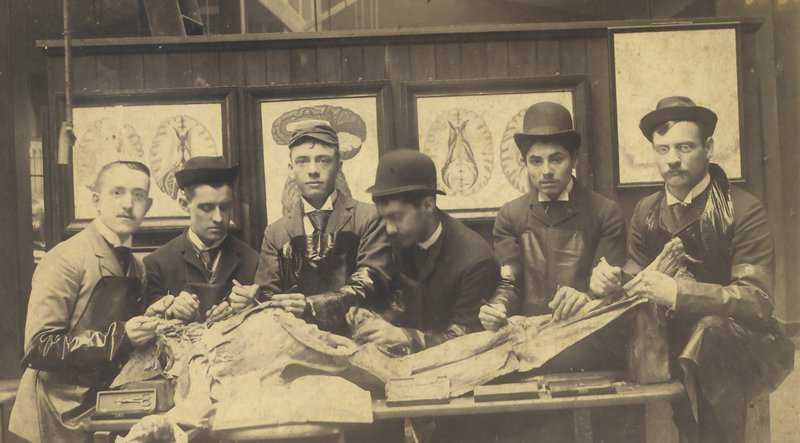
7 thoughts on “Radiology Thesis – More than 400 Research Topics (2022)!”
Amazing & The most helpful site for Radiology residents…
Thank you for your kind comments 🙂
Dr. I saw your Tips is very amazing and referable. But Dr. Can you help me with the thesis of Evaluation of Diagnostic accuracy of X-ray radiograph in knee joint lesion.
Wow! These are excellent stuff. You are indeed a teacher. God bless
Glad you liked these!
happy to see this
Glad I could help :).
Leave a Comment Cancel Reply
Your email address will not be published. Required fields are marked *
Get Radiology Updates to Your Inbox!
This site is for use by medical professionals. To continue, you must accept our use of cookies and the site's Terms of Use. Learn more Accept!
Wish to be a BETTER Radiologist? Join 14000 Radiology Colleagues !
Enter your email address below to access HIGH YIELD radiology content, updates, and resources.
No spam, only VALUE! Unsubscribe anytime with a single click.
An official website of the United States government
The .gov means it's official. Federal government websites often end in .gov or .mil. Before sharing sensitive information, make sure you're on a federal government site.
The site is secure. The https:// ensures that you are connecting to the official website and that any information you provide is encrypted and transmitted securely.
- Publications
- Account settings
- Browse Titles
NCBI Bookshelf. A service of the National Library of Medicine, National Institutes of Health.
StatPearls [Internet]. Treasure Island (FL): StatPearls Publishing; 2024 Jan-.

StatPearls [Internet].
Sonography physical principles and instrumentation.
Christopher S. Borowy ; Taif Mukhdomi .
Affiliations
Last Update: March 20, 2023 .
- Definition/Introduction
The development of sonography or medical ultrasound was built on the understanding and research of sound, which can be dated as far back as the 6th century. Specifically, sonography can be tied to as far back as the 1700s, an Italian physicist, Lazaro Spallanzani, who was researching the navigation of bats. [1] In the early 20th century, more applicable uses of ultrasound were studied by French physicist Paul Langevin who eventually laid the groundwork for SONOR (Sound Navigation and Ranging) as he was commissioned to investigate the sunken Titanic.
One of the first medical applications of ultrasound was by Ian Donald in the 1950s. As Ian Donald extrapolated ultrasound's ability to detect flaws and cracks in steel welds, he began to research and apply it to the field of obstetrics and gynecology as he published a fetus at 14 weeks gestational age in the prestigious academic journal, The Lancet .
Basic Ultrasound Physics
As the name implies, ultrasound uses sound waves at a higher frequency than what is audible for the human ear. For example, the average human ear hears frequencies from the 20 hertz to 20 kHz (kilohertz) range, whereas the typical clinical ultrasound uses frequencies from 1 to 20 MHz (megahertz).
To better understand ultrasound, a simple understanding of sinusoidal waves is useful. Wavelength ( λ ) is inversely proportional to frequency ( f ) and proportionate to the velocity ( v ), as depicted in the following equation.
- λ =v /f
Modern-day ultrasounds utilize piezoelectricity technology to convert electricity to sound waves and convert sound waves back into electricity through the use of lead zirconate titanate (PZT), which are human-made crystals (also called ceramic). The vibration occurs as these crystals are electrically stimulated, which produce longitudinal sound waves in the previously described frequency, thus producing ultrasound waves. [2]
PZT crystals are arranged in unique schematics within an ultrasound transducer (also known as a probe) to be clinically applied in a variety of situations. Further details on the variation of these transducers will be discussed later.
Similar to a rock being dropped into a still pond with the waves bouncing off the shore, the sound waves emitted by the transducer resonate and refract back to the transducer. When the ultrasound reverberates off tissue and returns, they induce vibration into the PZT crystals, converted back into an electric signal. The processing computer within the ultrasound machine then generates the signals into 2D (two dimensional) and, in more recent times, 3D (three dimensional) images.
The frequency (and inversely wavelength) of the sound waves produced can be modified to produce greater penetration through soft tissue by modifying the power or gain. The lower the frequency, the deeper or greater penetrance a sound wave has. Unfortunately, as more penetrance is obtained, the poorer the image quality typically is.
Lastly, some common terminology unique to the field of ultrasound will be discussed. Hyperechoic is defined by an object that is brighter than the surrounding tissue. Hypoechoic or anechoic is the opposite, by which an object is darker. Both of which are determined by the density of the object.
Instrumentation
The following section will focus on a variety of transducers. Each transducer is shaped uniquely and has a specific and intentional arrangement of PZT crystals to produce a distinct image. Each ceramic arrangement has a specific use for various clinical scenarios.
The phased array transducer is one of the physically smaller probes with a usefully small footprint. Specifically, the phased array probe’s footprint can view between ribs to better view the heart or lungs. Due to the probe's size, the periphery and superficial image have poor focus and detail compared to other transducers. In addition to the footprint, the phased array transducers utilize every crystal within the probe to produce the image. [3] (Media #3)
The linear transducer does not directly utilize every crystal, but each element is electrically stimulated or vibrated and subsequently excites adjacent crystals. Each crystal within the linear transducer is not given direct electrical stimulation. The advantage of the linear probe is a larger field of view and a higher superficial focus. Common clinical applications of this probe are within the soft tissue and more superficial structures. (Media #2)
Endocavity probes are one of the most unique in appearance. The wand-like shape and long neck allow sonographers to extend into orifices such as the oropharynx, rectum, or vagina (also called a transvaginal probe). Common clinical applications include surveillance of early pregnancies. Transabdominal imaging may not provide optimal imaging in smaller fetuses and diagnose and be used as a therapeutic tool in draining parapharyngeal abscesses. (Media #5)
Lastly, the curvilinear or curved-array transducer has a convex shape, where the two previously described transducers have a more linear shape. The curvilinear probe typically has the largest footprint of the three. Clinically this probe is utilized within the endocervical, intrauterine, or abdominal cavity. The image produced from the probe is “C” shaped, which is reflective of the probe’s shape. Superficial focus is poor, but the advantage is an overall wider field of view. [4] (Media #4)
With advances in ultrasonography, new research has brought products into the market that utilize a silicon chip in place of piezo crystals that have enhanced ultrasound machines' ability and access multiple images guiding care beyond PZT imaging.
- Issues of Concern
To obtain ideal images when scanning human tissue, a sonographer must understand how to optimize its resolution. The following section will focus on the properties of ultrasound to achieve such images and the common artifacts produced from imperfections in both the tissue and the ultrasound that may cause major issues in the final images.
A sound wave penetrates deeper into a particular cavity; more interference is met. As such, the reverberant sound waves become weaker as they reflect towards the transducer. To optimize image quality, one must alter the gain. Altering the gain does not affect the sound wave impulse sent from the transducer but amplifies what is echoed back and received by the PZT crystals. If the produced image is still suboptimal despite optimizing the gain, the intensity or power of the sound wave emitting can be changed.
If the image produced is too dim, then the strength of the sound waves may be changed by altering the power. The greater the power of the sound waves, the deeper the waves typically penetrate. Power is measured in decibels (dB). As described above, by increasing the frequency of PZT crystals, a louder sound wave, one with greater power, will penetrate deeper, but at the cost of more artifacts in the resulting image.
Despite proper optimization, several errors or artifacts can occur in the production of images. In a perfect world, there are some assumptions used to make these images. These assumptions are that sound waves travel in a straight line and reflect in that same line, sound travels at 1,540 m/s and that the only source of sound waves production is the transducer; with any deviation from these assumptions, artifacts are produced.
When a sound wave encounters a medium such as air, which sound waves poorly pass through compared to water-based tissue, the artifact is known as a shadow is produced. This appears as a hypoechoic or anechoic area seen distal to the area of gas or air, which obscures any views within the shadow . Besides air, a common cause of shadowing is gallstones, for which the shadow can be diagnostic in identifying gallstones within a gallbladder (Media #1).
Comet tails typically have the appearance opposite of a shadow. A comet tail has a hyperechoic, or white, presentation. Common causes of this type of artifact are two closely approximated objects, like a mechanical heart valve. With two objects close together, there is reverberation, which produces the hyperechoic streak.
The last artifact to be discussed is a mirror image. The artificial mirror image is always produced in parallel with the sound beam emitted from an ultrasound transducer. Gas or air is a common culprit in the production of various artifacts, and when a smooth and flat pocket of gas interacts with sound waves, it reflects in the same manner as a common household mirror. Sonographers can utilize the mirror artifact and reflective surfaces to obtain views to their advantage. [5] [6] [7]
- Clinical Significance
The clinical significance of sonography span across multiple medical specialties and fields. Equally important is the training and education surrounding ultrasound, as it is a heavily user-dependent skill and tool. A poor sonographer with the highest quality ultrasound can produce inadequate images. Competent training and experience are crucial. There are three regulatory organizations for sonographic certification; Cardiovascular Credentialing International, the American Registry of Radiologic Technologists, and the American Registry for Diagnostic Medical Sonography. Formal training takes 12-18 months and is combined with written and practical knowledge. [8]
Outside of ultrasound technician training, medical residents and fellows study ultrasound. Specifically, cardiology fellows focus on the cardiac application, nephrology on renal images, and formal fellowships for using ultrasound for emergency medicine residents and attendings. Lastly, radiology residency has formal training in obtaining and interpreting sonographic images. [9]
As described above, the training to properly utilize medical ultrasound has many different pathways. Collectively, technicians, nursing staff, and clinicians benefit from the information obtained from sonography. The coordination subsequently increases patient care and eventual outcomes.
In summary, from the foundation of Lazaro Spallanzani’s research of sonography in bats to Ian Donald extrapolating its use from iron welds to the field of obstetrics, ultrasound is now an immediately available and safe diagnostic and therapeutic tool. The utility and applicability of ultrasound span across outpatient obstetrical offices to obtaining critical information within intensive care units. As technology develops, ultrasound machines are becoming smaller and more portable. The future of ultrasound utility continues to expand and will likely span across almost all medical specialties.
- Nursing, Allied Health, and Interprofessional Team Interventions
Just as Ian Donald saw the use of ultrasound in the field of obstetrics, many have followed and seen its utility in other medical fields: cardiology, vascular surgery, anesthesiology, pain medicine, radiology, general surgery, nephrology, ophthalmology, urology, emergency medicine, obstetrics, and gynecology. [3] [10] Ultrasound is a quick, efficient, and safe diagnostic and therapeutic tool within the medical field. Team efforts can be taken to influence medical care and decision-making.
- Review Questions
- Access free multiple choice questions on this topic.
- Comment on this article.
Gallstone on Point-of-Care Ultrasound Contributed by Emory Emergency Medicine Ultrasound Section
Ultrasound Linear Probe Contributed by Chris Borowy, DO
Ultrasound Phased Array Probe Contributed by Chris Borowy, DO
Ultrasound Curvilinear Probe Contributed by Chris Borowy, DO
Endocavity Ultrasound Probe Contributed by Chris Borowy, DO
Disclosure: Christopher Borowy declares no relevant financial relationships with ineligible companies.
Disclosure: Taif Mukhdomi declares no relevant financial relationships with ineligible companies.
This book is distributed under the terms of the Creative Commons Attribution-NonCommercial-NoDerivatives 4.0 International (CC BY-NC-ND 4.0) ( http://creativecommons.org/licenses/by-nc-nd/4.0/ ), which permits others to distribute the work, provided that the article is not altered or used commercially. You are not required to obtain permission to distribute this article, provided that you credit the author and journal.
- Cite this Page Borowy CS, Mukhdomi T. Sonography Physical Principles And Instrumentation. [Updated 2023 Mar 20]. In: StatPearls [Internet]. Treasure Island (FL): StatPearls Publishing; 2024 Jan-.
In this Page
Bulk download.
- Bulk download StatPearls data from FTP
Related information
- PMC PubMed Central citations
- PubMed Links to PubMed
Similar articles in PubMed
- Jonas S. Friedenwald, man of science. [Invest Ophthalmol Vis Sci. 1980] Jonas S. Friedenwald, man of science. Patz A. Invest Ophthalmol Vis Sci. 1980 Oct; 19(10):1139-49.
- Sonography Female Pelvic Pathology Assessment, Protocols, and Interpretation. [StatPearls. 2024] Sonography Female Pelvic Pathology Assessment, Protocols, and Interpretation. Karena ZV, Mehta AD. StatPearls. 2024 Jan
- [History of the tuning fork. II: Evolution of the classical experiments by Weber, Rinne and Schwabach]. [Laryngorhinootologie. 1997] [History of the tuning fork. II: Evolution of the classical experiments by Weber, Rinne and Schwabach]. Feldmann H. Laryngorhinootologie. 1997 May; 76(5):318-26.
- Review [Reading Spallanzani today]. [Rev Chilena Infectol. 2020] Review [Reading Spallanzani today]. Ledermann D W. Rev Chilena Infectol. 2020 Feb; 37(1):64-68.
- Review [On the first studies of electrophysiology]. [Arch Cardiol Mex. 2011] Review [On the first studies of electrophysiology]. de Micheli A. Arch Cardiol Mex. 2011 Oct-Dec; 81(4):337-42.
Recent Activity
- Sonography Physical Principles And Instrumentation - StatPearls Sonography Physical Principles And Instrumentation - StatPearls
Your browsing activity is empty.
Activity recording is turned off.
Turn recording back on
Connect with NLM
National Library of Medicine 8600 Rockville Pike Bethesda, MD 20894
Web Policies FOIA HHS Vulnerability Disclosure
Help Accessibility Careers
- Type 2 Diabetes
- Heart Disease
- Digestive Health
- Multiple Sclerosis
- Diet & Nutrition
- Supplements
- Health Insurance
- Public Health
- Patient Rights
- Caregivers & Loved Ones
- End of Life Concerns
- Health News
- Thyroid Test Analyzer
- Doctor Discussion Guides
- Hemoglobin A1c Test Analyzer
- Lipid Test Analyzer
- Complete Blood Count (CBC) Analyzer
- What to Buy
- Editorial Process
- Meet Our Medical Expert Board
Sonography: How a Sonogram Test Works and What It Shows
How to prepare for the test and how the results can help diagnose conditions
Before the Test
During the test, interpreting the results, frequently asked questions.
Sonography is a diagnostic medical test that uses high-frequency sound waves, or ultrasound waves, to create images of tissues, glands, organs, and blood or fluid flow within the body. This test is also referred to as an ultrasound or sonogram.
Sonography uses a device called a transducer on the surface of the skin to send ultrasound waves and listen for an echo. A computer translates the ultrasound waves into an image. A trained technician can see, measure, and identify structures in the image. A healthcare provider then reads the images to help diagnose the issue or problem at hand.
This article explains the purpose and limitations of sonography. To demystify the test, this article also explains what to expect before and during the test.
Verywell / Emily Roberts
Purpose of the Test
A sonogram captures a live image of what's going on inside the body. It functions like a camera that takes pictures of body parts or processes in real time.
Sonography is useful for evaluating the size, shape, and density of tissues to help diagnose certain medical conditions. Traditionally, ultrasound imaging is great for looking into the abdomen without having to cut it open.
Abdominal ultrasound is often used to diagnose:
- Gallbladder disease or gallstones
- Kidney stones or kidney disease
- Liver disease
- Appendicitis
- Ovarian cysts
- Ectopic pregnancy
- Uterine growths or fibroids and other conditions
A sonogram is most commonly used to monitor the development of the uterus and fetus during pregnancy. It can also be used to evaluate glands, breast lumps , joint conditions , bone disease , testicular lumps , or to guide needles during biopsies.
Sonography can also recognize blood or fluid flow that moves toward or away from the transducer. It uses color overlays on the image to show the direction of the flow. Very hard and dense tissues or empty spaces, such as organs filled with gas, do not conduct ultrasound waves and therefore cannot be viewed on a sonogram.
Physicians often order a sonogram before moving on to imaging technologies that have more potential for complications. Computerized tomography (CT) , for example, exposes you to significant levels of radiation.
Magnetic resonance imaging (MRI) uses an extremely strong magnet to capture an image. The strength of an MRI magnet can limit its use in patients with metal or devices in their bodies, such as some pacemakers .
What Is the Difference Between Ultrasound and Sonography?
Ultrasound and sonogram are slightly different terms used to refer to the same procedure. Ultrasound imaging, based on sound waves, describes the process itself. The sonogram is the image that an ultrasound generates.
Precautions and Risks
A sonogram is a noninvasive imaging test that has no known complications. Ultrasound waves are thought to be harmless.
While the energy of the ultrasound waves could potentially irritate or disrupt tissues with prolonged exposure, the computer modulates the power of the sound. Also, a trained technician uses techniques to minimize exposure times and angles, making sonography the safest of all imaging tests.
Healthcare providers order sonography as a first-line test, usually together with blood tests. Make sure you ask your provider if you should follow any special instructions before your sonogram.
In an emergency setting, sonography will typically be performed right away. For a test on a future date, find out if you should or should not eat or drink anything before the test. The answer may change depending on the purpose of the test.
For example, healthcare providers often ask patients to fast (not eat or drink) for six hours before an abdominal ultrasound to look at the gallbladder. But they may tell you to drink several glasses of water and not urinate before a sonogram of the bladder.
A sonogram usually doesn’t take longer than 30 minutes. In most cases, it’s important to arrive about 15 minutes before the test to fill out forms and possibly answer other questions. If the test requires that you drink fluids to fill your bladder, you might need to drink water before the test.
Once the technician acquires all the pictures, they will check with the radiologist (a healthcare provider trained to read images) to make sure no other views are required. Medical protocols call for the radiologist to interpret the images from a sonogram before sending a report to the healthcare provider. The provider then shares the results with the patient.
Sonography is done at most imaging centers, hospitals, and some obstetrics offices. The sonography machine looks a bit like a computer with a microphone attached—almost like a Karaoke machine. Usually, the sonography machine is rolled right up to the bedside.
What to Wear
Wear something comfortable and easy to remove to your sonogram appointment. In most cases, you will have to expose only the skin that the technician needs access to. An abdominal ultrasound, for example, can be done while you wear pants and a shirt. You'll just have to pull your shirt up and away to expose your abdomen.
In the case of a transvaginal sonogram, you'll have to undress below the waist, including removing underwear.
Cost and Health Insurance
Sonography is a relatively inexpensive imaging test. It is covered by most insurance policies and might require pre-authorization, depending on the reason the healthcare provider ordered it in the first place.
A 3D or 4D sonogram is an elective test that some expectant parents get during pregnancy. The 3D image shows a three-dimensional rendering of the baby; 4D refers to an animated video rendering of the baby in utero, captured over time. These are known as entertainment tests and are not covered by most health insurance policies.
Is a Sonogram Safe for a Baby When You're Pregnant?
The Food and Drug Administration says ultrasound imaging has an excellent safety record and does not pose the same risks as other imaging tests (like X-rays). It is considered safe for use during pregnancy, but imaging for non-medical purposes is discouraged by the American College of Obstetricians and Gynecologists (ACOG).
In many cases, a sonogram is over before you know it. Here's what you can expect:
Throughout the Test
A sonogram is conducted by a single technician right at the bedside. The technician will ask you to undress enough to expose the area where the test will be performed and to lie down on the bed.
The technician will coat the transducer with conductive gel, which feels like lubricant jelly. If possible, depending on the tools and supplies available, the gel will be warm. Then the technician will slide the transducer over the skin, sometimes with firm pressure. Occasionally, the pressure can cause mild discomfort.
Using the transducer to point to areas of interest, the technician will use the computer to capture images and might use a mouse to drag lines across the screen. The lines help measure size, like a virtual yardstick. You should be able to watch the entire procedure and even ask questions throughout the procedure.
When the sonogram is over, the technician will usually provide a towel to wipe off the conductive gel. Once the technician confirms that all the necessary images have been captured, you will be free to get dressed. There are no special instructions or side effects to manage.
It often takes a radiologist only a few minutes to interpret a sonogram. Typically, sonogram results are sent to the healthcare provider to share with a patient. So if you don't hear from your provider within the promised time frame, be sure to follow up.
If necessary, you can also request a copy of the radiologist's report and a disc containing the original images. For many expectant parents, this makes the entire trip worthwhile.
A sonogram is used to evaluate, diagnose, and treat a wide range of medical conditions, from lumps to kidney stones. Its most common usage, by far, is to check the development of a fetus and hear its heartbeat during pregnancy.
The live image that a sonogram captures is a painless procedure as well as a quick one. In many cases, a sonogram takes no more than 30 minutes from start to finish.
Follow your provider's instructions on whether you should eat or drink before the test, wear comfortable clothing, and the test will probably be over before you have a chance to fully relax.
A Word From Verywell
Sonography is one of the most noninvasive diagnostic medical tests available. It is a safe option for patients who need to know what is going on inside their bodies. If images are necessary, ask your healthcare provider if ultrasound is an option for you.
Most sonographers complete a two-year degree in an accredited sonography training program. Additionally, they may complete the American Registry for Diagnostic Medical Sonography (ARDMS) certification exams.
No. Radiology professionals use imaging that relies on radiation, while sonography is a technology based on sound waves. Sonographers use this ultrasound imagery, with the opportunity to specialize in specific areas like breast ultrasound or diagnostic cardiac ultrasound.
Tomizawa M, Shinozaki F, Hasegawa R, et al. Abdominal ultrasonography for patients with abdominal pain as a first-line diagnostic imaging modality . Exp Ther Med . 2017;13(5):1932-1936. doi:10.3892/etm.2017.4209.
American Pregnancy Association. Ultrasound: Sonogram .
U.S. Food and Drug Administration. Ultrasound imaging.
Whitson MR, Mayo PH. Ultrasonography in the emergency department. Critical Care . 2016;20:227. doi:10.1186/s13054-016-1399-x.
American College of Obstetricians and Gynecologists. Ultrasound in Pregnancy .
American Registry for Diagnostic Medical Sonography. How to Become a Sonographer .
Yeo, L. and Romero, R. (2016), Intelligent navigation to improve obstetrical sonography. Ultrasound Obstet Gynecol, 47: 403-409. doi:10.1002/uog.12562.
By Rod Brouhard, EMT-P Rod Brouhard is an emergency medical technician paramedic (EMT-P), journalist, educator, and advocate for emergency medical service providers and patients.
- Bioimaging Sciences
- Bioimaging Sciences Division
- Clinical Trials Office
- TIMC & Imaging Support Services
- Interventional Oncology Research Lab
- Health Care Research
- Publications
- Imaging Informatics
- Abdominal Imaging
- Breast Imaging
- Cardiac Imaging
- Emergency Radiology
- Interventional Radiology
- Medical Physics
- Musculoskeletal Radiology
Neuroradiology
- Nuclear Cardiology
- Nuclear Medicine
- Pediatric Radiology
- Thoracic Imaging
- VA Services
- Clinical Locations
- CDS Presentation
- EPIC Decision Support FAQ
- Non-EPIC users FAQ
- CMS - CDS – Appropriate Use Criteria
- To Reach Radiology
- Contact for Various Services
- Questions about Contrast
- Difficult to Order Studies
- Protocolling
- Yale Health
- Further Resources
- Official Interpretations on Outside Imaging Exams
- Practice Guidelines
- For Patients
- Premedication Policy
- Oral Contrast Policies
- CT Policy Regarding a Patient with a Single Kidney
- CT Policy Regarding Contrast-Associated Acute Kidney Injury
- For Diabetic Patients on Glucophage
- Gadolinium Based Contrast Agents
- Gonadal shielding policy for and X-ray/CT
- Policy for Power Injection
- Breastfeeding Policies
- Low-Osmolar Iodinated Contrast and Myasthenia Gravis
- CT intraosseous needle iodinated contrast injection
- Policy Regarding Testing for Pregnancy
- YDR Oral Contrast Policy for Abdominal CT in ED Patients
- YDR Policy for Suspected Pulmonary Embolism in Pregnancy
- Insulin Pumps and Glucose Monitors
- Critical Result Guidelines
- Q & A on "Change order" button for CT and MRI protocols
- EpiPen How to & Safety
- Thoracic Radiology
- West Haven VA
- Emeritus Faculty
- Secondary Faculty Listing
- Voluntary Faculty Listing
- Guidelines for Appointment
- Voluntary Guidelines Clarification
- Application Requirements & Process
- Vol. Faculty Contact Info & Conference Scheduling
- Visiting IR Scholarships for Women and URiM
- Neuroimaging Sciences Training Program
- Why Yale Radiology?
- Applicant Information
- Selection Procedure
- EEO Statement
- Interview Information
- Resident Schedule
- Clinical Curriculum
- Being Well @Yale
- Contact Information & Useful Links
- IR Integrated Application Info
- IR Independent Application Info
- Meet the IR Faculty
- Meet the IR Trainees
- YDMPR Program Overview
- Program Statistics
- Applying to the Program
- YDMPR Eligibility
- Current Physics Residents
- Graduated Residents
- Meet the Faculty
- Residents Wellbeing Policy
- Breast Imaging Fellowship
- Body Imaging Fellowship
- Cardiothoracic Imaging Fellowship
- The ED Fellow Experience
- How to Apply
- Virtual Tour for Applicants
- Current ED Fellows
- Meet the ED Faculty
- Applying to the Fellowship
- Fellowship Origins
- Program Director
- Current Fellows
- Howie's OpEd
- Musculoskeletal Imaging Fellowship
- Neuroradiology Fellowship
- Nuclear Radiology Fellowship
- Pediatric Radiology Fellowship
- Tanzania IR Initiative
- Opportunities for Yale Residents
- Medical School Curriculum
- Elective in Diagnostic Radiology
- Elective Case Presentations
- Visiting Students
- Physician Assistant (PA) Radiology Course
Radiology Educational Videos
- Anatomy Presentations
- Visiting IR Scholarships
- Information Technology
- Graduate Students
- Yale-New Haven Hospital School of Diagnostic Ultrasound
- Radiologic Technology Opportunities
- Educational Resources
- CME & Conferences
- The Boroff-Forman Lecture Series
- Grand Rounds Schedule
- Yale Radiology & Biomedical Imaging Global Outreach Program Tanzania
- Contrast Policies
- MRI Safety Policies and Procedures
- Pregnancy related guidelines
- Radiation safety policies
- COVID19 related SBARS
- Other policies
- Peer Learning and Fellow recheck Program
- Attending Radiologists Resources and SOPs
- Related News and Articles
INFORMATION FOR
- Residents & Fellows
- Researchers
The following are free educational modules produced by our faculty. The “Anatomy” presentations are interactive Powerpoints while the videos are essential medical student level lectures which range from introductory material to Ultrasound guided biopsy techniques.
Educational Videos
Introductory videos.
Abdominal & Pelvic Imaging
Cardiothoracic Imaging
- U.S. Department of Health & Human Services
- National Institutes of Health

En Español | Site Map | Staff Directory | Contact Us
- Science Education
- Science Topics
What is medical ultrasound?
How does it work, what is ultrasound used for, are there risks, what are examples of nibib-funded projects using ultrasound.
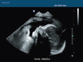
Medical ultrasound falls into two distinct categories: diagnostic and therapeutic.
Diagnostic ultrasound can be further sub-divided into anatomical and functional ultrasound. Anatomical ultrasound produces images of internal organs or other structures. Functional ultrasound combines information such as the movement and velocity of tissue or blood, softness or hardness of tissue, and other physical characteristics, with anatomical images to create “information maps.” These maps help doctors visualize changes/differences in function within a structure or organ.
Therapeutic ultrasound also uses sound waves above the range of human hearing but does not produce images. Its purpose is to interact with tissues in the body such that they are either modified or destroyed. Among the modifications possible are: moving or pushing tissue, heating tissue, dissolving blood clots, or delivering drugs to specific locations in the body. These destructive, or ablative, functions are made possible by use of very high-intensity beams that can destroy diseased or abnormal tissues such as tumors. The advantage of using ultrasound therapies is that, in most cases, they are non-invasive. No incisions or cuts need to be made to the skin, leaving no wounds or scars.
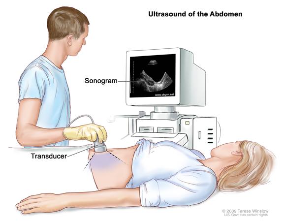
Ultrasound waves are produced by a transducer, which can both emit ultrasound waves, as well as detect the ultrasound echoes reflected back. In most cases, the active elements in ultrasound transducers are made of special ceramic crystal materials called piezoelectrics. These materials are able to produce sound waves when an electric field is applied to them, but can also work in reverse, producing an electric field when a sound wave hits them. When used in an ultrasound scanner, the transducer sends out a beam of sound waves into the body. The sound waves are reflected back to the transducer by boundaries between tissues in the path of the beam (e.g. the boundary between fluid and soft tissue or tissue and bone). When these echoes hit the transducer, they generate electrical signals that are sent to the ultrasound scanner. Using the speed of sound and the time of each echo’s return, the scanner calculates the distance from the transducer to the tissue boundary. These distances are then used to generate two-dimensional images of tissues and organs.
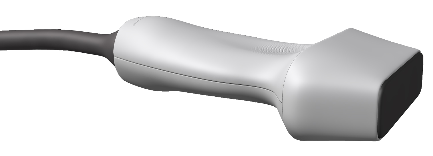
During an ultrasound exam, the technician will apply a gel to the skin. This keeps air pockets from forming between the transducer and the skin, which can block ultrasound waves from passing into the body.
Click here to watch a short video about how ultrasound works.
Diagnostic ultrasound. Diagnostic ultrasound is able to non-invasively image internal organs within the body. However, it is not good for imaging bones or any tissues that contain air, like the lungs. Under some conditions, ultrasound can image bones (such as in a fetus or in small babies) or the lungs and lining around the lungs, when they are filled or partially filled with fluid. One of the most common uses of ultrasound is during pregnancy, to monitor the growth and development of the fetus, but there are many other uses, including imaging the heart, blood vessels, eyes, thyroid, brain, breast, abdominal organs, skin, and muscles. Ultrasound images are displayed in either 2D, 3D, or 4D (which is 3D in motion).
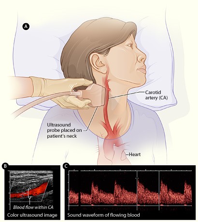
Functional ultrasound. Functional ultrasound applications include Doppler and color Doppler ultrasound for measuring and visualizing blood flow in vessels within the body or in the heart. It can also measure the speed of the blood flow and direction of movement. This is done using color-coded maps called color Doppler imaging. Doppler ultrasound is commonly used to determine whether plaque build-up inside the carotid arteries is blocking blood flow to the brain.
Another functional form of ultrasound is elastography, a method for measuring and displaying the relative stiffness of tissues, which can be used to differentiate tumors from healthy tissue. This information can be displayed as either color-coded maps of the relative stiffness; black-and white maps that display high-contrast images of tumors compared with anatomical images; or color-coded maps that are overlayed on the anatomical image. Elastography can be used to test for liver fibrosis, a condition in which excessive scar tissue builds up in the liver due to inflammation.
Ultrasound is also an important method for imaging interventions in the body. For example, ultrasound-guided needle biopsy helps physicians see the position of a needle while it is being guided to a selected target, such as a mass or a tumor in the breast. Also, ultrasound is used for real-time imaging of the location of the tip of a catheter as it is inserted in a blood vessel and guided along the length of the vessel. It can also be used for minimally invasive surgery to guide the surgeon with real-time images of the inside of the body.
Therapeutic or interventional ultrasound. Therapeutic ultrasound produces high levels of acoustic output that can be focused on specific targets for the purpose of heating, ablating, or breaking up tissue. One type of therapeutic ultrasound uses high-intensity beams of sound that are highly targeted, and is called High Intensity Focused Ultrasound (HIFU). HIFU is being investigated as a method for modifying or destroying diseased or abnormal tissues inside the body (e.g. tumors) without having to open or tear the skin or cause damage to the surrounding tissue. Either ultrasound or MRI is used to identify and target the tissue to be treated, guide and control the treatment in real time, and confirm the effectiveness of the treatment. HIFU is currently FDA approved for the treatment of uterine fibroids, to alleviate pain from bone metastases, and most recently for the ablation of prostate tissue. HIFU is also being investigated as a way to close wounds and stop bleeding, to break up clots in blood vessels, and to temporarily open the blood brain barrier so that medications can pass through.
Diagnostic ultrasound is generally regarded as safe and does not produce ionizing radiation like that produced by x-rays. Still, ultrasound is capable of producing some biological effects in the body under specific settings and conditions. For this reason, the FDA requires that diagnostic ultrasound devices operate within acceptable limits. The FDA, as well as many professional societies, discourage the casual use of ultrasound (e.g. for keepsake videos) and recommend that it be used only when there is a true medical need.
The following are examples of current research projects funded by NIBIB that are developing new applications of ultrasound that are already in use or that will be in use in the future:
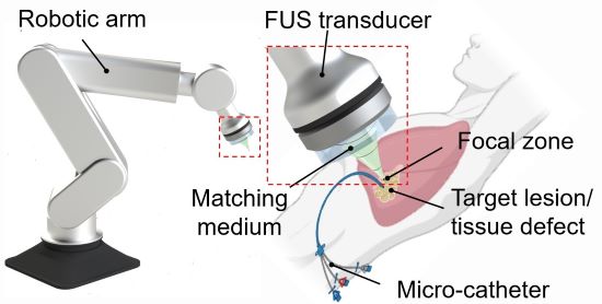
3D printing through the skin : Researchers at Duke University have developed a method to 3D print biocompatible structures through thick, multi-layered tissues. The approach entails using focused ultrasound to solidify a special ink that has been injected into the body to repair bone or repair soft tissues, for example. Initial experiments in animal tissue suggest the method could turn highly invasive surgical procedures into safer, less invasive ones. (Image on left courtesy of Junjie Yao (Duke University) and Yu Shrike Zhang (Harvard Medical School and Brigham and Women’s Hospital)).
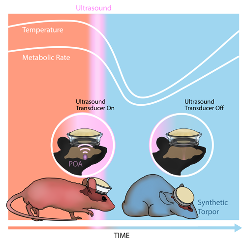
Inducing a hibernation-like state : Researchers at Washington University in St. Louis used ultrasound waves directed into the brain to lower the body temperature and metabolic rates of mice, inducing a hibernation-like state, called torpor. The researchers replicated some of these results in rats, which, like humans, don’t naturally enter torpor. Inducing torpor could help minimize damage from stroke or heart attack and buy precious time for patients in critical care. (Image on right courtesy of Yang et al./Washington University in St. Louis).
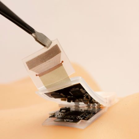
High-quality imaging at home : Brigham and Women’s Hospital researchers compared ultrasound scans acquired by experts with those taken by inexperienced volunteers, finding little difference in the image quality of the two groups. The unconventional approach of having patients take ultrasound images of themselves at home and share them with healthcare professionals could allow for remote monitoring and reduce the need for hospitalization. (Image on right courtesy of Duggan et al./Brigham and Women's Hospital).
Reviewed December 2023
Explore More
Heath Topics
- Breast Cancer
- Cardiovascular Disease
- Digestive Diseases
- Heart Disease
- Musculoskeletal Disease
- Reproductive Health
- Women's Health
Research Topics
- Optical Imaging
- Ultrasound (US) diagnostic
- Ultrasound (US) therapy
PROGRAM AREAS
- Ultrasound: Diagnostic and Interventional
Inside NIBIB
- Director's Corner
- Funding Policies
- NIBIB Fact Sheets
- Press Releases
- Search the site GO Please fill out this field.
- Newsletters
- Health Conditions A-Z
- Imaging and Procedures
What Is Sonography?
:max_bytes(150000):strip_icc():format(webp)/SB-headshot-Dotdash-9969efdd9a684f1ea4ff441ca264ce97.jpeg)
Risks and Precautions
- Preparation
FG Trade Latin / Getty Images
Sonography is a type of medical imaging test that uses high-frequency sound waves to create pictures of different internal parts of the body, like the uterus, heart, intestines, and other organs. Sonography uses ultrasound technology to create an image known as a sonogram. This technology is one of the most common imaging tests used to diagnose and treat medical conditions.
A radiologist may perform sonography, but there are also sonographers and ultrasound technicians who specialize in this form of medical imaging and receive more robust training to administer the test. Sonography is considered to be generally safe for most people. Additionally, the test doesn’t require much preparation and needs virtually no recovery time.
Sonography and ultrasound are often used interchangeably. However, there is a slight difference between these key terms. Here's what you need to know:
- Sonography: The scientific process of developing medical images
- Ultrasound: The technology that sonographers (people who specialize in sonography) use to generate medical images
- Sonogram: The medical image that is produced after the completion of the ultrasound
Healthcare providers use sonography to assess the health of your internal organs. If you’ve been injured, have an infection, or are experiencing symptoms suggestive of illness, your provider may schedule you for an ultrasound to get a better look at the area of concern.
During the test, it's common to check for inflammation , organ damage, changes in blood flow, and abnormal growths. If you’re expecting a baby, an ultrasound will examine the development of your baby and measure your amniotic fluid throughout your pregnancy. Since ultrasounds are safe and non-invasive (meaning, a provider doesn't cut into your skin to perform the test), people of all ages can get them.
Types of Sonography
Sonography is usually divided into two broad categories: ultrasounds for pregnancy and ultrasounds for diagnosing other types of medical conditions.
Pregnancy Ultrasound
A pregnancy ultrasound allows your provider to see how your baby is growing, estimate your gestational age, and identify birth defects, among other concerns. Most pregnant people have ultrasounds between 10 and 13 weeks of gestation and 18 and 22 weeks of gestation. You may have additional ultrasounds, too—especially if you’re not sure when you became pregnant or have a high-risk pregnancy.
Diagnostic Ultrasound
A healthcare provider can order a diagnostic ultrasound to identify a variety of medical conditions, including:
- Kidney stones
- Blood clots
- Thyroid nodules
- Uterine fibroids
- Ovarian cysts
- Enlarged spleen
- Breast lumps
How Does It Work?
Sonography is a fairly simple procedure that doesn’t require much from the person being tested. You will have to keep the part of your body being imaged relatively still, but ultrasounds are not as restrictive as other tests, like MRIs.
Before the Test
You should arrive at your ultrasound prepared to lie on a table. Depending on the part of your body that is being tested, your provider may ask you to lie on your back, stomach, or side.
Your sonographer should let you know in advance of any fasting requirements for your ultrasound. You may have no requirements, or you may need to avoid eating or drinking for a certain amount of time before the test. Sometimes you will need to have a full bladder for all or part of the test.
Most people don’t need to be sedated for an ultrasound unless they’re unable to remain still for the test. This typically only applies to very young children who may not be able to stay still for the duration of the exam. Ultrasounds generally last between 30 and 60 minutes.
During the Test
When the sonographer is ready to begin, they will apply a gel-based lubricant to your skin in the area being tested. Then they will use a wand called a transducer, which emits high-frequency sound waves that help take the images.
During external sonography (e.g., on your stomach), you can expect your sonographer to move the transducer over your skin outside the area being tested and apply small amounts of pressure in different positions to obtain the necessary images. For internal sonography (e.g., in your vagina or rectum), your sonographer will insert the transducer inside of you to capture the images.
The images will appear on a small screen in front of the sonographer. You may or may not be able to see them as you’re being tested. The sonographer will be seated right next to you, so you can ask them questions or communicate any concerns during the test. Ultrasounds aren’t painful, but can sometimes cause a little discomfort or pressure. If you’re uncomfortable, you can let the sonographer know and they may be able to modify the test or give you a short break.
After the Test
Once the ultrasound is complete, the sonographer will help you clean off the gel lubricant and move into a more comfortable seated position. If you need to change back into your clothes, they will leave the room so you can do so in private.
You can resume all your normal activities right after the ultrasound. You won't have to wait in the office or stay overnight and you can drive yourself home after the procedure. The sonographer may share the results of your ultrasound with you on the spot, or you may have to wait for your primary care provider to follow-up with you by phone or appointment.
For the most part, ultrasounds are extremely safe and don’t pose a health risk. They don’t use ionizing radiation like X-rays do, so there’s no need for you to worry about the effects of the ultrasound on your body. There is a slight chance you could be allergic to the gel used during the ultrasound procedure, which could result in contact dermatitis (skin rash) after the test and require minor treatment.
However, ultrasounds generate a small amount of heat that can affect your skin and tissues in the area where the test is being conducted. That said, experts recommend undergoing ultrasounds only when medically necessary. While anyone of any age can safely have an ultrasound—and they are considered safe even for pregnant women and unborn children—you shouldn’t have more ultrasounds than necessary during pregnancy and shouldn’t utilize an at-home imaging device to monitor your pregnancy on your own.
How to Prepare for Sonography
The good news: there isn’t much preparation needed for sonography. Depending on the test, your provider may recommend fasting for a certain amount of time before your test or refraining from emptying your bladder before you go. But many ultrasounds don’t require this.
Ultrasounds can be performed at a variety of locations, so it's best to know where your test will be performed well before your appointment. This can sometimes be at a hospital, clinic, or private doctor's office. In some cases, radiology satellite clinics can perform an ultrasound in your home.
Keep in mind: you may or may not need to change into a hospital gown, so plan to wear comfortable clothes that are easy to take off. You may also need to remove any jewelry and accessories that could interfere with the sonographer’s ability to image that area.
Bring your insurance card with you to the exam if you have insurance coverage and are planning to utilize it. If you have a copay, you should expect to pay for the procedure prior to the test, so it's best to have a form of payment ready to go. If you’re paying out-of-pocket, try to call the ultrasound location prior to the exam and ask how they prefer to receive payment.
Finally, in most ultrasound appointments, you are allowed to have one or two people attend with you. This is especially true for pregnancy ultrasounds. However, other diagnostic ultrasounds also tend to allow a guest to emotionally support you during the test. Ask your healthcare provider before your appointment about best practices for your guest(s).
How and when you get your results can vary based on the reason for your test and who is performing it. If you use an electronic program or app that maintains your medical information, you may sometimes see the results show up there before or after your provider contacts you.
With a diagnostic ultrasound, a radiologist (or, a doctor who specializes in medical imaging) will review the images before sending their assessment to your primary care provider. Your provider will then contact you to discuss your results or make a follow-up appointment, if necessary.
With a pregnancy ultrasound, your sonographer may share your results with you at the end of the appointment or have your primary care provider or obstetrician (a doctor who specializes in pregnancy) give you a call. If you're expecting and have concerns about your ultrasound, there may be a maternal health provider on site that can answer your questions. If there is no provider available on-site, your obstetrician can follow up on your results and discuss them with you in further detail.
Interpreting Your Results
If your results are positive for the issue or condition your primary care provider was trying to confirm, they may either officially diagnose you and begin treatment or order further testing to get more information. If the results from your ultrasound are negative, you will likely require additional testing to find out what is causing your symptoms.
Inconclusive results (results that are neither positive nor negative) can lead to several outcomes: your provider may order additional imaging tests or blood work or they may ask you to get a second ultrasound in a few months if the diagnosis isn’t time-sensitive.
A Quick Review
Sonography is a simple and safe diagnostic imaging process that helps healthcare providers identify and treat several medical conditions, including pregnancy, kidney stones, and tumors. Undergoing sonography may sound scary if you've never experienced one before, but rest assured, this medical imaging test is one of the safest exams available.
There is very little preparation required before going in for an ultrasound. You may have to refrain from eating or drinking before the exam and wear comfortable clothing that is easy to remove. If you have specific questions before, during, or after the exam, talk to your primary care provider or sonographer who will be conducting the test for more information.
Frequently Asked Questions
The term “ultrasound” can be used interchangeably with the term “sonography,” but an ultrasound is not the same thing as a sonogram. An ultrasound is the procedure used to capture an image and a sonogram is the image itself.
Both sonography and radiology are branches of medicine that use diagnostic imaging to diagnose and treat diseases, but they require different types of equipment. Sonography uses high-frequency sound waves to collect images while radiology uses X-ray beams.
There are times when an ultrasound isn’t the best way for your provider to diagnose or evaluate a medical condition. The lungs and bowels can’t be well-studied with an ultrasound, and there are limitations to how far into bone an ultrasound can penetrate. Ultrasounds also can’t differentiate between benign and malignant tumors.
U.S. Food and Drug Administration. Ultrasound Imaging .
Society of Diagnostic Medical Sonography. Understanding Sonography .
American Registry for Diagnostic Medical Sonography. How to Become a Sonographer .
Radiological Society of North America, Inc. General Ultrasound .
MedlinePlus. Ultrasound .
Ulrich CC, Dewald O. Pregnancy Ultrasound Evaluation . In: StatPearls . StatPearls Publishing; 2023.
American College of Obstetricians and Gynecologists. Ultrasound Exams .
American Cancer Society. Ultrasound for Cancer .
Related Articles
Presentations
Got any suggestions?
We want to hear from you! Send us a message and help improve Slidesgo
Top searches
Trending searches

memorial day
12 templates

21 templates

summer vacation
23 templates

17 templates

20 templates

11 templates
Ultrasound Technician and Sonographer CV
Ultrasound technician and sonographer cv presentation, free google slides theme and powerpoint template.
Don't wait any longer – this ultrasound technician and sonographer CV is the perfect one-stop shop for all your job hunting needs! No more worry of a long hunt trying to design the perfect resume or dreading the flight through application forms. This complete professional template provides everything you need to make a great impression, including customized cover letter forms, modern slides and an awesome resume example that you can fill with your own information. Plus it's 100% printer ready so you're on your way faster than ever!
Features of this template
- 100% editable and easy to modify
- 6 different slides to impress your audience
- Contains easy-to-edit graphics such as graphs, maps, tables, timelines and mockups
- Includes 500+ icons and Flaticon’s extension for customizing your slides
- Designed to be used in Google Slides and Microsoft PowerPoint
- A4 format optimized for printing
- Includes information about fonts, colors, and credits of the resources used
How can I use the template?
Am I free to use the templates?
How to attribute?
Attribution required If you are a free user, you must attribute Slidesgo by keeping the slide where the credits appear. How to attribute?
Related posts on our blog.

How to Add, Duplicate, Move, Delete or Hide Slides in Google Slides

How to Change Layouts in PowerPoint

How to Change the Slide Size in Google Slides
Related presentations.

Premium template
Unlock this template and gain unlimited access


IMAGES
VIDEO
COMMENTS
Lecture Session 3: Professional Topics - Clinical Supervisors, May 28, 2021, 3:00 PM - 5:00 PM ... This presentation will explore the ultrasound appearances of the normal pancreas and demonstrate the changes associated with acute and chronic pancreatitis. The role of ultrasound in various treatment options is also discussed.
Relationship Between Ultrasound Viewing and Proceeding to Abortion. Transrectal Ultrasound of the Prostate With a Biopsy. Iron-Based Catalysts Used in Water Treatment Assisted by Ultrasound. 88 Tourism Management Essay Topic Ideas & Examples 64 Virtual Team Essay Topic Ideas & Examples.
Some other topics in my class were: travel sonography vs. traditional sonography, patient safety in kidney stone diagnosis, mammograms vs. breast ultrasounds, colonoscopy vs. ultrasound, sonographers and scope of practice, etc. Mine only had to be five pages, so I would definitely make sure that there is a lot of information backing whatever ...
We trust that you will find a few clinical pearls or reminders that you could apply to your patients that you care for in your emergency department or other health setting. Search by keywords; disease process; condition; eponym or clinical features…. Ultrasound Quiz Library 111. Ultrasound Quiz Library 110. Ultrasound Quiz Library 109.
UCSF Ultrasound Lecture Series. A monthly one-hour lecture series for sonographers, radiologists, physicians, and other medical professionals involved with the scanning and interpretation of ultrasound images in patient care. This series is approved by the SDMS for CME credit for licensed sonographers (RDMS). A monthly one-hour lecture series ...
As an adjunct to mammography, ultrasound can be a primary breast imaging application for women under the age of 40 who have dense breasts, or breast implants, or breastfeed. Ultrasound can also help doctors assess masses that are detected in mammograms, aid in the detection of cysts and guide breast biopsies.
<i>Ultrasound Imaging - Current Topics</i> presents complex and current topics in ultrasound imaging in a simplified format. It is easy to read and exemplifies the range of experiences of each contributing author. Chapters address such topics as anatomy and dimensional variations, pediatric gastrointestinal emergencies, musculoskeletal and nerve imaging as well as molecular sonography. The ...
Workshop Session 5E: Prof topics, May 28, 2022, 11:15 AM-11:55 AM. Ultrasound of wrist and carpal tunnel syndrome Lisa Howell. ... This presentation is for the sonographer with little experience in ultrasound examination of the ankle. During the session, we will go through how to complete a basic ankle ultrasound protocol. ...
ASRT strives to be the premier professional association for the medical imaging and radiation therapy community through education, advocacy, research and innovation. 15000 Central Ave. SE. Albuquerque, NM 87123-3909. [email protected]. This list of study guides is for ultrasound review for sonography certification.
Sonography is a diagnostic test that uses high-frequency sound waves to create detailed images of different parts of the body to detect anomalies and make diagnoses. Without sonography, it would be much harder to treat a range of conditions. The sonography field has a lot to offer students who have the desire to make a difference in the medical ...
Introduction. A thesis or dissertation, as some people would like to call it, is an integral part of the Radiology curriculum, be it MD, DNB, or DMRD. We have tried to aggregate radiology thesis topics from various sources for reference. Not everyone is interested in research, and writing a Radiology thesis can be daunting.
Disney Templates with your favorite Disney and Pixar characters Slidesclass Ready-to-go classes on many topics for everyone Editor's Choice Our favorite slides Teacher Toolkit Content for teachers Multi-purpose Presentations that suit any project Interactive & Animated Templates to create engaging presentations
The development of sonography or medical ultrasound was built on the understanding and research of sound, which can be dated as far back as the 6th century. Specifically, sonography can be tied to as far back as the 1700s, an Italian physicist, Lazaro Spallanzani, who was researching the navigation of bats.[1] In the early 20th century, more applicable uses of ultrasound were studied by French ...
Sonography is a non-invasive medical procedure that uses the echoes of high-frequency sound waves (ultrasound) to construct an image (sonogram) of internal organs or body structures. In sonography, a transmitting device (the transducer) sends out high-frequency ultrasound waves. Harmless sound waves, which contain no radiation, bounce off the ...
Frequently Asked Questions. Sonography is a diagnostic medical test that uses high-frequency sound waves, or ultrasound waves, to create images of tissues, glands, organs, and blood or fluid flow within the body. This test is also referred to as an ultrasound or sonogram. Sonography uses a device called a transducer on the surface of the skin ...
Radiology Educational Videos. The following are free educational modules produced by our faculty. The "Anatomy" presentations are interactive Powerpoints while the videos are essential medical student level lectures which range from introductory material to Ultrasound guided biopsy techniques.
One of the most common uses of ultrasound is during pregnancy, to monitor the growth and development of the fetus, but there are many other uses, including imaging the heart, blood vessels, eyes, thyroid, brain, breast, abdominal organs, skin, and muscles. Ultrasound images are displayed in either 2D, 3D, or 4D (which is 3D in motion).
The equipment uses high-frequency sound waves to create an image of what's going on internally. To capture the images, the sonographer uses a transducer, which is placed on the area of the body that's being imaged. The device sends pulses of sound waves into the body, which create echoes as they bounce back.
Sonography is a type of medical imaging test that uses high-frequency sound waves to create pictures of different internal parts of the body, like the uterus, heart, intestines, and other organs ...
Ultrasound works by using sound waves to create an image of organs and structures inside the body without having to open it up. It can be hard to understand for high school students, but not if you use this creative presentation design full of graphs, charts, illustrations and infographics!
Photoacoustic Imaging in a Multimodal Onco-Pharmacology Approach. Watch this very interesting presentation (by Dr. Florian Raes) that shares research aimed at assessing tumor oxygenation to validate non-invasive imaging measurements of tumor hypoxia. Get preclinical research ultrasound presentations from the experts at VisualSonics.
Free Google Slides theme and PowerPoint template. Don't wait any longer - this ultrasound technician and sonographer CV is the perfect one-stop shop for all your job hunting needs! No more worry of a long hunt trying to design the perfect resume or dreading the flight through application forms. This complete professional template provides ...
To investigated morphologic and clinical presentations of mpox in diverse populations, researchers conducted a review of the records of 54 individuals (mean age, 42.4 years) diagnosed with mpox at ...