Appointments at Mayo Clinic
- Pregnancy week by week
- Fetal presentation before birth
The way a baby is positioned in the uterus just before birth can have a big effect on labor and delivery. This positioning is called fetal presentation.
Babies twist, stretch and tumble quite a bit during pregnancy. Before labor starts, however, they usually come to rest in a way that allows them to be delivered through the birth canal headfirst. This position is called cephalic presentation. But there are other ways a baby may settle just before labor begins.
Following are some of the possible ways a baby may be positioned at the end of pregnancy.

Head down, face down
When a baby is head down, face down, the medical term for it is the cephalic occiput anterior position. This the most common position for a baby to be born in. With the face down and turned slightly to the side, the smallest part of the baby's head leads the way through the birth canal. It is the easiest way for a baby to be born.
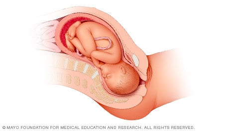
Head down, face up
When a baby is head down, face up, the medical term for it is the cephalic occiput posterior position. In this position, it might be harder for a baby's head to go under the pubic bone during delivery. That can make labor take longer.
Most babies who begin labor in this position eventually turn to be face down. If that doesn't happen, and the second stage of labor is taking a long time, a member of the health care team may reach through the vagina to help the baby turn. This is called manual rotation.
In some cases, a baby can be born in the head-down, face-up position. Use of forceps or a vacuum device to help with delivery is more common when a baby is in this position than in the head-down, face-down position. In some cases, a C-section delivery may be needed.
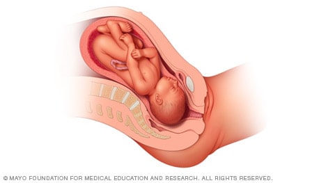
Frank breech
When a baby's feet or buttocks are in place to come out first during birth, it's called a breech presentation. This happens in about 3% to 4% of babies close to the time of birth. The baby shown below is in a frank breech presentation. That's when the knees aren't bent, and the feet are close to the baby's head. This is the most common type of breech presentation.
If you are more than 36 weeks into your pregnancy and your baby is in a frank breech presentation, your health care professional may try to move the baby into a head-down position. This is done using a procedure called external cephalic version. It involves one or two members of the health care team putting pressure on your belly with their hands to get the baby to roll into a head-down position.
If the procedure isn't successful, or if the baby moves back into a breech position, talk with a member of your health care team about the choices you have for delivery. Most babies in a frank breech position are born by planned C-section.

Complete and incomplete breech
A complete breech presentation, as shown below, is when the baby has both knees bent and both legs pulled close to the body. In an incomplete breech, one or both of the legs are not pulled close to the body, and one or both of the feet or knees are below the baby's buttocks. If a baby is in either of these positions, you might feel kicking in the lower part of your belly.
If you are more than 36 weeks into your pregnancy and your baby is in a complete or incomplete breech presentation, your health care professional may try to move the baby into a head-down position. This is done using a procedure called external cephalic version. It involves one or two members of the health care team putting pressure on your belly with their hands to get the baby to roll into a head-down position.
If the procedure isn't successful, or if the baby moves back into a breech position, talk with a member of your health care team about the choices you have for delivery. Many babies in a complete or incomplete breech position are born by planned C-section.

When a baby is sideways — lying horizontal across the uterus, rather than vertical — it's called a transverse lie. In this position, the baby's back might be:
- Down, with the back facing the birth canal.
- Sideways, with one shoulder pointing toward the birth canal.
- Up, with the hands and feet facing the birth canal.
Although many babies are sideways early in pregnancy, few stay this way when labor begins.
If your baby is in a transverse lie during week 37 of your pregnancy, your health care professional may try to move the baby into a head-down position. This is done using a procedure called external cephalic version. External cephalic version involves one or two members of your health care team putting pressure on your belly with their hands to get the baby to roll into a head-down position.
If the procedure isn't successful, or if the baby moves back into a transverse lie, talk with a member of your health care team about the choices you have for delivery. Many babies who are in a transverse lie are born by C-section.
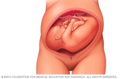
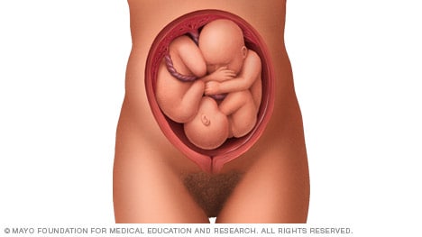
If you're pregnant with twins and only the twin that's lower in the uterus is head down, as shown below, your health care provider may first deliver that baby vaginally.
Then, in some cases, your health care team may suggest delivering the second twin in the breech position. Or they may try to move the second twin into a head-down position. This is done using a procedure called external cephalic version. External cephalic version involves one or two members of the health care team putting pressure on your belly with their hands to get the baby to roll into a head-down position.
Your health care team may suggest delivery by C-section for the second twin if:
- An attempt to deliver the baby in the breech position is not successful.
- You do not want to try to have the baby delivered vaginally in the breech position.
- An attempt to move the baby into a head-down position is not successful.
- You do not want to try to move the baby to a head-down position.
In some cases, your health care team may advise that you have both twins delivered by C-section. That might happen if the lower twin is not head down, the second twin has low or high birth weight as compared to the first twin, or if preterm labor starts.
- Landon MB, et al., eds. Normal labor and delivery. In: Gabbe's Obstetrics: Normal and Problem Pregnancies. 8th ed. Elsevier; 2021. https://www.clinicalkey.com. Accessed May 19, 2023.
- Holcroft Argani C, et al. Occiput posterior position. https://www.updtodate.com/contents/search. Accessed May 19, 2023.
- Frequently asked questions: If your baby is breech. American College of Obstetricians and Gynecologists https://www.acog.org/womens-health/faqs/if-your-baby-is-breech. Accessed May 22, 2023.
- Hofmeyr GJ. Overview of breech presentation. https://www.updtodate.com/contents/search. Accessed May 22, 2023.
- Strauss RA, et al. Transverse fetal lie. https://www.updtodate.com/contents/search. Accessed May 22, 2023.
- Chasen ST, et al. Twin pregnancy: Labor and delivery. https://www.updtodate.com/contents/search. Accessed May 22, 2023.
- Cohen R, et al. Is vaginal delivery of a breech second twin safe? A comparison between delivery of vertex and non-vertex second twins. The Journal of Maternal-Fetal & Neonatal Medicine. 2021; doi:10.1080/14767058.2021.2005569.
- Marnach ML (expert opinion). Mayo Clinic. May 31, 2023.
Products and Services
- A Book: Obstetricks
- A Book: Mayo Clinic Guide to a Healthy Pregnancy
- 3rd trimester pregnancy
- Fetal development: The 3rd trimester
- Overdue pregnancy
- Pregnancy due date calculator
- Prenatal care: 3rd trimester
Mayo Clinic does not endorse companies or products. Advertising revenue supports our not-for-profit mission.
- Opportunities
Mayo Clinic Press
Check out these best-sellers and special offers on books and newsletters from Mayo Clinic Press .
- Mayo Clinic on Incontinence - Mayo Clinic Press Mayo Clinic on Incontinence
- The Essential Diabetes Book - Mayo Clinic Press The Essential Diabetes Book
- Mayo Clinic on Hearing and Balance - Mayo Clinic Press Mayo Clinic on Hearing and Balance
- FREE Mayo Clinic Diet Assessment - Mayo Clinic Press FREE Mayo Clinic Diet Assessment
- Mayo Clinic Health Letter - FREE book - Mayo Clinic Press Mayo Clinic Health Letter - FREE book
- Healthy Lifestyle
Your gift holds great power – donate today!
Make your tax-deductible gift and be a part of the cutting-edge research and care that's changing medicine.
Enter search terms to find related medical topics, multimedia and more.
Advanced Search:
- Use “ “ for exact phrases.
- For example: “pediatric abdominal pain”
- Use – to remove results with certain keywords.
- For example: abdominal pain -pediatric
- Use OR to account for alternate keywords.
- For example: teenager OR adolescent
Fetal Presentation, Position, and Lie (Including Breech Presentation)
, MD, Children's Hospital of Philadelphia
Variations in Fetal Position and Presentation
- 3D Models (0)
- Calculators (0)
- Lab Test (0)

Presentation refers to the part of the fetus’s body that leads the way out through the birth canal (called the presenting part). Usually, the head leads the way, but sometimes the buttocks (breech presentation), shoulder, or face leads the way.
Position refers to whether the fetus is facing backward (occiput anterior) or forward (occiput posterior). The occiput is a bone at the back of the baby's head. Therefore, facing backward is called occiput anterior (facing the mother’s back and facing down when the mother lies on her back). Facing forward is called occiput posterior (facing toward the mother's pubic bone and facing up when the mother lies on her back).
Lie refers to the angle of the fetus in relation to the mother and the uterus. Up-and-down (with the baby's spine parallel to mother's spine, called longitudinal) is normal, but sometimes the lie is sideways (transverse) or at an angle (oblique).
For these aspects of fetal positioning, the combination that is the most common, safest, and easiest for the mother to deliver is the following:
Head first (called vertex or cephalic presentation)
Facing backward (occiput anterior position)
Spine parallel to mother's spine (longitudinal lie)
Neck bent forward with chin tucked
Arms folded across the chest
If the fetus is in a different position, lie, or presentation, labor may be more difficult, and a normal vaginal delivery may not be possible.
Variations in fetal presentation, position, or lie may occur when
The fetus is too large for the mother's pelvis (fetopelvic disproportion).
The fetus has a birth defect Overview of Birth Defects Birth defects, also called congenital anomalies, are physical abnormalities that occur before a baby is born. They are usually obvious within the first year of life. The cause of many birth... read more .
There is more than one fetus (multiple gestation).

Position and Presentation of the Fetus
Some variations in position and presentation that make delivery difficult occur frequently.
Occiput posterior position
In occiput posterior position (sometimes called sunny-side up), the fetus is head first (vertex presentation) but is facing forward (toward the mother's pubic bone—that is, facing up when the mother lies on her back). This is a very common position that is not abnormal, but it makes delivery more difficult than when the fetus is in the occiput anterior position (facing toward the mother's spine—that is facing down when the mother lies on her back).
Breech presentation
In breech presentation, the baby's buttocks or sometimes the feet are positioned to deliver first (before the head).
When delivered vaginally, babies that present buttocks first are more at risk of injury or even death than those that present head first.
The reason for the risks to babies in breech presentation is that the baby's hips and buttocks are not as wide as the head. Therefore, when the hips and buttocks pass through the cervix first, the passageway may not be wide enough for the head to pass through. In addition, when the head follows the buttocks, the neck may be bent slightly backwards. The neck being bent backward increases the width required for delivery as compared to when the head is angled forward with the chin tucked, which is the position that is easiest for delivery. Thus, the baby’s body may be delivered and then the head may get caught and not be able to pass through the birth canal. When the baby’s head is caught, this puts pressure on the umbilical cord in the birth canal, so that very little oxygen can reach the baby. Brain damage due to lack of oxygen is more common among breech babies than among those presenting head first.
Breech presentation is more likely to occur in the following circumstances:
Labor starts too soon (preterm labor).
Sometimes the doctor can turn the fetus to be head first before labor begins by doing a procedure that involves pressing on the pregnant woman’s abdomen and trying to turn the baby around. Trying to turn the baby is called an external cephalic version and is usually done at 37 or 38 weeks of pregnancy. Sometimes women are given a medication (such as terbutaline ) during the procedure to prevent contractions.
Other presentations
In face presentation, the baby's neck arches back so that the face presents first rather than the top of the head.
In brow presentation, the neck is moderately arched so that the brow presents first.
Usually, fetuses do not stay in a face or brow presentation. These presentations often change to a vertex (top of the head) presentation before or during labor. If they do not, a cesarean delivery is usually recommended.
In transverse lie, the fetus lies horizontally across the birth canal and presents shoulder first. A cesarean delivery is done, unless the fetus is the second in a set of twins. In such a case, the fetus may be turned to be delivered through the vagina.
Drugs Mentioned In This Article

Was This Page Helpful?

Test your knowledge
Brought to you by Merck & Co, Inc., Rahway, NJ, USA (known as MSD outside the US and Canada)—dedicated to using leading-edge science to save and improve lives around the world. Learn more about the Merck Manuals and our commitment to Global Medical Knowledge .
- Permissions
- Cookie Settings
- Terms of use
- Veterinary Edition

- IN THIS TOPIC
- Type 2 Diabetes
- Heart Disease
- Digestive Health
- Multiple Sclerosis
- COVID-19 Vaccines
- Occupational Therapy
- Healthy Aging
- Health Insurance
- Public Health
- Patient Rights
- Caregivers & Loved Ones
- End of Life Concerns
- Health News
- Thyroid Test Analyzer
- Doctor Discussion Guides
- Hemoglobin A1c Test Analyzer
- Lipid Test Analyzer
- Complete Blood Count (CBC) Analyzer
- What to Buy
- Editorial Process
- Meet Our Medical Expert Board
What Is Cephalic Position?
The ideal fetal position for labor and delivery
- Why It's Best
Risks of Other Positions
- Determining Position
- Turning a Fetus
The cephalic position is when a fetus is head down when it is ready to enter the birth canal. This is one of a few variations of how a fetus can rest in the womb and is considered the ideal one for labor and delivery.
About 96% of babies are born in the cephalic position. Most settle into it between the 32nd and 36th weeks of pregnancy . Your healthcare provider will monitor the fetus's position during the last weeks of gestation to ensure this has happened by week 36.
If the fetus is not in the cephalic position at that point, the provider may try to turn it. If this doesn't work, some—but not all—practitioners will attempt to deliver vaginally, while others will recommend a Cesarean (C-section).
Getty Images
Why Is the Cephalic Position Best?
During labor, contractions dilate the cervix so the fetus has adequate room to come through the birth canal. The cephalic position is the easiest and safest way for the baby to pass through the birth canal.
If the fetus is in a noncephalic position, delivery becomes more challenging. Different fetal positions have a range of difficulties and varying risks.
A small percentage of babies present in noncephalic positions. This can pose risks both to the fetus and the mother, and make labor and delivery more challenging. It can also influence the way in which someone can deliver.
A fetus may actually find itself in any of these positions throughout pregnancy, as the move about the uterus. But as they grow, there will be less room to tumble around and they will settle into a final position.
It is at this point that noncephalic positions can pose significant risks.
Cephalic Posterior
A fetus may also present in an occiput or cephalic posterior position. This means they are positioned head down, but they are facing the abdomen instead of the back.
This position is also nicknamed "sunny-side up."
Presenting this way increases the chance of a painful and prolonged delivery.
There are three different types of breech fetal positioning:
- Frank breech: The legs are up with the feet near the head.
- Footling breech: One or both legs is lowered over the cervix.
- Complete breech: The fetus is bottom-first with knees bent.
A vaginal delivery is most times a safe way to deliver. But with breech positions, a vaginal delivery can be complicated.
When a baby is born in the breech position, the largest part—its head—is delivered last. This can result in them getting stuck in the birth canal (entrapped). This can cause injury or death.
The umbilical cord may also be damaged or slide down into the mouth of the womb, which can reduce or cut off the baby's oxygen supply.
Some providers are still comfortable performing a vaginal birth as long as the fetus is doing well. But breech is always a riskier delivery position compared with the cephalic position, and most cases require a C-section.
Likelihood of a Breech Baby
You are more likely to have a breech baby if you:
- Go into early labor before you're full term
- Have an abnormally shaped uterus, fibroids , or too much amniotic fluid
- Are pregnant with multiples
- Have placenta previa (when the placenta covers the cervix)
Transverse Lie
In transverse lie position, the fetus is presenting sideways across the uterus rather than vertically. They may be:
- Down, with the back facing the birth canal
- With one shoulder pointing toward the birth canal
- Up, with the hands and feet facing the birth canal
If a transverse lie is not corrected before labor, a C-section will be required. This is typically the case.
Determining Fetal Position
Your healthcare provider can determine if your baby is in cephalic presentation by performing a physical exam and ultrasound.
In the final weeks of pregnancy, your healthcare provider will feel your lower abdomen with their hands to assess the positioning of the baby. This includes where the head, back, and buttocks lie
If your healthcare provider senses that the fetus is in a breech position, they can use ultrasound to confirm their suspicion.
Turning a Fetus So They Are in Cephalic Position
External cephalic version (ECV) is a common, noninvasive procedure to turn a breech baby into cephalic position while it's still in the uterus.
This is only considered if a healthcare provider monitors presentation progress in the last trimester and notices that a fetus is maintaining a noncephalic position as your delivery date approaches.
External Cephalic Version (ECV)
ECV involves the healthcare provider applying pressure to your stomach to turn the fetus from the outside. They will attempt to rotate the head forward or backward and lift the buttocks in an upward position. Sometimes, they use ultrasound to help guide the process.
The best time to perform ECV is about 37 weeks of pregnancy. Afterward, the fetal heart rate will be monitored to make sure it’s within normal levels. You should be able to go home after having ECV done.
ECV has a 50% to 60% success rate. However, even if it does work, there is still a chance the fetus will return to the breech position before birth.
Natural Methods For Turning a Fetus
There are also natural methods that can help turn a fetus into cephalic position. There is no medical research that confirms their efficacy, however.
- Changing your position: Sometimes a fetus will move when you get into certain positions. Two specific movements that your provider may recommend include: Getting on your hands and knees and gently rocking back and forth. Another you could try is pushing your hips up in the air while laying on your back with your knees bent and feet flat on the floor (bridge pose).
- Playing stimulating sounds: Fetuses gravitate to sound. You may be successful at luring a fetus out of breech position by playing music or a recording of your voice near your lower abdomen.
- Chiropractic care: A chiropractor can try the Webster technique. This is a specific chiropractic analysis and adjustment which enables chiropractors to establish balance in the pregnant person's pelvis and reduce undue stress to the uterus and supporting ligaments.
- Acupuncture: This is a considerably safe way someone can try to turn a fetus. Some practitioners incorporate moxibustion—the burning of dried mugwort on certain areas of the body—because they believe it will enhance the chances of success.
A Word From Verywell
While most babies are born in cephalic position at delivery, this is not always the case. And while some fetuses can be turned, others may be more stubborn.
This may affect your labor and delivery wishes. Try to remember that having a healthy baby, and staying well yourself, are your ultimate priorities. That may mean diverting from your best laid plans.
Speaking to your healthcare provider about turning options and the safest route of delivery may help you adjust to this twist and feel better about how you will move ahead.
Glezerman M. Planned vaginal breech delivery: current status and the need to reconsider . Expert Rev Obstet Gynecol. 2012;7(2):159-166. doi:10.1586/eog.12.2
Cleveland Clinic. Fetal positions for birth .
MedlinePlus. Breech birth .
UT Southwestern Medical Center. Can you turn a breech baby around?
The American College of Obstetricians and Gynecologists. If your baby is breech .
Roecker CB. Breech repositioning unresponsive to Webster technique: coexistence of oligohydramnios . Journal of Chiropractic Medicine . 2013;12(2):74-78. doi:10.1016/j.jcm.2013.06.003
By Cherie Berkley, MS Cherie Berkley is an award-winning journalist and multimedia storyteller covering health features for Verywell.
- Find in topic
INTRODUCTION
Diagnosis and management of face and brow presentations will be reviewed here. Other cephalic malpresentations are discussed separately. (See "Occiput posterior position" and "Occiput transverse position" .)
Prevalence — Face and brow presentation are uncommon. Their prevalences compared with other types of malpresentations are shown below [ 1-9 ]:
● Occiput posterior – 1/19 deliveries
● Breech – 1/33 deliveries
To continue reading this article, you must sign in . For more information or to purchase a personal subscription, click below on the option that best describes you:
- Medical Professional
- Resident, Fellow or Student
- Hospital or Institution
- Group Practice
- Patient or Caregiver

Print Options

An official website of the United States government
Here’s how you know
Official websites use .gov A .gov website belongs to an official government organization in the United States.
Secure .gov websites use HTTPS A lock ( Lock Locked padlock icon ) or https:// means you’ve safely connected to the .gov website. Share sensitive information only on official, secure websites.

- Health Topics
- Drugs & Supplements
- Medical Tests
- Medical Encyclopedia
- About MedlinePlus
- Customer Support
Your baby in the birth canal
During labor and delivery, your baby must pass through your pelvic bones to reach the vaginal opening. The goal is to find the easiest way out. Certain body positions give the baby a smaller shape, which makes it easier for your baby to get through this tight passage.
The best position for the baby to pass through the pelvis is with the head down and the body facing toward the mother's back. This position is called occiput anterior.
Information
Certain terms are used to describe your baby's position and movement through the birth canal.
FETAL STATION
Fetal station refers to where the presenting part is in your pelvis.
- The presenting part. The presenting part is the part of the baby that leads the way through the birth canal. Most often, it is the baby's head, but it can be a shoulder, the buttocks, or the feet.
- Ischial spines. These are bone points on the mother's pelvis. Normally the ischial spines are the narrowest part of the pelvis.
- 0 station. This is when the baby's head is even with the ischial spines. The baby is said to be "engaged" when the largest part of the head has entered the pelvis.
- If the presenting part lies above the ischial spines, the station is reported as a negative number from -1 to -5.
In first-time moms, the baby's head may engage by 36 weeks into the pregnancy. However, engagement may happen later in the pregnancy, or even during labor.
This refers to how the baby's spine lines up with the mother's spine. Your baby's spine is between their head and tailbone.
Your baby will most often settle into a position in the pelvis before labor begins.
- If your baby's spine runs in the same direction (parallel) as your spine, the baby is said to be in a longitudinal lie. Nearly all babies are in a longitudinal lie.
- If the baby is sideways (at a 90-degree angle to your spine), the baby is said to be in a transverse lie.
FETAL ATTITUDE
The fetal attitude describes the position of the parts of your baby's body.
The normal fetal attitude is commonly called the fetal position.
- The head is tucked down to the chest.
- The arms and legs are drawn in towards the center of the chest.
Abnormal fetal attitudes include a head that is tilted back, so the brow or the face presents first. Other body parts may be positioned behind the back. When this happens, the presenting part will be larger as it passes through the pelvis. This makes delivery more difficult.
DELIVERY PRESENTATION
Delivery presentation describes the way the baby is positioned to come down the birth canal for delivery.
The best position for your baby inside your uterus at the time of delivery is head down. This is called cephalic presentation.
- This position makes it easier and safer for your baby to pass through the birth canal. Cephalic presentation occurs in about 97% of deliveries.
- There are different types of cephalic presentation, which depend on the position of the baby's limbs and head (fetal attitude).
If your baby is in any position other than head down, your doctor may recommend a cesarean delivery.
Breech presentation is when the baby's bottom is down. Breech presentation occurs about 3% of the time. There are a few types of breech:
- A complete breech is when the buttocks present first and both the hips and knees are flexed.
- A frank breech is when the hips are flexed so the legs are straight and completely drawn up toward the chest.
- Other breech positions occur when either the feet or knees present first.
The shoulder, arm, or trunk may present first if the fetus is in a transverse lie. This type of presentation occurs less than 1% of the time. Transverse lie is more common when you deliver before your due date, or have twins or triplets.
CARDINAL MOVEMENTS OF LABOR
As your baby passes through the birth canal, the baby's head will change positions. These changes are needed for your baby to fit and move through your pelvis. These movements of your baby's head are called cardinal movements of labor.
- This is when the widest part of your baby's head has entered the pelvis.
- Engagement tells your health care provider that your pelvis is large enough to allow the baby's head to move down (descend).
- This is when your baby's head moves down (descends) further through your pelvis.
- Most often, descent occurs during labor, either as the cervix dilates or after you begin pushing.
- During descent, the baby's head is flexed down so that the chin touches the chest.
- With the chin tucked, it is easier for the baby's head to pass through the pelvis.
Internal Rotation
- As your baby's head descends further, the head will most often rotate so the back of the head is just below your pubic bone. This helps the head fit the shape of your pelvis.
- Usually, the baby will be face down toward your spine.
- Sometimes, the baby will rotate so it faces up toward the pubic bone.
- As your baby's head rotates, extends, or flexes during labor, the body will stay in position with one shoulder down toward your spine and one shoulder up toward your belly.
- As your baby reaches the opening of the vagina, usually the back of the head is in contact with your pubic bone.
- At this point, the birth canal curves upward, and the baby's head must extend back. It rotates under and around the pubic bone.
External Rotation
- As the baby's head is delivered, it will rotate a quarter turn to be in line with the body.
- After the head is delivered, the top shoulder is delivered under the pubic bone.
- After the shoulder, the rest of the body is usually delivered without a problem.
Alternative Names
Shoulder presentation; Malpresentations; Breech birth; Cephalic presentation; Fetal lie; Fetal attitude; Fetal descent; Fetal station; Cardinal movements; Labor-birth canal; Delivery-birth canal

Barth WH. Malpresentations and malposition. In: Landon MB, Galan HL, Jauniaux ERM, et al, eds. Gabbe's Obstetrics: Normal and Problem Pregnancies . 8th ed. Philadelphia, PA: Elsevier; 2021:chap 17.
Kilpatrick SJ, Garrison E, Fairbrother E. Normal labor and delivery. In: Landon MB, Galan HL, Jauniaux ERM, et al, eds. Gabbe's Obstetrics: Normal and Problem Pregnancies . 8th ed. Philadelphia, PA: Elsevier; 2021:chap 11.
Review Date 11/10/2022
Updated by: John D. Jacobson, MD, Department of Obstetrics and Gynecology, Loma Linda University School of Medicine, Loma Linda, CA. Also reviewed by David C. Dugdale, MD, Medical Director, Brenda Conaway, Editorial Director, and the A.D.A.M. Editorial team.
Related MedlinePlus Health Topics
- Childbirth Problems
Delivery, Face and Brow Presentation
Affiliations.
- 1 Vilnius University, Lithuania, Imperial London Healthcare NHS Trust
- 2 University of Health Sciences, Rawalpindi Medical College
- PMID: 33620804
- Bookshelf ID: NBK567727
The term presentation describes the leading part of the fetus or the anatomical structure closest to the maternal pelvic inlet during labor. The presentation can roughly be divided into the following classifications: cephalic, breech, shoulder, and compound. Cephalic presentation is the most common and can be further subclassified as vertex, sinciput, brow, face, and chin. The most common presentation in term labor is the vertex, where the fetal neck is flexed to the chin, minimizing the head circumference.
Face presentation – an abnormal form of cephalic presentation where the presenting part is mentum. This typically occurs because of hyperextension of the neck and the occiput touching the fetal back. Incidence of face presentation is rare, accounting for approximately 1 in 600 of all presentations.
In brow presentation, the neck is not extended as much as in face presentation, and the leading part is the area between the anterior fontanelle and the orbital ridges. Brow presentation is considered the rarest of all malpresentation with a prevalence of 1 in 500 to 1 in 4000 deliveries.
Both face and brow presentations occur due to extension of the fetal neck instead of flexion; therefore, conditions that would lead to hyperextension or prevent flexion of the fetal neck can all contribute to face or brow presentation. These risk factors may be related to either the mother or the fetus. Maternal risk factors are preterm delivery, contracted maternal pelvis, platypelloid pelvis, multiparity, previous cesarean section, black race. Fetal risk factors include anencephaly, multiple loops of cord around the neck, masses of the neck, macrosomia, polyhydramnios.
These malpresentations are usually diagnosed during the second stage of labor when performing a digital examination. It is possible to palpate orbital ridges, nose, malar eminences, mentum, mouth, gums, and chin in face presentation. Based on the position of the chin, face presentation can be further divided into mentum anterior, posterior, or transverse. In brow presentation, anterior fontanelle and face can be palpated except for the mouth and the chin. Brow presentation can then be further described based on the position of the anterior fontanelle as frontal anterior, posterior, or transverse.
Diagnosing the exact presentation can be challenging, and face presentation may be misdiagnosed as frank breech. To avoid any confusion, a bedside ultrasound scan can be performed. The ultrasound imaging can show a reduced angle between the occiput and the spine or, the chin is separated from the chest. However, ultrasound does not provide much predicting value in the outcome of the labor.
Copyright © 2024, StatPearls Publishing LLC.
- Continuing Education Activity
- Introduction
- Anatomy and Physiology
- Indications
- Contraindications
- Preparation
- Technique or Treatment
- Complications
- Clinical Significance
- Enhancing Healthcare Team Outcomes
- Review Questions
Publication types
- Study Guide
We have a new app!
Take the Access library with you wherever you go—easy access to books, videos, images, podcasts, personalized features, and more.
Download the Access App here: iOS and Android . Learn more here!
- Remote Access
- Save figures into PowerPoint
- Download tables as PDFs

Chapter 15: Abnormal Cephalic Presentations
Jessica Dy; Darine El-Chaar
- Download Chapter PDF
Disclaimer: These citations have been automatically generated based on the information we have and it may not be 100% accurate. Please consult the latest official manual style if you have any questions regarding the format accuracy.
Download citation file:
- Search Book
Jump to a Section
Malpresentations.
- TRANSVERSE POSITIONS OF THE OCCIPUT
- POSTERIOR POSITIONS OF THE OCCIPUT
- BROW PRESENTATIONS
- MEDIAN VERTEX PRESENTATIONS: MILITARY ATTITUDE
- FACE PRESENTATION
- SELECTED READING
- Full Chapter
- Supplementary Content
The fetus enters the pelvis in a cephalic presentation approximately 95 percent to 96 percent of the time. In these cephalic presentations, the occiput may be in the persistent transverse or posterior positions. In about 3 percent to 4 percent of pregnancies, there is a breech-presenting fetus (see Chapter 25 ). In the remaining 1 percent, the fetus may be either in a transverse or oblique lie (see Chapter 26 ), or the head may be extended with the face or brow presenting.
Predisposing Factors
Maternal and uterine factors.
Contracted pelvis: This is the most common and important factor
Pendulous maternal abdomen: If the uterus and fetus are allowed to fall forward, there may be difficulty in engagement
Neoplasms: Uterine fibromyomas or ovarian cysts can block the entry to the pelvis
Uterine anomalies: In a bicornuate uterus, the nonpregnant horn may obstruct labor in the pregnant one
Abnormalities of placental size or location: Conditions such as placenta previa are associated with unfavorable positions of the fetus
High parity
Fetal Factors
Errors in fetal polarity, such as breech presentation and transverse lie
Abnormal internal rotation: The occiput rotates posteriorly or fails to rotate at all
Fetal attitude: Extension in place of normal flexion
Multiple pregnancy
Fetal anomalies, including hydrocephaly and anencephaly
Polyhydramnios: An excessive amount of amniotic fluid allows the baby freedom of activity, and he or she may assume abnormal positions
Prematurity
Placenta and Membranes
Placenta previa
Cornual implantation
Premature rupture of membranes
Effects of Malpresentations
Effects on labor.
The less symmetrical adaptation of the presenting part to the cervix and to the pelvis plays a part in reducing the efficiency of labor.
The incidence of fetopelvic disproportion is higher
Inefficient uterine action is common. The contractions tend to be weak and irregular
Prolonged labor is seen frequently
Pathologic retraction rings can develop, and rupture of the lower uterine segment may be the end result
The cervix often dilates slowly and incompletely
The presenting part stays high
Premature rupture of the membranes occurs often
The need for operative delivery is increased
Effects on the Mother
Because greater uterine and intraabdominal muscular effort is required and because labor is often prolonged, maternal exhaustion is common
There is more stretching of the perineum and soft parts, and there are more lacerations
Tears of the uterus, cervix, and vagina
Uterine atony from prolonged labor
Early rupture of the membranes
Excessive blood loss
Tissue damage
Frequent rectal and vaginal examinations
Prolonged labor
Pop-up div Successfully Displayed
This div only appears when the trigger link is hovered over. Otherwise it is hidden from view.
Please Wait
- TheFreeDictionary
- Word / Article
- Starts with
- Free toolbar & extensions
- Word of the Day
- Free content
cra·ni·al
Patient discussion about cephalic.
Q. I need help with a delicate topic. My neice was diagnoised with Cranial Transannular Where he forehead was once as normal, now it has a forming point in the center to make it look as though her skull is shrinking inward. Please anyone help with any information you may have A. your question troubled me... from what i know of bone development - what you say can very much happen but i never heard of a case like that.and i looked a bit about maybe some information about it, but i'm pretty sure that the name you gave is not the disease that she has, it's just a description. Cranial means skull, Trans means cross over and Annular means ring. but if you'll find the right name, or if it is really the real name, here is a bit of places you might find information- http://www.merck.com/mmhe/sec05.html http://www.nlm.nih.gov/medlineplus/bonediseases.html
Q. Is there any problem, if an arachnoid cyst ,2cmx1.5cm size, rostral to cerebellar region left untreated? symptoms: repeated headaches, twitching of muscles, tiredness A. An arachnoid cyst that leads to symptoms usually needs treatment. Mild symptoms as you suggested are ok to left untreated however gradual onset of new symptoms may arise such as seizures, paralysis and other complications, therefore once symptoms occur one should consider treatment.
- abnormal presentation
- accessory cephalic vein
- angular vein
- appendix of testis
- appendix testis
- arch of cephalic vein
- basilic vein
- bimanual version
- bipolar version
- brachycephalic
- brachycephaly
- brachycranic
- breech presentation
- cardinal veins
- cephalic ganglion
- cephalic index
- cephalic malpresentation
- centrum tendineum
- Centruroides
- centruroides immune F2 Iinjection
- cephal-, caphalo-
- cephalad march
- cephalalgia
- cephalea agitata
- cephaledema
- cephalhaematoma
- cephalhematocele
- cephalhematoma
- cephalhydrocele
- cephalic angle
- cephalic arterial rami
- cephalic curve
- cephalic flexure
- cephalic phase response
- cephalic pole
- cephalic presentation
- cephalic replacement
- cephalic suspension
- cephalic tetanus
- cephalic triangle
- cephalic vein
- cephalic vein of forearm
- cephalic version
- cephalin-cholesterol flocculation test
- cephalization
- cephalocaudal
- cephalocaudal axis
- Cephalaspidomorpha
- Cephalaspidomorphi
- Cephalaspis
- cephalea attonita
- Céphalées Chroniques Quotidiennes
- Cephaleonomancy
- Cephalhaematoma
- cephalhydrocele traumatica
- Cephalic Blood Lacuna
- Cephalic disorder
- cephalic indexes
- Cephalic Limit of the Pericardial Reflection
- cephalic module
- Cephalic Phase Insulin Release
- Facebook Share
An official website of the United States government
The .gov means it’s official. Federal government websites often end in .gov or .mil. Before sharing sensitive information, make sure you’re on a federal government site.
The site is secure. The https:// ensures that you are connecting to the official website and that any information you provide is encrypted and transmitted securely.
- Publications
- Account settings
Preview improvements coming to the PMC website in October 2024. Learn More or Try it out now .
- Advanced Search
- Journal List
- Geburtshilfe Frauenheilkd
- v.76(3); 2016 Mar
Language: English | German
Foetal Gender and Obstetric Outcome
Fetales geschlecht und geburtshilfliches outcome, b. schildberger.
1 FH Gesundheitsberufe OÖ, Linz, Österreich
2 Institut für klinische Epidemiologie der Tirol Kliniken, Innsbruck, Österreich
Associated Data
Introduction: Data on specific characteristics based on the gender of the unborn baby and their significance for obstetrics are limited. The aim of this study is to analyse selected parameters of obstetric relevance in the phases pregnancy, birth and postpartum period in dependence on the gender of the foetus. Materials and Methods: The selected study method comprised a retrospective data acquisition and evaluation from the Austrian birth register of the Department of Clinical Epidemiology of Tyrolean State Hospitals. For the analysis all inpatient singleton deliveries in Austria during the period from 2008 to 2013 were taken into account (live and stillbirths n = 444 685). The gender of the baby was correlated with previously defined, obstetrically relevant parameters. Results: In proportions, significantly more premature births and sub partu medical interventions (vaginal and abdominal surgical deliveries. episiotomies) were observed for male foetuses (p < 0.001). The neonatal outcome (5-min Apgar score, umbilical pH value less than 7.1, transfer to a neonatal special unit) is significantly poorer for boys (p < 0.001). Discussion: In view of the vulnerability of male foetuses and infants, further research is needed in order to be able to react appropriately to the differing gender-specific requirements in obstetrics.
Zusammenfassung
Einleitung: Die Studienlage über die vom Geschlecht des ungeborenen Kindes ausgehenden Spezifika und deren Bedeutung in der Geburtshilfe ist limitiert. Ziel der Arbeit ist, anhand ausgewählter, geburtshilflich relevanter Parameter die Phasen Schwangerschaft, Geburt und Wochenbett in Abhängigkeit zum fetalen Geschlecht zu analysieren. Material und Methoden: Als Methode wurde eine retrospektive Datenerhebung und -auswertung aus dem Geburtenregister Österreich des Instituts für klinische Epidemiologie der Tirol Kliniken gewählt. Zur Analyse wurden alle stationären Einlingsgeburten in Österreich im Zeitraum von 2008–2013 (Lebend- und Totgeburten n = 444 685) herangezogen. Das Geschlecht des Kindes wurde mit vorab definierten, geburtshilflich relevanten Variablen in Beziehung gesetzt. Ergebnisse: Im Verhältnis sind bei männlichen Feten signifikant mehr Frühgeburten und medizinische Interventionen sub partu (vaginal und abdominal operativer Entbindungsmodus, Episiotomie) zu verzeichnen (p < 0,001). Das neonatale Outcome (5-min-Apgar-Wert, Na-pH-Wert unter 7,1, Verlegung auf neonatologische Abteilung) ist bei Knaben signifikant schlechter (p < 0,001). Diskussion: Im Hinblick auf die Vulnerabilität von männlichen Feten und Neugeborenen ist weitere Forschung notwendig, um in der Geburtshilfe den geschlechtsspezifisch unterschiedlichen Bedarfen entsprechend agieren zu können.
Introduction
The available studies on foetal gender as a specific influencing variable during pregnancy and birth are limited. In comparison our knowledge on the progress of infantile growth is relatively certain. In 280 days the unborn baby achieves a length of ca. 50 centimetres, whereby infant boys have on average a body length 1 centimetre and a head circumference of ca. 5 millimetres more than infant girls. At all times during pregnancy female foetuses have larger length-weight and head circumference-weight ratios than males. Already in the second trimester girls exhibit a shorter femur length and a smaller biparietal head diameter. Although girls show a lower intrauterine growth in comparison to boys they exhibit an accelerated maturation of about 4–6 weeks 1 .
Various studies have shown that placental dysfunctions, especially severe pre-eclampsia and intrauterine growth retardation, occur significantly more frequently in pregnancies involving a male foetus. The placentas of male foetuses exhibit significantly higher rates of deciduitis and velamentous navel insertions as well as a significantly lower incidence of placental infarction than the placenta of female foetuses 2 , 3 , 4 , 5 , 6 .
In the mothers of male infants, in comparison to the mothers of female babies, higher incidences of premature rupture of membranes and premature births can be observed 7 . The rates of gestational diabetes mellitus, macrosomia, protracted opening and expulsion phases, umbilical cord prolapses, umbilical cord looping and genuine umbilical cord knots are significantly elevated. Furthermore, male babies are more frequently delivered by Caesarean section than female babies 7 , 8 , 9 .
Also, the rates of vaginal surgical deliveries are higher, and the indication for birth completion is more frequently given with the diagnosis of “threatening intrauterine asphyxia” in the case of male babies. The rate of sonographically diagnosed growth retardation, however, is higher for female infants 10 .
Several studies have recognised male gender as being a risk factor during pregnancy and birth. The biological mechanisms of this gender-specific difference are, however, still virtually unknown even though various theories discuss the influence of hormonal, physiological or genetic factors 8 , 11 , 12 , 13 , 14 .
Although new management strategies have led to better and better therapeutic results for very preterm infants, boys still exhibit higher mortality and morbidity than girls. For boys in the group of very preterm infants significant differences can be seen in the criteria higher birth weight, oxygen dependency, hospital stay, pulmonary bleeding, treatment with steroids, skull anomalies and mortality. These differences also remain significant in the subsequent course. The authors conclude that male gender per se represents a risk factor for the poor general condition of very premature babies as well as for their poorer developmental course 15 , 16 , 17 .
The accelerated maturation for girls is well known and, in the neonatal period and infancy, results in girls having a markedly greater ability to form a primary relationship, being emotional stable, easier to calm down and being less restless. The perception of social and other interactions starts earlier in girls. In boys, on the other hand, the early ability to form relationships and coping strategies, for example, in cases of pain and discomfort are influenced by the greater restlessness, the delayed development of sleep rhythm and the vulnerability. “In unfavourable circumstances this leads to a higher degree of psychophysical stress in the male child” 18 .
The consideration of gender-specific components already in the perinatal phase should, in the sense of the aims of gender medicine, help to optimise the preventative, diagnostic, therapeutic and rehabilitation processes of general and health care from the very beginning “to act as an important step and bridge towards personalised medicine” 19 .
The aim of this study is to consider with the help of selected obstetrically relevant parameters the phases pregnancy, birth and postpartum period in relation to foetal gender. On the basis of the above-mentioned considerations, we posed the following guiding question for our research: what influence does foetal gender have on selected obstetric parameters?
Materials and Methods
For this publication we have obtained a positive vote from the Ethics Commission of Upper Austria and permission for the data analysis from the board of the Austrian birth register at the Department of Clinical Epidemiology of Tyrolean State Hospitals in Innsbruck.
We have decided upon a retrospective data acquisition and evaluation from the Austrian birth register as method for this study. The Austrian birth register contains the results of all births occurring in hospitals in Austria since 2008 in an epidemiological data base.
Samples and variables
For the analysis, all inpatient singleton births in Austria in the period from 2008 to 2013 (n = 444 685 live births) were included. The gender of the baby was correlated with the following, previously defined and obstetrically relevant variables: parity, duration of pregnancy, birth weight, tocolysis, position of the baby at birth, drug-induced labour, micro blood gas analysis sub partu, peridural or spinal anaesthesia, delivery mode, delivery position, duration of delivery, performance of episiotomy, perineal trauma, 5-min Apgar score, umbilical cord pH, disorder of placenta separation, transfer of the infant post partum to neonatal department, perinatal mortality.
The parameters duration of delivery, episiotomy, 5-min Apgar score, umbilical cord pH under 7.1 and transfer of the baby to a neonatal department were additionally analysed with the specific sample “live birth at term” (gestational weeks 36 + 6–42 + 0) (n = 411 380), in order to enable a differentiated presentation of the results.
Statistical evaluation
The χ2 test was used for the statistical evaluation and presentation of the results. As a consequence of the large sample size statistically significant results are possible already for small differences and have to be considered for their clinical relevance.
Among the total number of all inpatient live births from singleton pregnancies in Austria in the period 2008–2013 (n = 444 685) 51.5 % were boys and 48.5 % were girls ( Table 1 ).
Table 1 Representation of the proportions of boys and girls arranged according to the obstetric parameters. The influence of the foetal gender on selected obstetric parameters.
On analysis of the parity and its relationship to the gender of the baby, at first, no significant differences can be determined. Only after the motherʼs 7th delivery does a difference become visible: the relationship for boys of 56 % is significantly higher than that for girls with 44 %.
Duration of pregnancy and birth weight
The proportion of male infants is markedly higher than that for female infants not only for the extremely preterm births (weeks of gestation < 27 + 6) and the very preterm births (weeks of gestation 28 + 0–31 + 6) but also for the late preterm births (weeks of gestation 32 + 0–36 + 6) (55.1 : 44.9 %; 56.3 : 43.8 %; 55.2 : 44.8 %). For term births, the ratio of girls to boys is balanced. Significantly more boys (53.4 %) than girls (46.6 %) are born after completion of the 42nd week of pregnancy (p < 0.001).
For the birth weights of the babies, it is seen that in the category under 500 g the ratio of male infants with 47.1 % is markedly lower than that of female infants with 52.9 %. In the category between 500 g and 750 g the ratio of boys to girls is balanced. The ratio of male to female infants is significantly higher in the categories 750–999 g (55.3 % boys and 44.7 % girls) and 1000–1499 g (52.6 % boys and 47.4 % girls) (p < 0.001). In the category 1500–2499 g the ratio is reversed (46.5 % boys and 53.5 % girls), in the category 2500–3999 g it is balanced and in the category 4000–6500 g again markedly elevated in favour of boys (66.4 % boys and 33.6 % girls).
The proportion of pregnancies and births in which tocolysis was applied is markedly higher for male foetuses with 55.4 % as compared to females with 44.6 %.
Sub partu interventions and delivery mode
For the parameters position of the baby at delivery, the ratio of boys to girls is relatively balanced for the proper cephalic position. On the other hand, there are marked differences with higher proportions for boys in cases with anomalous cephalic presentation (52.7 % boys, 47.3 % girls) and transverse presentation (53.8 % boys, 46.2 % girls) and there are significantly more girls with breech presentations at birth (45.6 % boys, 54.4 % girls) (p < 0.001).
With regard to drug-induced labour and foetal gender no differences could be found.
Micro blood gas analyses during birth for evaluation of the general condition of the infant were employed significantly more often for boys (55 %) than for girls (45 %) (p < 0.001).
Use of peridural or, respectively, spinal anaesthesia as pain therapy during birth was requested similarly independent of the gender of the unborn baby.
With regard to the mode of delivery, there were no significant differences between the genders of the babies in the categories “spontaneous birth” and “primary Caesarean section”. The proportion of boys is markedly higher than that of girls in the vaginal surgical delivery modes (vacuum: 56.8 % boys, 43.2 % girls; forceps: 62.4 % boys, 37.6 % girls) and also in the category “secondary Caesarean section” (55.7 % boys, 44.3 % girls).
For the parameter position of the baby in vaginal deliveries, the Austrian birth register records the categories “delivery room bed”, “stool delivery”, “water birth” and “others”. In the context of these categories no relevant differences between the foetal genders could be demonstrated.
For the duration of delivery, no relationships with foetal gender could be derived. In the case of duration of delivery in excess of 24 hours, the proportion of girls is slightly elevated as compared to boys, and this is also the case in the sample of term births.
With 54.6 %, episiotomies are performed significantly more frequently in the births of boys than in the births of girls (p < 0.001). If we consider only the sample of term births (weeks of gestation 36 + 6 to 42 + 0), the result is very similar. In this subgroup the ratio is 54.4 % for the boys and 45.6 % for the girls.
Similarly, perineal traumata, especially severe perineal traumata are more frequent in the course of birth of boys than during the birth of girls.
Disorders of placental separation are significantly less frequent with 47.2 % in the placentas of male foetuses than in the placentas of female foetuses with 52.8 %.
Perinatal outcome
In the case of the 5-min Apgar score as instrument to evaluate the clinically identifiable general condition of the new born baby we find a significantly higher proportion especially of low scores for the boys. For all new born infants exhibiting an Apgar score of 9–10 after 5 minutes, the ratio between boys and girls is balanced.
This tendency can also be seen when the reduced sample of live births at term is used in the analysis (weeks of gestation 36 + 6–42 + 0).
The proportion of boys with an umbilical cord pH value of less than 7.1 is 53.9 % which is significantly higher than that for the girls with 46.1 %. Also in the sample of live births at term, boys show a significantly higher proportion of 53.6 % than girls with 46.4 % (p < 0.001).
Transfer of the new born baby to a neonatal department between birth and the 7th day after birth in all categories is significantly more frequently necessary for boys than for girls. The same result is found on evaluating the sample of live births at term.
The obstetric parameter mortality prior to birth (n = 2214) does not exhibit any differences with regard to the gender of the foetus. During delivery the relative proportion of male foetuses with 63.3 % as compared to 36.7 % for the female foetuses is markedly elevated as it is also after birth with 63.3 % for the boys and 36.7 % for the girls.
An analysis of selected, obstetrically relevant parameters in relation to the gender of the foetus provides a contribution to the gender-oriented optimisation of general and health-care management. Our results are, in principle, in accord with the results of studies by Di Renzo et al. (2007), Aibar et al. (2012) and Khalil (2013) and demonstrate that male foetuses have a higher vulnerability in the perinatal phase and a high obstetric risk 7 , 10 , 11 .
The high tendency towards premature births or, respectively, the higher rate of preterm births for boys revealed by these data confirms the results of previous studies 2 , 3 , 6 , 7 . Di Renzo et al. assumed that the higher incidences of premature rupture of membranes and preterm births among boys can be attributed to their relatively higher weights and lower gestational ages 7 . However, this assumption contradicts the results of Challis et al. (2013), who demonstrated in their study that chorionic trophoblast cells from pregnancies with a male foetus possess the potential to generate a pro-inflammatory environment by which the significantly higher rate of male preterm births than female preterm births can be explained 20 . This further substantiates the results of Zeitlin et al. (2004) that the tendency for preterm births among boys is significantly more frequently due to the spontaneous onset of contractions and less often due to a medically indicated and drug-induced completion of the pregnancy 21 . This generally high rate of preterm births requires more research initiatives on the pathophysiological processes. For this there is a need to acquire basic knowledge about the intrinsic and extrinsic triggers of the threatening preterm birth as a complex event under consideration of gender-specific characteristics.
We can also confirm the high rates of interventions among males foetuses sub partu (vaginal surgical conclusion of the birth process, Caesarean section) demonstrated in the studies of Di Renzo et al., Sheiner et al. and Dunn et al. 7 , 8 , 14 . The relatively higher rate of micro blood gas analyses (MBA) employed during the birth of boys allows the conclusion of a higher rate of at least suspected hypoxia in the foetal metabolism. In this context it is still not clear whether the physiological characteristics of boys lead more often to stress phases during birth or if the interpretation of findings (above all of CTG and MBA) require a gender-specific differentiation. In order to more exactly evaluate the foetal condition sub partu, we need a further specialisation of the possible monitoring and diagnostic options as well as their adaptation to gender-specific peculiarities.
The higher birth weight of boys may be a factor to help explain the higher rates of vaginal surgical deliveries and Caesarean sections. However, it has not been possible to confirm a relationship between higher birth weight and longer duration of birth. At duration of birth in excess of 24 hours the proportion of girls is even higher than that of boys.
As can be seen from the parameters 5-min Apgar score, umbilical pH value and transfer of the infant post partum, the higher intervention rate sub partu does not lead to a better neonatal outcome for boys in comparison to girls. Dunn et al. reported similar results where, in spite of a higher rate of interventions, lower Apgar scores, more reanimation procedures and a higher rate of respiratory distress were demonstrated for male babies 14 . Also in this context the question must be posed as to how reliable are the instruments used during birth to monitor the foetus and how valid is the interpretation of the so obtained data.
One limitation of this study is the lacking analysis of multifactorial events. This would have been suitable to more precisely describe indications, interventions and neonatal outcome in relation to foetal gender.
According to these results male foetuses and babies exhibit a vulnerable constitution not only in the prepartal phase but also sub partu and post partum. On the basis of this finding, it is essential to generate further basic knowledge about the physiological and pathophysiological processes during pregnancy and birth in dependence on the gender of the foetus.
Then it would be possible to derive preventative, diagnostic and therapeutic measures according to the foetal gender and to apply them accordingly. This knowledge could contribute to an optimisation of obstetric management, especially with regard to a reduction in the rates of preterm births and preterm interventions.
Conflict of Interest None.
Supporting Information

IMAGES
VIDEO
COMMENTS
Occiput or cephalic anterior: This is the best fetal position for childbirth. It means the fetus is head down, facing the birth parent's spine (facing backward). Its chin is tucked towards its chest. The fetus will also be slightly off-center, with the back of its head facing the right or left. This is called left occiput anterior or right ...
A cephalic presentation or head presentation or head-first presentation is a situation at childbirth where the fetus is in a longitudinal lie and the head enters the pelvis first; the most common form of cephalic presentation is the vertex presentation, where the occiput is the leading part (the part that first enters the birth canal). All other presentations are abnormal (malpresentations ...
This position is called cephalic presentation. But there are other ways a baby may settle just before labor begins. ... When a baby is head down, face up, the medical term for it is the cephalic occiput posterior position. In this position, it might be harder for a baby's head to go under the pubic bone during delivery. That can make labor take ...
There are medical terms that describe precisely how the fetus is positioned, and identifying the fetal position helps doctors to anticipate potential difficulties during labor and delivery. Presentation refers to the part of the fetus's body that leads the way out through the birth canal (called the presenting part).
The term presentation describes the leading part of the fetus or the anatomical structure closest to the maternal pelvic inlet during labor. The presentation can roughly be divided into the following classifications: cephalic, breech, shoulder, and compound. Cephalic presentation is the most common and can be further subclassified as vertex, sinciput, brow, face, and chin.
Turning a Fetus. The cephalic position is when a fetus is head down when it is ready to enter the birth canal. This is one of a few variations of how a fetus can rest in the womb and is considered the ideal one for labor and delivery. About 96% of babies are born in the cephalic position. Most settle into it between the 32nd and 36th weeks of ...
Head Down, Facing Down (Cephalic Presentation) This is the most common position for babies in-utero. In the cephalic presentation, the baby is head down, chin tucked to chest, facing their mother's back. ... The LG HealthHub features breaking medical news and straightforward advice to help individuals of all ages make healthy choices and ...
cephalic presentation: [ prez″en-ta´shun ] that part of the fetus lying over the pelvic inlet; the presenting body part of the fetus. See also position and lie . breech presentation presentation of the fetal buttocks, knees, or feet in labor; the feet may be alongside the buttocks (complete breech presentation); the legs may be extended ...
The vast majority of fetuses at term are in cephalic presentation. Approximately 5 percent of these fetuses are in a cephalic malpresentation, such as occiput p ... Oxorn-Foote Human Labor and Birth, 6th ed, Posner G (Ed), McGraw-Hill Medical, New York 2013. Levy DL. Persistent brow presentation: a new approach to management. South Med J 1976 ...
Cephalic presentation means head first. This is the normal presentation. Breech ... Whenever a fetal transverse lie is encountered near term or in labor, evaluate the patient carefully with ultrasound to determine if there are any predisposing factors, such as a placenta previa or pelvic kidney that could modify your management of the patient ...
Cephalic presentation occurs in about 97% of deliveries. There are different types of cephalic presentation, which depend on the position of the baby's limbs and head (fetal attitude). If your baby is in any position other than head down, your doctor may recommend a cesarean delivery. Breech presentation is when the baby's bottom is down ...
The long axis of the fetus is parallel to the long axis of the mother. The head is at or in the pelvis. The back is on the left and anterior and is palpated easily except in obese women. The small parts are on the right and are not felt clearly. The breech is in the fundus of the uterus. The cephalic prominence (in this case the forehead) is on ...
The most common presentation in term labor is the vertex, where the fetal neck is flexed to the chin, minimizing the head circumference. Face presentation - an abnormal form of cephalic presentation where the presenting part is mentum. This typically occurs because of hyperextension of the neck and the occiput touching the fetal back.
Malpresentation (i.e. noncephalic presentation) complicates approximately 5% of pregnancies at term. 1 Diagnosis is typically made by ultrasound, preferably before the onset of labor. Although abdominal palpation or vaginal exam may suggest malpresentation, the diagnosis should be confirmed by ultrasound.
The global cesarean section rate has increased from approximately 23% to 34% in the past decade. Fetal malpresentation is now the third-most common indication for cesarean delivery, encompassing nearly 17% of cases. Almost one-fourth of all fetuses are in a breech presentation at 28 weeks gestational age; this number decreases to between 3% and 4% at term. In current clinical practice, most ...
The fetus enters the pelvis in a cephalic presentation approximately 95 percent to 96 percent of the time. In these cephalic presentations, the occiput may be in the persistent transverse or posterior positions. In about 3 percent to 4 percent of pregnancies, there is a breech-presenting fetus (see Chapter 25).
Neuraxial block reduces maternal discomfort and improves success rates of ECV, probably by improving the mother's ability to tolerate ECV, and facilitating ECV by relaxation of the abdominal wall musculature. The term neuraxial block includes both surgical anaesthesia and analgesia (for pain relief). A meta-analysis of six RCTs by Lavoie and ...
zeteki (Figure 1), in having long cephalic and caudal waxy tufts, with the caudal tuft being longer than the cephalic tuft, and with the more caudal lateral waxy filaments longer than the rest of the marginal filaments (Hempel, 1900, 1918; Kondo et al., 2016).
The gender of the baby was correlated with previously defined, obstetrically relevant parameters. Results: In proportions, significantly more premature births and sub partu medical interventions (vaginal and abdominal surgical deliveries. episiotomies) were observed for male foetuses (p < 0.001). The neonatal outcome (5-min Apgar score ...