Thank you for visiting nature.com. You are using a browser version with limited support for CSS. To obtain the best experience, we recommend you use a more up to date browser (or turn off compatibility mode in Internet Explorer). In the meantime, to ensure continued support, we are displaying the site without styles and JavaScript.
- View all journals
- My Account Login
- Explore content
- About the journal
- Publish with us
- Sign up for alerts
- Open access
- Published: 26 April 2024

Phyco-synthesis of silver nanoparticles by environmentally safe approach and their applications
- Sunita Choudhary 1 ,
- Geetanjali Kumawat 1 ,
- Manisha Khandelwal 2 ,
- Rama Kanwar Khangarot 2 ,
- Vinod Saharan 3 ,
- Subhasha Nigam 4 &
Scientific Reports volume 14 , Article number: 9568 ( 2024 ) Cite this article
84 Accesses
Metrics details
- Microbiology
- Nanoscience and technology
- Pathogenesis
In recent years, there has been an increasing interest in the green synthesis of metallic nanoparticles, mostly because of the evident limitations associated with chemical and physical methods. Green synthesis, commonly referred to as "biogenic synthesis," is seen as an alternative approach to produce AgNPs (silver nanoparticles). The current work focuses on the use of Asterarcys sp. (microalga) for biological reduction of AgNO 3 to produce AgNPs. The optimal parameters for the reduction of AgNPs were determined as molarity of 3 mM for AgNO 3 and an incubation duration of 24 h at pH 9, using a 20:80 ratio of algal extract to AgNO 3 . The biosynthesized Ast -AgNPs were characterised using ultraviolet–visible spectroscopy (UV–Vis), zeta potential, scanning electron microscopy (SEM), Fourier-transform infrared spectroscopy (FTIR), X-ray diffraction (XRD), energy-dispersive X-ray spectroscopy (EDX), and high-resolution transmission electron microscopy (HR-TEM) with selected area electron diffraction (SAED) patterns. The nanoparticles exhibited their highest absorption in the UV–visible spectra at 425 nm. The X-ray diffraction (XRD) investigation indicated the presence of characteristic peaks at certain angles: 38.30° (1 1 1), 44.40° (2 0 0), 64.64° (2 2 0), and 77.59° (3 1 1) according to the JCPDS file No. 04-0783. Based on SEM and TEM, the Ast -AgNPs had an average size of 35 nm and 52 nm, respectively. The zeta potential was determined to be − 20.8 mV, indicating their stability. The highest antibacterial effectiveness is shown against Staphylococcus aureus , with a zone of inhibition of 25.66 ± 1.52 mm at 250 μL/mL conc. of Ast -AgNPs. Likewise, Ast -AgNPs significantly suppressed the growth of Fusarium sp. and Curvularia sp. by 78.22% and 85.05%, respectively, at 150 μL/mL conc. of Ast -AgNPs. In addition, the Ast -AgNPs exhibited significant photocatalytic activity in degrading methylene blue (MB), achieving an 88.59% degradation in 120 min, revealing multiple downstream applications of Ast -AgNPs.
Removal of heavy metal ions from wastewater: a comprehensive and critical review

Electricity generation from carbon dioxide adsorption by spatially nanoconfined ion separation

Enhancing photocatalytic H2O2 production with Au co-catalysts through electronic structure modification
Introduction.
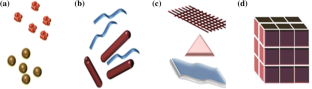
New vistas in fundamental and applied nanotechnology are being opened up by the production and the utilisation of nanoscale matter for distinctive physicochemical and optoelectronic applications 1 . The two main types of nanoparticles (NPs) in the classification are organic (made from dendrimers, micelles, liposomes, and compact polymers) and inorganic (made from noble metals like silver, gold, copper, zinc, titanium, and palladium, etc.). Because of their high surface-to-volume ratio, they have the ability to drastically modify biological, chemical, and physical aspects 2 . Silver nanoparticles (AgNPs), which are among the metal nanoparticles, are particularly important in the domains of therapeutics and biology 3 . The antibacterial effects of AgNPs against many pathogens, such as bacteria, viruses, and fungus, are well established 4 . Nanoparticles (NPs) can be manufactured using physical, chemical, and green method 5 . Simple biosynthesised nanoparticles have recently been identified as significant nanomedicines for a wide range of biomedical uses, such as antimicrobial activity and cytotoxic action against different cancer cells. Among all the metal oxide nanoparticles, their composite has found application in pharmacology and medicine 6 . Sustainable (green) synthesis is an alternative to physical and chemical processes that utilise hazardous substances, surfactants and adverse conditions, such as high temperatures or excess energy. Green nanoparticle synthesis is relatively energy-efficient, sustainable, affordable, simple, and scalable for industrial production 7 . In order to sustain the value of green synthesised NP S , there are three prerequisites. (i) Opting for environmentally favourable systems of solvents (ii) A sustainable reducing agent and (iii) an appropriate capping agent for stabilising nanoparticles. A variety of fungus, plants, algae, and other microbes are utilised for green synthesis 8 . Seaweeds are the most promising source of bioactive metabolites and have been used in food products, wastewater remediation, and medicinal potential. An excellent source of bioactive metabolites with abundant secondary components that have a wide range of biological actions are seaweed-mediated nanoparticles. The biological activities of seaweeds, such as green, brown, and red algae, include antibacterial, antiviral, anthelminthic, spermicidal, anticoagulant, antioxidant, antithrombotic, immuno-inflammatory, and cytotoxic effects 6 . Among these algae are aquatic organisms that can perform photosynthetic reactions to fulfil their nutritional requirements 9 . Algal synthesis of AgNPs is especially intriguing due to algae's exceptional capacity to assimilate metals and reduce metal ions. These can be used as effective bioagents to remove heavy metal pollution because of their capacity to endure a wide range of harsh environmental conditions 10 . Algae is a plentiful and widely dispersed organism; their ability to thrive in a lab setting is an additional benefit. These organisms offer low-cost assistance in large-scale production. Cellular reductase, which results in a reduction during the synthesis of AgNPs, is the primary component that aids in the synthesis of AgNPs via algae. Researchers reported that algae can produce silver nanoparticles both intracellularly and extracellularly 11 . Proteins, lipids, carbohydrates, carotenoids, vitamins, and secondary metabolites are among the bioactive compounds found in green algae (Terpenoids, phenols, flavonoids, and alkaloids), stabilise the created nanoparticles by serving as both capping and reductant 7 , 8 . Numerous scientists have asserted the sustainable synthesis of AgNPs and their multiple uses using a variety of algal extracts, including Sargassum myriocystum 6 , Chlorella pyrenoidosa 12 , Chlorella vulgaris , Chaetoceros calcitrans 13 , Scenedesmus abundans 14 , Caulerpa serrulate 15 etc.
Silver nanoparticles have numerous biological applications, including bio-imaging, bio-sensors, gene transport, photocatalysis, anti-microbial, anti-oxidant, and anti-cancer agents 7 , 16 . The antimicrobial activity of AgNPs is increased by interactions between Ag + and bacterial cell walls, the inactivation of enzymes which linked to membranes, the assembly of bacterial cells, the impairment of vital bacterial biomolecules, the breakdown of the cell envelope, and the generation of Reactive Oxygen Species (ROS) 17 . AgNPs are employed by pharmaceutical firms because of their minimal toxicity to human cells and stability across a wide temperature range 18 .
Currently, the aquatic ecosystem is exposed to a significant amount of industrial effluent, involving hazardous dyes from the textile, printing, and paper industries, which poses a significant threat to ecosystems. Resistant dyes in effluent can be dissolved or absorbed using AgNPs. When AgNPs absorb visible solar light, the outermost electron is promoted to an increased energy level during the breakdown of a dye. Furthermore, the radicals produced by the oxygen molecule and hydroxyl ion accepting the excited electron contribute to decomposition of dye molecules adsorbed on the surface of AgNP. By accepting an electron from the dye, the hole generated on the AgNP orbital facilitates the dye's continued degradation 9 . Recent research has focused on the investigation of photocatalytic breakdown utilising biological metallic NPs by numerous scientists 19 . The photocatalytic elimination of organic pollutants by algal silver nanoparticles has recently attracted the attention of researchers 20 .
In the present study, aqueous extract of Asterarcys (microalgae) was utilised for the biological reduction of AgNO 3 for green synthesis of AgNPs after thoroughly optimized parameters. The Asterarcys -mediated synthesis of silver nanoparticle denoted as “ Ast- AgNPs” were characterized by UV–Vis spectroscopy (Ultraviolet–visible spectroscopy), Fourier-Transform Infrared Spectroscopy (FTIR), X-ray diffraction (XRD), Scanning Electron Microscopy (SEM), Energy-dispersive X-ray (EDX), High-Resolution Transmission Electron Microscopy (HR-TEM), and Zeta potential. The synthesized Ast- AgNPs were examined for their effectiveness against gram positive and gram-negative bacteria as well as fungal strains. Additionally, the photocatalytic activities of Ast -AgNPs against methylene blue (MB), was also examined.
Materials and methods
The sample of algae was collected from Lake in Udaipur. The chemicals used in the BG-11 medium were obtained from Hi-Media and Sigma-Aldrich. The following items were also obtained like Agar–agar from Hi-Media, Antibiotics (Penicillin G, Chloramphenicol, and Streptomycin sulphate) from Hi-Media, Silver nitrate from Merck, Muller-Hinton Agar (MHA) from Hi-Media, Potato Dextrose Agar from Hi-Media, and Methyl blue (MB) from LOBA Chemie. The Microbial Research Laboratory, Department of Botany at Mohanlal Sukhadia University, Udaipur, Rajasthan, India, provided several strains for evaluating the antibacterial and antifungal properties. These strains include Staphylococcus aureus , Bacillus subtilis , Proteus vulgaris , Klebsiella pneumoniae , Fusarium sp., and Curvularia sp.
Collection of samples
Microalgal samples were collected from many places with fresh water bodies, including Fatehsagar Lake, Pichola Lake and Kukhadashwar temple pond from the Udaipur Region of Rajasthan, India. Water samples with a noticeable algal component were collected in labelled sampling polybags and Teflon bottles, then subsequently transferred them to the lab for additional research.
Isolation, purification and establishment of axenic culture
To separate single strain of algae from mixed cultures, BG-11 medium was added to the collected samples. The enriched algal cultures in BG-11 media were carefully examined to confirm the presence of several species 21 . Serial dilution was used to isolate a single algal strain from the collected sample. The image was then examined under a light microscope (Olympus CH20i), and then striking was performed on culture plates with 20 mL of the solidified Agar-BG-11 growth media at the laminar air flow clean bench. It took several iterations of this procedure to obtain only one strain of algae. The isolated strain was treated to a triple antibiotic solution technique in order to generate axenic culture 22 . After two to three weeks of incubation, the colonies emerged, and they were separated and put into liquid media. Algal species were cultured in growth chamber with the temperature of 25 °C under a white, cold fluorescent light with 16:8 h light:dark period at pH 7.4 of media for additional studies. Centrifugation process at 4000 rpm (20 min, 4 °C) was employed to harvest axenic microalgal cultures. To prevent contamination, the pelleted biomass was rinsed thrice with sterile water (deionized). The harvested wet biomass of algae was dried in oven (Yorco Scientific industries) at room temperature (RT) for 24 h 23 .
Identification of isolated algae
Under a calibrated compound light microscope (Olympus CH20i) with 10 ×, 40 ×, and 100 × immersion lenses, the main morphological characteristics of the isolated algal strain were examined and digital photomicrographs of the specimens were taken. For molecular identification, the genomic DNA extraction method (CTAB) was used to characterize the algal strain, which was then followed by PCR, gel electrophoresis, and algal identification was done by using rbcL (Ribulose bisphosphate Carboxylase Large subunit) gene sequencing. In addition, that sequence was uploaded to the NCBI database to obtain an accession number.
Algal extract preparation
The algal extract was prepared following slightly modification of previously published protocol 24 . The oven dried algal biomass of Asterarcys sp. was pulverized into a fine powder. The algal extract was made by combining dried algal biomass and 100 mL Deionized water (DI water) in a 150 mL beaker and incubating it at 60 °C (30 min). The extract was centrifuged at 5000 revolutions per minute (RPM) for 15 min. Following centrifugation, the filtrate (after passing through Whatman 1 filter paper (grade B59501, 125 mm) was employed in the biosynthesis of AgNPs as a reducing and capping agent. The purified algal extract was refrigerated at 4 °C for further use.
AgNO 3 solution preparation
By weighing AgNO 3 (Silver nitrate) in a specific quantity according to molarity and dissolving it in DI water, different concentrations of AgNO 3 solution were obtained. After being thoroughly dissolved, the mixture was then employed for additional experiments.
Synthesis of Ast -AgNPs
Optimization of synthesis of ast -agnps.
Optimizing factors such pH, reaction mixture temperature, the ratio of biomass to metallic ions, reaction time, and precursor concentration is crucial. Individual parameters were optimized one at a time while the other parameters kept constant. Changing the morphology of the end product, various reaction settings can have a significant influence on the reduction procedure. The "one factor at a time" method and the factor based experimental design were utilized throughout the course of the experiment 25 .
Effect of weight of algal biomass
To investigate the optimal conditions for AgNPs production, algal extract was prepared using various amounts of dried algal biomass (1, 3 and 5 gm).
Effect of molarity of AgNO 3
Algal extract was mixed with solutions containing varying amounts of AgNO 3 (1, 2, 3, 4, and 5 mM). The mixture was kept at the ambient temperature. A spectrophotometer was used to measure the color change (Shimadzu UV-1900).
Effect of algal extract and AgNO 3 ratio
Algal extract and AgNO 3 solution were added in five different ratios (5:95, 10:90, 15:85, 20:80, and 25:75). The reaction solution was allowed to incubate for 24 h at the ambient temperature. The peak of UV–Vis absorbance was examined in order to detect the synthesis of nanoparticles.
Effect of pH
Algal extract's pH could be adjusted to look at how pH affects the production of silver nanoparticles. Analytically graded 0.1 N NaOH and 0.1 N HCl standard solutions were added dropwise to adjust the pH of algal extract, within the range of 7, 8, 9, 10, and 11. The 20 mL of algal extract was added to 80 mL of 3 mM AgNO 3 , and the reaction solution was left for 24 h at ambient temperature. Utilising a UV–vis spectrophotometry with a visible wavelength (300–700 nm), the pH effect on the bio-fabrication process was determined.
Effect of temperature
The 80 mL of a 3 mM AgNO 3 solution was combined with 20 mL of algal extract before being incubated at room temperature (25 °C), 40, 50, and 60 °C for 24 h. By varying color, the absorbance spectra were produced.
Effect of incubation time
The 80 mL of AgNO 3 solution and 20 mL of algal extract were combined at various intervals throughout the incubation period. At specific intervals of 3, 6, 12, 24, and 48 h was used to analyze color fluctuations using UV–vis spectrophotometer.
Phyco‑synthesis of Ast ‑AgNPs after optimization
In a 150 mL conical flask, 20 mL of algal extract at pH 9 was combined with 80 mL of 3 mM AgNO 3 solution to produce silver nanoparticles. The mixture was subsequently kept for 24 h at ambient temperature 15 . After process was complete, the AgNPs were centrifuged (15,000 rpm) for 20 min at a temperature of 4 °C. The biogenic Ast ‑AgNPs were repeatedly centrifuged with DI water to eliminate contaminants before being transferred to a beaker and lyophilized. For further investigations, dried particles were gathered and preserved (Fig. 1 ).

Schematic representation of green synthesis of Silver nanoparticles from Asterarcys extract.
Characterization of Ast- AgNPs
Uv–visible spectra analysis (uv–vis).
The phyco-synthesis of AgNPs is confirmed with the help of UV–vis spectrophotometer at wavelengths between 300 and 700 nm. AgNPs (0.50 mL) were taken in a quartz cuvette and 2.50 mL of deionized water were added to dilute them for the UV–Vis study. The reaction solution that was anticipated to produce nanoparticles was exposed to wavelengths from 400 to 500 nm in order to identify the suitable wavelengths for nanoparticles production 26 .
Scanning electron microscopy (SEM) analysis
The surface morphology of the fabricated AgNPs was examined by SEM (EVO 18, Zeiss, Germany). Just a tiny amount of the sample was used to produce the thin films on the carbon-coated gold grids. With the help of paper, excess solution was blotted off., and the grids were then dried under a mercury lamp for five minutes before the photos were taken 26 .
Energy-dispersive X-ray spectroscopy (EDX) analysis
EDX (EVO 18, Zeiss, Germany) carried out an elemental analysis of silver. The drop coating procedure was applied to prepare the sample of aqueous AgNPs suspension for the analysis of EDX. At several locations on the sample, EDAX analysis was done in the spot profile mode with a beam diameter of 1 µm.
High-resolution transmission electron microscopy (HR-TEM) analysis
The interior structure of AgNPs was examined using TEM (Tecnai, G-20 FEI, USA). Specimens were supplied in a copper frame and given a 30 min drying period and further images were captured.
Selected area electron diffraction (SAED) patterns analysis
Using the SAED approach, multiple lattice parameters were examined from a range of diffraction patterns in order to assess the crystalline structure of the nanoparticles 27 . Using a parallel stream of intense electrons, this method focuses attention on a small sample. The XRD results were verified by comparing the d-spacing of the circular rings in the diffraction patterns with the standard JCPDS databases 28 .
Zeta potential analysis
The NP’s negative electrostatic charges can reveal NP’s stability 29 . Using the Zeta-sizer Nano, (ZS90) Malvern, the consistency of the generated nanoparticles, which were colloid in nature, was subjected to zeta potential analysis to examine stability of synthesized AgNPs.
X-ray diffraction (XRD) analysis
The dried AgNPs were analysed through an X-ray diffractometer to identify their crystal structure. The XRD patterns were captured using CuK radiation with a wavelength of 1.5406 Å using a Rigaku Ultima IV X-ray diffractometer (Japan). The diffractometer was operated at 40 kV and 44 mA with a step size of 0.02° and a speed of scanning of 4°/min, scanning in the range of 2θ = 20°–80°. The crystalline nature of the nanomaterial has been determined by comparing the obtained results with the standard JCPDS database. The characteristic crystalline nature of nanomaterials has been determined using the Debye–Scherrer formula 30 .
where D is the coherent scattering length (crystalline size in nm), K is the Scherrer's constant (0.98), λ is the wavelength of the X-ray source, β is the angular FWHM of the XRD diffraction peak and θ is the Bragg angle.
Using Origin Pro 2023 software, the FWHM was computed from the Gaussian function.
Fourier-transform infrared spectroscopy (FTIR) analysis
The functional groups of the algal constituents for the stabilisation of AgNPs and a reduction of Ag + ions were identified using FTIR analysis. The Ast -AgNPs that had been biosynthesized were mixed with potassium bromide (Kbr) to create a pellet, which was subsequently tested for the presence of infrared spectral bands with a resolution of 4 cm and wavelengths between 4000 and 400 cm −1 using FTIR spectrophotometer (Bruker Alpha, USA) 26 .
Applications of Ast ‑AgNPs
- Antibacterial activity
The bacterial strains like Staphylococcus aureus , Bacillus subtilis , Klebsiella pneumoniae and Proteus vulgaris were taken from the Microbial Research Laboratory at the Mohanlal Sukhadia University in Udaipur, Rajasthan, India. The obtained bacterial culture was sub cultured and placed in Muller-Hinton nutrient agar media (Hi-Media) in a Petri dish for further experiments.
Preparation of inoculums
To create bacterial inoculums, a loopful of bacterial culture was transferred from new agar plates to tubes containing 10 mL of Nutrient Broth (Hi-media), where it was cultured for 24 h at 37 °C. Occasionally, the culture tubes were shaken to aerate the contents and encourage growth 31 .
Following Kathiraven et al. and Sinha et al. with a few minor modifications 24 , 32 , the anti-bacterial activity was performed on Muller-Hinton Agar (MHA) plates, the antibacterial efficacy of biologically produced silver nanoparticles against Staphylococcus aureus, Bacillus subtilis, Proteus vulgaris , and Klebsiella pneumoniae was examined by the agar well diffusion procedure. The sterilized MHA (25 mL/plate) was added to petri dishes and allowed for a while until it solidified (at least 15–20 min). Using a flame-sterile glass spreader, overnight broth cultures of each strain of microbes (100 μL) was evenly distributed on an MHA plate and given some time to fully absorb the inoculums. Each of these solidified MHA medium plates was hollowed out to a diameter of 6 mm using a sterilized cork borer. Using a micropipette, the following samples were added to each well of all the plates: different concentration of AgNPs, pure algal extract, AgNO 3 solution, DI water (as a negative control), and streptomycin sulphate (as a positive control). The substances were then put in the wells and allowed to diffuse for 30 min at ambient temperature. Streptomycin, algal extract, AgNO 3 and DI water are used at the same concentration (100 µL/mL), however silver nanoparticles are employed at varied concentrations (100–250 µL/mL). The petridishes were incubated for 24 h at 37 °C. A distinct inhibition zone created around each well after incubation, indicating antimicrobial activity. The zones of inhibition were assessed by taking measurements of the width of the inhibition zones and it proved that bacteria were being inhibited by nanoparticles. Three replicates of each experiment were carried out simultaneously.
- Antifungal activity
The antifungal activity of Ast -AgNPs, against Fusarium sp. and Curvularia sp. were examined using the poison food method 33 . Silver nanoparticle stock solution (5 mg/mL) was prepared and placed at sonicator (Ningbo Sjialab Eqipment Co., Ltd) for 1 h homogenization process by probe-3. The 50 µL/mL, 100 µL/mL, and 150 µL/mL solutions were made from this stock solution. Additionally, a bavistin solution was made and used as a control. Algal extract was also employed in an antifungal assay to compare the extract's antifungal activity to that of synthesized Ast- AgNPs. The nine millilitres of sterilized PDA and 1 mL of AgNPs solution were mixed together and after that, the liquid was put into petri dishes and let to set for at least 15–20 min. Additional 6-mm diameter wells were created from 7-days old culture using a sterilized cork borer, put aseptically in the centre of solidified agar and maintained at 25 ± 2 °C in the incubator. After seven days, the average diameter of the growth was calculated. The % of mycelium growth inhibition used to measure the antifungal activity of each plate sample was calculated as follows 34 .
- Photocatalytic activity
A number of oxidation reactions utilise AgNPs as a catalyst because of their high surface-to-volume ratio 35 . Methyl blue (MB) is from the member of the phenothiazine-family solid brown dye colourant with a molecular weight of 319.85 g/mole and the formula C 37 H 27 N 3 Na 2 O 9 S 3 . It is frequently employed as a colourant in numerous applications. Methylene blue, a cationic dye, was used to assessed the dye degradation potential of green synthesised AgNPs. The % of decolorization measured by the formula given 25 .
where, C 0 = dye initial optical density, C = dye optical density after the photocatalytic decolorization.
Photocatalytic degradation of dye: operational parameters
The spectra at 664 nm demonstrate the photocatalytic breakdown of MB dye by AgNPs. Colour change is the indicator of Dye degradation 6 . The declining absorbance value, which occurs every 15 min, signals MB deterioration. The photocatalytic activity of Ast -AgNPs was examined in the presence of natural sunlight by adjusting the test solution's pH, initial dye concentration, catalyst concentration and reaction time 20 .
Effect of pH: The initial pH of the reaction solution affects the production of active radicals and the characteristics of the photocatalyst in the photocatalytic degradation process 36 . Studies were conducted utilising silver nanoparticle nano catalyst by calibrating the pH of the methylene blue dye solution to 3, 5, 7, 9, and 11 by adding standard solutions of 1N HCl or 1N NaOH, as pH plays important role in dye degradation.
Effect of MB dye concentration: The concentration of MB dye altered from 20 to 100 ppm while holding all other variables constant in order to determine the effect on photocatalytic activity (one factor at a time).
Effect of catalyst dosage: By shifting the catalyst concentration, the effect of catalyst dosage on MB dye degradation was investigated from 5 to 25 mg at an optimal pH of 11 and a dye concentration of 20 ppm.
Effect of light and catalyst: UV irradiation activated the catalyst, causing *OH radicals to appear on the surface 36 . We conducted three sets of reactions: (1) with the catalyst in the dark (2) with the catalyst in light (3) without the catalyst in light, in order to determine if the degradation happened through adsorption, photolysis or photocatalysis. In both the presence of the catalyst in the dark and the absence of the catalyst in the light, we found that there was barely any dye degradation. The catalyst and light were the conditions where dye degradation happened at the highest rate.
Statistical analysis
The entire set of data were statistically analysed using GraphPad Prism (version 3.02) software. The statistically significant difference is calculated with a one-way ANOVA, and the p-value denotes the likelihood of error.
Results and discussion
Algal identification.
Microscopic images of isolated microalga demonstrated that the algal cells were non-motile and spherical in shape. Nonetheless, it was extremely challenging to properly categorise the isolate based solely on morphological characteristics. However, the rbcL sequence found to have the greatest degree of similarity to the alga Asterarcys quadricellulare (accession number MW560279).
The Asterarcys sp. algal extract was investigated as a possible substitute for harsh chemical reagents such sodium borohydride as a reducing, capping, and stabilizing agent. The AgNPs' sizes and morphologies are influenced by a variety of variables, including pH, AgNO 3 and reducing agent concentrations, incubation duration, temperature and preparation techniques 37 . To generate NPs that are smaller and more stable, numerous variables, including metallic concentrations, the ratio of algal extract and AgNO 3 , temperature, pH, light, and duration of incubation, were investigated (Fig. 2 A–F). In this study varied weights of dried algal biomass were used for optimisation to synthesize AgNPs. The findings showed that biomass had a significant influence on the formation of AgNPs. Algal extract with 5 gm of dried algal biomass resulted in a narrow band with a high intensity peak at 430 nm (Fig. 2 A) suggesting this more appropriate conc. over 1 and 3 gm of algal dry wt. Optimising the AgNO 3 molarity is essential for producing appropriate size of AgNPs needed for subsequent studies since it significantly affects particle size 38 . When the quantity of AgNO 3 was rise-up from 1 to 3 mM, a large amount of AgNPs were generated, and a higher UV–Vis absorbance peak (428 nm) was observed (Fig. 2 B). But from 3 to 5 mM of AgNO 3 , it revealed the lower peak. Thus, the best optimized concentration of AgNO 3 solution was determined to be 3 mM. Similar outcomes were also observed when AgNPs were synthesised using a green technique 39 . Asterarcys extract concentrations (up to 20%) were added to a silver nitrate (AgNO 3 ) solution, and it was found that this increased the absorbance band intensity. The red shift in the synthesised AgNPs is related to the increased concentration of algal extract and AgNO 3 ratio (from 20 to 25%). The highest absorbance peak (428 nm) was achieved at a 20:80 (algal extract: silver nitrate) ratio (Fig. 2 C). A comparable investigation on Chlorella vulgaris was also carried out by Rajkumar and associates. Similar results were seen when AgNPs were produced using the Chlorella vulgaris algal extract serves as a reducing and capping agents. The results showed that AgNPs were produced at the: 8:2 v/v extract ratio 40 . Both the colour of the reaction solution and the UV–Vis spectrum have been reported to be affected by change of the pH of the solution. The effects of different pH levels on the fabrication of AgNPs were investigated (Fig. 2 D). The highest absorption peak was seen at 425 nm at pH 9 in this experiment. Small-sized nanoparticles were produced in an alkaline pH environment, whereas large-sized nanoparticles were produced in an acidic pH environment. Numerous functional groups are said to be ionised at an alkaline pH, making them available for reduction and facilitating the fabrication of NPs of small size 41 . The synthesis of AgNPs at various temperatures (RT °C, 40 °C, 50 °C, and 60 °C) is also investigated and results are shown in Fig. 2 E. The silver nanoparticles synthesized at RT °C exhibited highest peak at 426 nm. The optimal temperature for generating NPs was determined to be ambient temperature. As the temperature changes, bands also become wider 40 . The reaction time when the silver nitrate reacts with the extract also determines the quantity of nanoparticles produced 42 . As reaction time increased, absorbance spectra at 425 nm increased, and colour intensity enhanced over the duration of the incubation period. The greatest absorption was seen 24 h after the incubation period (Fig. 2 F). Dashora et al. also made similar observations regarding incubation time 33 . The UV–Vis spectra measurements revealed that the colour intensity increased over the course of the different time intervals up to 24 h, proving that AgNPs were synthesised without agglomeration. Comparable investigation has been carried out on the generation of AgNPs employing olive oil leaf extract 43 .

UV–Visible spectroscopic graphs of optimized parameters for AgNPs synthesis likewise, ( A ) algal biomass, ( B ) conc. of AgNO 3 , ( C ) AE:AgNO 3 , ( D ) pH, ( E ) temperature, ( F ) time interval.
The best optimised conditions for the production of Ast- AgNPs were discovered to be as incubation temperature (RT °C), extract: silver nitrate ratio (20:80), ionic strength of extract (pH-9), AgNO 3 molarity (3 mM), and maximum incubation length (24 h). The conversion of Ag + ions into AgNPs by biomolecules in the supernatant was verified by the presence of a significant UV–Vis spectra peak at 425 nm (Fig. 1 ). Similarly, AgNPs produced by Palmaria decipiens exhibited an absorption maximum at 425 nm 44 . The silver nanoparticles mediated by Sargassum myriocystum , a marine algae, showed characteristics peak at 420 nm 6 . On the other hand, the UV–Vis peak for AgNPs produced by Chlorella ellipsoidea algal extract was detected at 436 nm 45 . Previous studies have already shown that various silver nanoparticles from microalgae exhibited a peak in the wavelength region of 410–450 nm 46 .
The size of Ast -AgNPs has been found to be within 10 and 90 nm, with an approximate mean size of 35 nm (Fig. 3 A,B). These SEM findings were corroborated by previous published research 47 . The AgNPs with a spherical shape were found in C. vulgaris and C. calcitrans , with dimensions of 50–70 and 30–35 nm, respectively. The EDX observation that Ast- AgNPs contained 60.54 weight percent of silver implies a pure synthesis of silver nanoparticle (Fig. 3 C). The nanoparticle’s EDX analysis revealed that silver (Ag + ) atoms accounted for the greatest percentage of the peak intensity 13 . The EDX investigation for silver particles revealed a high signal in between 2.8 and 3.4 keV region 48 . The TEM micrograph (Fig. 3 D) of the AgNPs revealing that Ast- AgNPs are polyform, quasi spherical, rectangular and triangular in shapes. The 52 nm was found to be the average size of nanoparticles ranging in size from 20 to 100 nm (Fig. 3 E). The shape of the nanoparticles was described by other researchers as spherical, hexagonal and of a moderately wide range of sizes 46 . The crystal structure of NPs supported by selected area emission diffraction (Fig. 3 F) The SAED pattern generated the interplanar d-spacings of 2.29, 2.10, 1.45, and 1.23. The diffraction rings with the characteristic patterns were indexed (1 1 1), (2 0 0), (2 2 0), and (3 1 1) and are compatible with the FCC (Face-centered cubic) lattice structure often seen in AgNPs Joint Committee on Powder Diffraction Standards (JCPDS) file no. 00-004-0783. Result are in parity with previous reports 49 . The XRD pattern of synthesised AgNPs was analysed and compared to the standard powder diffraction card designed by the JCPDS. According to the FCC structure of AgNPs, intense diffraction peaks attributable to AgNPs are plainly observed at 38.30, 44.40, 64.64, and 77.59, which correspond to the (1 11), (2 0 0), (2 2 0), and (3 1 1) Bragg's reflection planes (Fig. 4 A) 32 . It was noted that reflections were sharp and intense, demonstrating the highly crystalline character of the produced AgNPs. When leaf extracts from Ficus virens were used to facilitate the synthesis of AgNPs, comparable outcomes have been found 50 . The synthesised nanoparticle’s Zeta potential values were determined to be -20.8 mV with a single peak (Fig. 4 B), suggesting moderately stable dispersion in the solution. The negative potential value of biosynthesized AgNPs indicates the existence of bio-organic constituents serving as a capping agent in the extract 48 . Due to the negative value, synthesised Ast -AgNPs did not aggregate and remained in suspension 33 . Understanding the functional groups involved in the interactions between metal particles and biomolecules is made possible with the use of FTIR analysis. In this investigation, the Asterarcys sp . algal extract that stabilises and caps the AgNPs was identified using FTIR spectrum. In order to establish the functional groups, the measured intensity bands were assessed against standard values absorption bands in the FTIR spectrum at 3217, 2926, 1650, 1400, 1068, and 563 cm −1 indicate the presence of a capping agent on the NPs (Fig. 4 C,D). Bands in the spectrum at 3217 cm −1 correspond to O–H stretching vibrations, indicating the presence of phenol and alcohol. The C–H stretching of the aromatic compound was observed to produce bands at 2926 cm −1 . The 1650 cm −1 spectral band, which corresponds to C–N and C–C stretching, indicates the presence of proteins. The band at 1400 cm −1 is attributed to the N–H stretch vibration in protein amide bonds. Numerous studies indicate that these functional groups contribute to the stability and capping of AgNPs. The bands at 1068 cm −1 correspond to the protein's C–N (amines) stretch vibration. Alkyl halide’s typical C–Br stretching may be responsible for the band at 563 cm −1 area. The FTIR spectroscopic analysis may lead to the conclusion that interactions between proteins and Ag + ions or nanoparticles in the algal extract have no impact on the proteins' secondary structure. In a related work, silver nanoparticles made from Urtica dioica leaves were also reported 51 . The wavelength absorption bands observed and presented as well as the FTIR spectrum of the algal extract and Ast -AgNPs, correspond with prior research 52 .

Structural characterization of Ast -AgNPs; ( A ) SEM micrograph (scale bar-100 nm), ( B ) SEM histogram, ( C ) EDX, ( D ) TEM micrograph (scale bar 100 nm), ( E ) TEM histogram, ( F ) SAED pattern (Scale bar-5 1/nm).

Characterization of Ast -AgNPs; ( A ) XRD, ( B ) zeta potential, ( C , D ) FTIR of Algal extract and AgNPs.
Applications of Ast- AgNPs
Antibacterial studies.
In the present study, the antibacterial activity of phyco-fabricated Ast -AgNPs was investigated against Staphylococcus aureus (gram + ve) (Fig. 5 A), Bacillus subtilis (gram + ve) (Fig. 5 B), Proteus vulgaris (gram-ve) (Fig. 5 C), and Klebsiella pneumoniae (gram-ve) (Fig. 5 D), with the results demonstrated that maximum antibacterial activity is reported against Staphylococcus aureus with a zone of inhibition as 25.66 ± 1.52 mm at 250 μL/mL, and lowest antibacterial activity is reported against Proteus vulgaris , measured at 19.33 ± 1.52 mm at 250 μL/mL (Fig. 5 E). This could be explained by the perception that the NPs enter within the bacterium and cling to the cell membrane, preferably, target the respiratory chain, where they cause cell division and ultimately apoptosis. In the bacterial cells, the NPs cause the release of silver ions, which improves the bactericidal activity. Multiple studies suggest that AgNPs may adhere to the plasma membrane's surface and interfere with the cell's permeability and respiration processes 32 .

Anti-bacterial activity of Ast -AgNPs against different bacterial strains ( A ) Staphylococcus aureus ( B ) Bacillus subtilis ( C ) Proteus vulgaris and ( D ) Klebsiella pneumoniae in both control and treatment conditions by agar well diffusion method, ( E ) Graphical representation of zone of inhibition of all four bacterial strain against different concentration of AGNPs. Anti-fungal activity of Ast -AgNPs against different fungal strains ( F ) Fusarium and ( G ) Curvularia , ( H ) Graphical representation of percentage growth inhibition of two fungal strains against different concentration of AgNPs.
In this study, different concentrations (50, 100, and 150 μL/mL) of Ast -AgNPs against pathogenic fungi were tested for their antifungal efficiency. Remarkable antifungal activity was shown by Ast- AgNPs against fungi Fusarium sp. and Curvularia sp (Fig. 5 F,G). Curvularia sp showed the highest percentage growth suppression by Ast- AgNPs at 150 μL/mL, at a rate of 85.05%, followed by 81.62% at 100 μL/mL and 69.66% at 50 μL/mL (Fig. 5 H). The results demonstrates that as silver nanoparticle concentration was increased, growth inhibitory percentage increased as well. At 150 μL/mL, the AgNPs had a 78.22% effectiveness rate against Fusarium sp. growth inhibition, followed by 65.78% at 100 μL/mL and 63.12% at 50 μL/mL (Fig. 5 H). The antifungal activity is caused by Ag + , leading to in membrane depolarization, pits in the cell wall, and the creation of plasma membrane apertures, thereby disrupting the integrity of the fungal membrane and obstructing the fungal cell cycle 53 . The algal extract of Asterarcys sp. lacked antibacterial properties. The extract exhibited minimal or no susceptibility in microbial strains, but the green synthesised AgNPs shown potent antibacterial activity against the pathogens 54 .
Phyco-synthesized AgNPs were used to investigate the photocatalytic dye degradation of methylene blue. The early indication that the dye was degrading came from a change in colour in the dye solution. In the meanwhile, AgNP concentrations, MB dye, and time intervals were found to influence photocatalytic activity. Silver nanoparticle plays a potential role in photocatalytic activity. Due to the SPR action of synthesised AgNPs, solar radiation transports e- from the valence band to the conduction band. The *O −2 is created when the excited electrons interact with O 2 species. O 2 + 2H ++ 2e− → H 2 O 2 is formed as a result of the reaction between *O −2 and H+. All varieties of cationic dye can be degraded by the *OH radicals produced when H 2 O 2 and H+ react 55 .
The initial pH of the reaction solution affects the generation of active species (radicals) and the characteristics of the photocatalyst in the photocatalytic system 56 . The results demonstrated in Fig. 6 A showed that the solution pH had a substantial impact on the photodegradation rate of MB dye, with 11.0 being the ideal pH. It is clear that lowering the MB working solution’s pH caused the decrease in decolorization efficiency of Ast- AgNPs from 59.78% and 42.95%, at neutral pH and 5 pH, respectively. Contrarily, as the MB solution's alkalinity rose, its decolorization effectiveness increased, reaching 77.67% and 83.9% at respective pH levels of 9 and 11. The results demonstrated that the photocatalytic efficiency increased as the pH increased. With optimum degradation at pH 11, alkaline conditions were found to promote better photocatalytic degradation. The alkaline pH of the MB solution enhanced the generation of OH*, thus speeding up the reaction rate and enhancing the solution's degradation efficiency. Several researchers used AgNPs under UV/solar light to degrade rhodamine B as well as MB and observed comparable outcomes 36 .

Optimized parameters for MB degradation; ( A ) pH, ( B ) MB Conc., ( C ) catalyst dosage, ( D ) WC (with catalyst); W/o (without catalyst), L (Light), D (Dark).
Effect of MB dye concentration
It was found that the 20 ppm dye concentration show maximum 87.92% of degradation at 120 min. Figure 6 B displayed the final results of effect of dye concentration on catalytic activity. On increasing the dye concentration of MB, the percentage degradation drastically decreased and reached 45.19% at 120 min (100 ppm). As a result, 20 ppm of dye concentration was found to be optimal. The efficiency of Ast -AgNPs' decolorization reduced when the original dye concentration was increased significantly. It is widely accepted that MB decolorization is sensitive to light process. Multiple-layer adsorption of dye molecules across the catalytic surface reduces UV light's ability to promote photo-oxidation as dye concentration increases. This would result in an abrupt decrease in the number of OH* assaulting MB molecules, thereby reducing the decolorization efficiency. It suggests that in order to have a same efficacy, the quantity of hydroxyl free radicals should rise proportionally to MB concentration. However, the photocatalytic degradation efficiency decreased after reaching the optimal initial dye concentration level 57 .
Effect of catalyst dosage
By adjusting the catalyst loading of Ast- AgNPs from 5 to 25 mg at an optimised 11 pH and a dye concentration of 20 ppm, catalyst dosage effect on the degradation % of MB dye was examined. It was noted that after 120 min, the 10 mg dosage of the catalyst showed a maximum 87.96% of degradation (Fig. 6 C). However, after the optimum dose (10 mg), the reaction mixture have turned turbid due to an increase in the amount of Ast- AgNPs that blocked UV light, which inhibited photochemical activation and produced OH radicals 36 . This occurred when numerous light backscattering at the fluid's interface appeared to reduce overall light absorption, preventing light from penetrating deeply into a solution with a high concentration of NPs 58 . The effect of light exposure duration on decolorization effectiveness was investigated for a maximum of 120 min.
Optimized protocol for MB dye decolorization
The purpose of the optimisation was to address the highest starting MB concentration while minimising irradiation duration at a pH and catalyst concentration that is practically applicable. To establish a optimized protocol for MB dye decolorization, these four crucial conditions were taken into consideration. Under the experimental conditions specified, the maximum amount of MB decolorization (88.59%) was anticipated for a photocatalytic mechanism in which an initial MB concentration of 20 ppm was subjected to treatment with Ast -AgNPs at 10 mg concentration, calibrated to 11 pH, and exposed to radiation for an overall exposure time of approximately 120 min (Fig. 6 D). The distinctive MB dye absorbance peak (664 nm) was observed dropping continuously with irradiation time, indicating that the dye was eliminated either by adsorption on the surface of the AgNPs or by catalytic degradation 20 . When it comes to the photodegradation of MB, light is crucial. With an increase in light exposure time, the photodegradation activity of AgNPs, TiO 2 , and Ag/TiO 2 nanomaterials increases. It is so because the photoelectron is produced when visible light is used to excite the valence electron. OH * generated by these highly powerful photoelectrons cause photodegradation of MB 57 .
The current study is significant since first time isolated microalga Asterarcys sp. was utilized in the biological reduction of AgNPs in a safer and more environmentally friendly manner. The AgNO 3 molarity (3 mM) with maximum incubation period of 24 h at pH 9 were observed to be the best optimized parameters for the AgNPs synthesis. AgNPs was determined with an estimated mean size of 35 nm and 52 nm by SEM and TEM, respectively. The EDX analysis confirmed 60.54 of Ag in AgNPs. Zeta potential value for the synthesized nanoparticle were found to be − 20.8 mV with a single peak suggesting that synthesised Ast -AgNPs did not aggregate and remained suspended. Utilising FTIR spectra, the biomolecular functional groups required for the bio reduction of Ag + and the capping/stabilization of AgNPs were identified. Staphylococcus aureus , Bacillus subtilis , Klebsiella pneumoniae , and Proteus vulgaris were among the Gram positive and Gram-negative bacteria that were significantly inhibited by Ast -AgNPs. In terms of antibacterial activity, Staphylococcus aureus had the highest levels of ZOI. An antifungal assay for biosynthesized nanoparticles was conducted utilising the poison food approach on Fusarium sp. and Curvularia sp. Curvularia sp. showed the highest growth inhibition percentage in antifungal activity. Furthermore, it was demonstrated that AgNPs have the ability to photocatalytically degrade hazardous dye methylene blue, in an aqueous solution. Methylene blue (MB) was potentially photocatalyzed by the Ast -AgNPs, with 88.59% of it being degraded in 120 min (Supplementary Information S1 ).
Data availability
The datasets generated during and/or analysed during the current study are available from the corresponding author on reasonable request.
Pei, J., Fu, B., Jiang, L. & Sun, T. Biosynthesis, characterization, and anticancer effect of plant-mediated silver nanoparticles using Coptis chinensis. Int. J. Nanomed. 14 , 1969 (2019).
Article CAS Google Scholar
Nagaraja, S. K., Niazi, S. K., Bepari, A., Assiri, R. A. & Nayaka, S. Leonotis nepetifolia flower bud extract mediated green synthesis of silver nanoparticles, their characterization, and in vitro evaluation of biological applications. Materials (Basel). 15 , 8990 (2022).
Murugesan, S., Bhuvaneswari, S. & Sivamurugan, V. Green synthesis, characterization of silver nanoparticles of a marine red alga Spyridia fusiformis and their antibacterial activity. Int. J. Pharm. Pharm. Sci 9 , 192–197 (2017).
Said, A., Abu-Elghait, M., Atta, H. M. & Salem, S. S. Antibacterial activity of green synthesized silver nanoparticles using lawsonia inermis against common pathogens from urinary tract infection. Appl. Biochem. Biotechnol. 196 , 85–98 (2024).
Article CAS PubMed Google Scholar
Soliman, M. K. Y., Salem, S. S., Abu-Elghait, M. & Azab, M. S. Biosynthesis of silver and gold nanoparticles and their efficacy towards antibacterial, antibiofilm, cytotoxicity, and antioxidant activities. Appl. Biochem. Biotechnol. 195 , 1158–1183 (2023).
Balaraman, P. et al. Phyco-synthesis of silver nanoparticles mediated from marine algae Sargassum myriocystum and its potential biological and environmental applications. Waste Biomass Valor. 11 , 5255–5271 (2020).
Nindawat, S. & Agrawal, V. Fabrication of silver nanoparticles using Arnebia hispidissima (Lehm.) A. DC. root extract and unravelling their potential biomedical applications. Artif. Cells Nanomed. Biotechnol. 47 , 166–180 (2019).
Mahajan, A., Arya, A. & Chundawat, T. S. Green synthesis of silver nanoparticles using green alga (Chlorella vulgaris) and its application for synthesis of quinolines derivatives. Synth. Commun. 49 , 1926–1937 (2019).
Garg, D. et al. Synthesis of silver nanoparticles utilizing various biological systems: mechanisms and applications—a review. Prog. Biomater. 9 , 81–95 (2020).
Article CAS PubMed PubMed Central Google Scholar
Hamida, R. S. et al. Green synthesis of hexagonal silver nanoparticles using a novel microalgae coelastrella aeroterrestrica strain BA_Chlo4 and resulting anticancer, antibacterial, and antioxidant activities. Pharmaceutics 14 , 2002 (2022).
Khanna, P., Kaur, A. & Goyal, D. Algae-based metallic nanoparticles: Synthesis, characterization and applications. J. Microbiol. Methods 163 , 105656 (2019).
Aziz, N. et al. Facile algae-derived route to biogenic silver nanoparticles: Synthesis, antibacterial, and photocatalytic properties. Langmuir 31 , 11605–11612 (2015).
Karthikeyan, P., Mohan, D., Abishek, G. & Priya, R. Synthesis of silver nanoparticles using Phytoplankton and its characteristics. Int. J. Fish. Aquat. Stud. 2 , 398–401 (2015).
Google Scholar
Aziz, N., Fatma, T., Varma, A. & Prasad, R. Biogenic synthesis of silver nanoparticles using Scenedesmus abundans and evaluation of their antibacterial activity. J. Nanoparticles 2014 , 1–6 (2014).
Article Google Scholar
Aboelfetoh, E. F., El-Shenody, R. A. & Ghobara, M. M. Eco-friendly synthesis of silver nanoparticles using green algae (Caulerpa serrulata): Reaction optimization, catalytic and antibacterial activities. Environ. Monit. Assess. 189 , 349 (2017).
Article PubMed Google Scholar
Rahmani, S. et al. Probing the interaction of silver nanoparticles with tau protein and neuroblastoma cell line as nervous system models. J. Biomol. Struct. Dyn. 36 , 4057–4071 (2018).
Aref, M. S. & Salem, S. S. Bio-callus synthesis of silver nanoparticles, characterization, and antibacterial activities via Cinnamomum camphora callus culture. Biocatal. Agric. Biotechnol. 27 , 101689 (2020).
Chugh, D., Viswamalya, V. S. & Das, B. Green synthesis of silver nanoparticles with algae and the importance of capping agents in the process. J. Genet. Eng. Biotechnol. 19 , (2021).
Bhagat, M., Anand, R., Datt, R., Gupta, V. & Arya, S. Green synthesis of silver nanoparticles using aqueous extract of Rosa brunonii Lindl and their morphological, biological and photocatalytic characterizations. J. Inorg. Organomet. Polym. Mater. 29 , 1039–1047 (2019).
Agnihotri, S., Sillu, D., Sharma, G. & Arya, R. K. Photocatalytic and antibacterial potential of silver nanoparticles derived from pineapple waste: Process optimization and modeling kinetics for dye removal. Appl. Nanosci. 8 , 2077–2092 (2018).
Article ADS CAS Google Scholar
Rippka, R., Deruelles, J., Waterbury, J. B., Herdman, M. & Stanier, R. Y. Generic assignments, strain histories and properties of pure cultures of cyanobacteria. Microbiology 111 , 1–61 (1979).
Kaushik, B. D. Laboratory methods for blue green algae (Associated Publishing Company, 1987).
Jena, J. et al. Microalga Scenedesmus sp.: A potential low-cost green machine for silver nanoparticle synthesis. J. Microbiol. Biotechnol. 24 , 522–533 (2014).
Sinha, S. N., Paul, D., Halder, N., Sengupta, D. & Patra, S. K. Green synthesis of silver nanoparticles using fresh water green alga Pithophora oedogonia (Mont.) Wittrock and evaluation of their antibacterial activity. Appl. Nanosci. 5 , 703–709 (2015).
Saranya, V. & Gowrie, S. U. Green synthesis, optimization, characterization of silver nanoparticles using Casuarina equisetifolia bark extract and its assays. World J. Pharm. Res. 3 , 797–814 (2016).
Choudhary, S. et al. Sustainable phyco-fabrication of silver nanoparticles using Coelastrella terrestris and their multiple downstream applications. Biocatal. Agric. Biotechnol. 53 , 102854 (2023).
Mourdikoudis, S., Pallares, R. M. & Thanh, N. T. K. Characterization techniques for nanoparticles: comparison and complementarity upon studying nanoparticle properties. Nanoscale 10 , 12871–12934 (2018).
Gupta, A., Koirala, A. R., Gupta, B. & Parajuli, N. Improved method for separation of silver nanoparticles synthesized using the Nyctanthes arbor-tristis shrub. Acta Chem. Malaysia 3 , 35–42 (2019).
Suresh, P. K. & Sharma, S. Formulation and in-vitro characterization of selfnanoemulsifying drug delivery system of cinnarizine. Drugs 11 , 12 (2011).
Krishnaraj, C., Ramachandran, R., Mohan, K. & Kalaichelvan, P. T. Optimization for rapid synthesis of silver nanoparticles and its effect on phytopathogenic fungi. Spectrochim. Acta Part A Mol. Biomol. Spectrosc. 93 , 95–99 (2012).
Pandithurai, M., Murugesan, S. & Sivamurugan, V. Antibacterial activity of various solvent extracts of marine brown alga Spatoglossum asperum. Int. J. Pharmacol. Res. 5 , 133–138 (2015).
Kathiraven, T., Sundaramanickam, A., Shanmugam, N. & Balasubramanian, T. Green synthesis of silver nanoparticles using marine algae Caulerpa racemosa and their antibacterial activity against some human pathogens. Appl. Nanosci. 5 , 499–504 (2015).
Dashora, A., Rathore, K., Raj, S. & Sharma, K. Synthesis of silver nanoparticles employing Polyalthia longifolia leaf extract and their in vitro antifungal activity against phytopathogen. Biochem. Biophys. Rep. 31 , 101320 (2022).
CAS PubMed PubMed Central Google Scholar
Al-Otibi, F. et al. Biosynthesis of silver nanoparticles using Malva parviflora and their antifungal activity. Saudi J. Biol. Sci. 28 , 2229–2235 (2021).
Joshi, N. et al. Biosynthesis of silver nanoparticles using Carissa carandas berries and its potential antibacterial activities. J. Sol-Gel Sci. Technol. 86 , 682–689 (2018).
Shaikh, W. A., Chakraborty, S. & Islam, R. U. Photocatalytic degradation of rhodamine B under UV irradiation using Shorea robusta leaf extract-mediated bio-synthesized silver nanoparticles. Int. J. Environ. Sci. Technol. 17 , 2059–2072 (2020).
Kummara, S., Patil, M. B. & Uriah, T. Synthesis, characterization, biocompatible and anticancer activity of green and chemically synthesized silver nanoparticles–a comparative study. Biomed. Pharmacother. 84 , 10–21 (2016).
Periaswamy Sivagnanam, S. et al. Green synthesis of silver nanoparticles from deoiled brown algal extract via Box-Behnken based design and their antimicrobial and sensing properties. Green Process. Synth. 6 , 147–160 (2017).
Chouhan, S. & Guleria, S. Green synthesis of AgNPs using Cannabis sativa leaf extract: Characterization, antibacterial, anti-yeast and α-amylase inhibitory activity. Mater. Sci. Energy Technol. 3 , 536–544 (2020).
CAS Google Scholar
Rajkumar, R., Ezhumalai, G. & Gnanadesigan, M. A green approach for the synthesis of silver nanoparticles by Chlorella vulgaris and its application in photocatalytic dye degradation activity. Environ. Technol. Innov. 21 , 101282 (2021).
Aslam, M. et al. Phyto-extract-mediated synthesis of silver nanoparticles using aqueous extract of Sanvitalia procumbens, and characterization, optimization and photocatalytic degradation of azo dyes Orange G and Direct Blue-15. Molecules 26 , 6144 (2021).
Article ADS CAS PubMed PubMed Central Google Scholar
Prathna, T. C., Chandrasekaran, N., Raichur, A. M. & Mukherjee, A. Biomimetic synthesis of silver nanoparticles by Citrus limon (lemon) aqueous extract and theoretical prediction of particle size. Colloids Surf. B Biointerfaces 82 , 152–159 (2011).
Khalil, M. M. H., Ismail, E. H., El-Baghdady, K. Z. & Mohamed, D. Green synthesis of silver nanoparticles using olive leaf extract and its antibacterial activity. Arab. J. Chem. 7 , 1131–1139 (2014).
González-Ballesteros, N., González-Rodríguez, J. B., Rodríguez-Argüelles, M. C. & Lastra, M. New application of two Antarctic macroalgae Palmaria decipiens and Desmarestia menziesii in the synthesis of gold and silver nanoparticles. Polar Sci. 15 , 49–54 (2018).
Article ADS Google Scholar
Borah, D. et al. Alga-mediated facile green synthesis of silver nanoparticles: Photophysical, catalytic and antibacterial activity. Appl. Organomet. Chem. 34 , e5597 (2020).
Soleimani, M. & Habibi-Pirkoohi, M. Biosynthesis of silver nanoparticles using Chlorella vulgaris and evaluation of the antibacterial efficacy against Staphylococcus aureus. Avicenna J. Med. Biotechnol. 9 , 120 (2017).
PubMed PubMed Central Google Scholar
Arya, A., Gupta, K., Chundawat, T. S. & Vaya, D. Biogenic synthesis of copper and silver nanoparticles using green alga Botryococcus braunii and its antimicrobial activity. Bioinorg. Chem. Appl. 2018 , (2018).
Ravichandran, V. et al. Green synthesis, characterization, antibacterial, antioxidant and photocatalytic activity of Parkia speciosa leaves extract mediated silver nanoparticles. Res. Phys. 15 , 102565 (2019).
Mavaei, M., Chahardoli, A., Shokoohinia, Y., Khoshroo, A. & Fattahi, A. One-step synthesized silver nanoparticles using isoimperatorin: Evaluation of photocatalytic, and electrochemical activities. Sci. Rep. 10 , 1762 (2020).
Salar, R. K., Sharma, P. & Kumar, N. Enhanced antibacterial activity of streptomycin against some human pathogens using green synthesized silver nanoparticles. Resour. Technol. 1 , 106–115 (2015).
Jyoti, K., Baunthiyal, M. & Singh, A. Characterization of silver nanoparticles synthesized using Urtica dioica Linn. leaves and their synergistic effects with antibiotics. J. Radiat. Res. Appl. Sci. 9 , 217–227 (2016).
Bhuyar, P. et al. Synthesis of silver nanoparticles using marine macroalgae Padina sp. and its antibacterial activity towards pathogenic bacteria. Beni-Suef Univ. J. Basic Appl. Sci. 9 , 1–15 (2020).
Pethakamsetty, L. et al. Green synthesis, characterization and antimicrobial activity of silver nanoparticles using methanolic root extracts of Diospyros sylvatica. J. Environ. Sci. 55 , 157–163 (2017).
Danaei, M., Motaghi, M. M., Naghmachi, M., Amirmahani, F. & Moravej, R. Green synthesis of silver nanoparticles (AgNPs) by filamentous algae extract: Comprehensive evaluation of antimicrobial and anti-biofilm effects against nosocomial pathogens. Biologia (Bratisl). 76 , 3057–3069 (2021).
Kanniah, P. et al. Green synthesis of multifaceted silver nanoparticles using the flower extract of Aerva lanata and evaluation of its biological and environmental applications. Chem. Select 5 , 2322–2331 (2020).
Wahab, H. S. & Hadi, H. M. Visible light N-TiO 2-induced photodegradation of Congo red: Characterization, kinetics and mechanistic study. Int. J. Environ. Sci. Technol. 14 , 2135–2148 (2017).
Tahir, K. et al. Visible light photo catalytic inactivation of bacteria and photo degradation of methylene blue with Ag/TiO2 nanocomposite prepared by a novel method. J. Photochem. Photobiol. B Biol. 162 , 189–198 (2016).
Hogan, N. J. et al. Nanoparticles heat through light localization. Nano Lett. 14 , 4640–4645 (2014).
Article ADS CAS PubMed Google Scholar
Download references
Acknowledgements
Sunita Choudhary and Geetanjali Kumawat wish to acknowledge the support of the Council of Scientific and Industrial Research CSIR New Delhi, (Ref No. 09/172(0089)/2019-EMR-I) and the University Grant Commission UGC New Delhi, (Ref No. 999/CSIR-UGC NET JUNE 2019) respectively. Funding support from Department of Science and Technology, New Delhi is also acknowledged for laboratory infrastructure (SERB File Number: EEQ/2020/000011). AIIMS SAIF, New Delhi, is thanked for the HR-TEM and SEM- EDAX. Prof. N. Laxmi and Dr. Prabhat K. Baroliya are acknowledged for their assistance with the XRD and FTIR analyses, respectively. Special thanks to Dr. Mukesh Meena and Dr. Tripta Jain for providing fungal and bacterial strains.
Author information
Authors and affiliations.
Department of Botany, Mohanlal Sukhadia University, Udaipur, 313001, India
Sunita Choudhary, Geetanjali Kumawat & Harish
Department of Chemistry, Mohanlal Sukhadia University, Udaipur, 313001, India
Manisha Khandelwal & Rama Kanwar Khangarot
Department of Molecular Biology and Biotechnology, Rajasthan College of Agriculture, Maharana Pratap University of Agriculture and Technology, Udaipur, 313001, Rajasthan, India
Vinod Saharan
Amity Institute of Biotechnology, Amity University, Noida, 201313, Uttar Pradesh, India
Subhasha Nigam
You can also search for this author in PubMed Google Scholar
Contributions
Sunita Choudhary, experimentation, methodology, literature survey, writing—original draft; Geetanjali Kumawat validation, writing—review and editing; Manisha Khandelwal, validation, methodology; Rama Kanwar Khangarot, validation, methodology; Vinod Saharan, writing—review and editing; Subhasha Nigam, writing—review and editing Harish, conceptualization, supervision, writing—review and editing.
Corresponding author
Correspondence to Harish .
Ethics declarations
Competing interests.
The authors declare no competing interests.
Additional information
Publisher's note.
Springer Nature remains neutral with regard to jurisdictional claims in published maps and institutional affiliations.
Supplementary Information
Supplementary information., rights and permissions.
Open Access This article is licensed under a Creative Commons Attribution 4.0 International License, which permits use, sharing, adaptation, distribution and reproduction in any medium or format, as long as you give appropriate credit to the original author(s) and the source, provide a link to the Creative Commons licence, and indicate if changes were made. The images or other third party material in this article are included in the article's Creative Commons licence, unless indicated otherwise in a credit line to the material. If material is not included in the article's Creative Commons licence and your intended use is not permitted by statutory regulation or exceeds the permitted use, you will need to obtain permission directly from the copyright holder. To view a copy of this licence, visit http://creativecommons.org/licenses/by/4.0/ .
Reprints and permissions
About this article
Cite this article.
Choudhary, S., Kumawat, G., Khandelwal, M. et al. Phyco-synthesis of silver nanoparticles by environmentally safe approach and their applications. Sci Rep 14 , 9568 (2024). https://doi.org/10.1038/s41598-024-60195-3
Download citation
Received : 16 August 2023
Accepted : 07 March 2024
Published : 26 April 2024
DOI : https://doi.org/10.1038/s41598-024-60195-3
Share this article
Anyone you share the following link with will be able to read this content:
Sorry, a shareable link is not currently available for this article.
Provided by the Springer Nature SharedIt content-sharing initiative
- Silver nanoparticles
- Methylene blue
By submitting a comment you agree to abide by our Terms and Community Guidelines . If you find something abusive or that does not comply with our terms or guidelines please flag it as inappropriate.
Quick links
- Explore articles by subject
- Guide to authors
- Editorial policies
Sign up for the Nature Briefing: Microbiology newsletter — what matters in microbiology research, free to your inbox weekly.


We apologize for the inconvenience...
To ensure we keep this website safe, please can you confirm you are a human by ticking the box below.
If you are unable to complete the above request please contact us using the below link, providing a screenshot of your experience.
https://ioppublishing.org/contacts/

- ‹ Previous Chapter
- Next Chapter ›
2. Literature Review
- Published: 2018
- Split-Screen
- Open the PDF for in another window
- Cite Icon Cite
- Permissions
- Search Site
Rajawat, S, & Mailk, M. "Literature Review." Silver Nanoparticles: Properties, Synthesis Techniques, Characterizations, Antibacterial and Anticancer Studies. Ed. Rajawat, S, & Mailk, M. ASME Press, 2018.
Download citation file:
- Ris (Zotero)
- Reference Manager
In nanotechnology, nanomaterials are created at molecular level which exhibit qualities quite different from the bulk material. Bulk silver, when reduced to nano-level, shows remarkable changes in the properties making it more environments friendly and useful. Nano-silver can be defined as a group of micro-sized (nano) bits of silver that are either covering or suspended in a medium. Nanosilver exhibits unique physical and chemical properties compared to “conventional” silver (e.g., macro scale “bulk” silver). Nano silver of different shapes can be synthesized using various synthesis processes.
Email alerts
Related chapters, related articles, related proceedings papers, asme journals.
- About ASME Journals
- Information for Authors
- Submit a Paper
- Call for Papers
- Title History
ASME Conference Proceedings
- About ASME Conference Publications and Proceedings
- Conference Proceedings Author Guidelines
ASME eBooks
- About ASME eBooks
- ASME Press Advisory & Oversight Committee
- Book Proposal Guidelines
- Frequently Asked Questions
- Publication Permissions & Reprints
- ASME Membership
Opportunities
- Faculty Positions
- ASME Community

- Accessibility
- Privacy Statement
- Terms of Use
- Get Adobe Acrobat Reader
This Feature Is Available To Subscribers Only
Sign In or Create an Account
Advertisement
Silver nanoparticles: various methods of synthesis, size affecting factors and their potential applications–a review
- Review Article
- Published: 13 March 2020
- Volume 10 , pages 1369–1378, ( 2020 )
Cite this article

- Asim Ali Yaqoob 1 ,
- Khalid Umar 1 &
- Mohamad Nasir Mohamad Ibrahim 1
8676 Accesses
299 Citations
4 Altmetric
Explore all metrics
Currently, synthesis of nanoparticles from several noble metals like palladium, tin, copper, silver and gold etc. has received more attention because of their unique properties as well as their application in different fields. Furthermore, silver nanoparticles play an important role in pharmaceutical industries because they function like antibacterial agents which carry less toxic effects. In case of industrial applications, silver particles (inkjet inks) having regular dispersions are helpful in making different electronic circuits. Over the period, various synthetic methods for the synthesis of silver nanoparticles were reported i.e. physical, chemical, and photochemical. However, most of the available techniques are expensive and not eco-friendly i.e. environmentally harmful. There are various factors such as the methods of synthesis, temperature, dispersing agent, surfactant etc. which greatly influence the quality and quantity of the synthesized nanoparticles and ultimately affect their properties. It is also pertinent to mention here that the main target for these silver nanoparticles was not only to synthesize in nano range, but also require easy, eco-friendly and economical synthesis of the nanoparticles. Therefore, this review mainly goes through the several methods of synthesis of nanoparticles which should be based on the green approach, and easy to be synthesized at low cost. In addition, we also discussed some approaches to fabricate silver-based nanoparticles, their enhanced properties and their different type of applications such as electrical conductivity, antibacterial, optical, photocatalytic properties.
This is a preview of subscription content, log in via an institution to check access.
Access this article
Price includes VAT (Russian Federation)
Instant access to the full article PDF.
Rent this article via DeepDyve
Institutional subscriptions
Recent advances in synthetic methods and applications of silver nanostructures.

An Introduction to Different Methods of Nanoparticles Synthesis

Synthesis of copper nanoparticles : An overview of the various methods
Albrecht M, Janke V, Sievers S, Siegner U, Schüler D, Heyen U (2005) Scanning force microscopy study of biogenic nanoparticles for medical applications. J Magn Magn Mater 290:269–271
Google Scholar
Anna Z, Ewa S, Adriana Z, Maria G, Jan H (2009) Preparation of silver nanoparticles with controlled particle size. Procedia Chem 1:1560–1566
Banerjee P, Satapathy M, Mukhopahayay A, Das P (2014) Leaf extract mediated green synthesis of silver nanoparticles from widely available Indian plants: synthesis, characterization, antimicrobial property and toxicity analysis. Bioresour Bioprocess 1(1):3. https://doi.org/10.1186/s40643-014-0003-y
Article Google Scholar
Boddu SR, Gutti VR, Ghosh TK, Tompson RV, Loyalka SK (2011) Gold, silver, and palladium nanoparticle/nano-agglomerate generation, collection, and characterization. J Nanopart Res 13:6591–6601
CAS Google Scholar
Bose D, Chatterjee S (2016) Biogenic synthesis of silver nanoparticles using guava ( Psidium guajava ) leaf extract and its antibacterial activity against Pseudomonas aeruginosa . Appl Nanosci 6:895–901. https://doi.org/10.1007/s13204-015-0496-5
Article CAS Google Scholar
Bosetti M, Masse A, Tobin E, Cannas M (2002) Silver coated materials for external fixation devices: in vitro biocompatibility and genotoxicity. Biomaterials 23:887–892
Bushra R, Shahadat M, Ahmad A, Nabi SA, Umar K, Oves M, Raeissi AS, Muneer M (2014) Synthesis, characterization, antimicrobial activity and applications of polyaniline Ti(IV) arsenophosphate adsorbent for the analysis of organic and inorganic pollutants. J Hazard Mater 264:481–489
Chen M, Feng YG, Wang X, Li TC, Zhang JY, Qian DJ (2007) Silver nanoparticles capped by oleylamine: formation, growth, and self-organization. Langmuir 23:5296–5304
Chen X, Schluesener H (2008) Nanosilver: a Nano product in medical application. Toxicol Lett 176:1–12
Chen SF, Zhang H (2012) Aggregation kinetics of nanosilver in different water condition. Adv Nat Sci Nanosci Nanotechnol 3(035006):1–7
Chou KS, Lai YS (2004) Effect of polyvinyl pyrrlidone molecular weights on the formation of nanosized silver colloids. Mater Chem Phys 83:82–88
Cho M, Chung H, Choi W, Yoon J (2005) Different inactivation behaviors of MS-2 phage and Escherichia coli in TiO 2 photocatalytic disinfection. Appl Environ Microbiol 71:270–275
Dalzon B, Torres A, Diemer H, Ravanel S, Collin-Faure V, Pernet-Gallay K, Jouneau PH, Bourguignon J, Cianférani S, Carrière M, Aude-Garcia C (2019) How reversible are the effects of silver nanoparticles on macrophages? A proteomic-instructed view. Environ Sci Nano 6(10):3133–3157
Dang TMD, Le TTT, Blance EF, Dang MC (2012) Influence of surfactant on the preparation of silver nanoparticles by polyol method. Adv Nat Sci Nanosci Nanotechnol 3(035004):1–4
Dar AA, Umar K, Mir NA, Haque MM, Muneer M, Boxall C (2011) Photocatalysed degradation of a herbicide derivative, Dinoterb, in aqueous suspension. Res Chem Intermediat 37:567–578
Darr JA, Zhang J, Makwana NM, Weng X (2017) Continuous hydrothermal synthesis of inorganic nanoparticles: applications and future directions. Chemical Rev 117(17):11125–11238
Das N, Karar A, Vasiliev M, Tan CL, Alameh K, Lee YT (2011) Analysis of nano-grating-assisted light absorption enhancement in metal–semiconductor–metal photodetectors patterned using focused ion-beam lithography. Opt Commun 284(6):1694–1700
Dolgaev I, Simakin AV, Voronov VV, Shafeev GA, Verduraz FB (2002a) Nanoparticles produced by laser ablation of solids in liquid environments. J Biomedical Optics 186:546–551
Dolgaev SI, Simakin AV, Voronov VV, Shafeev GA, Bozon-Verduraz F (2002b) Nanoparticles produced by laser ablation of solids in liquid environment. Appl Surf Sci 186(4):546–551
Duran N, Marcarto PD, Souza GHD, Alves OL, Esposito Ev (2007) Antibacterial effect of silver nanoparticles produced by fungal process on textile fabrics and their effluent treatment. J Biomed Nanotechnol 3:203–208
El-Nour KMM, Eftaiha A, Al-Reda A, Ammar AA (2004) Synthesis and applications of silver nanoparticles. Arabian J Chem 3:135–140
El-Nour KMMA, Eftaiha AA, Al-Warthan A, Ammar RA (2010a) Synthesis and applications of silver nanoparticles. Arab J Chem 3:135–140. https://doi.org/10.1016/j.arabjc.2010.04.008
Faisal M, Tariq MA, Khan A, Umar K, Muneer M (2011) Photochemical reactions of 2, 4-dichloroaniline and 4-nitroanisole in aqueous suspension of titanium dioxide. Sci Adv Mater 3:269–275
Ge J, Huynh T, Hu Y, Yin Y (2008) Hierarchical magnetite/silica Nano assemblies as magnetically recoverable catalyst–supports. Nano Lett 3(8):931–934
Guimarães ML, da Silva FA, da Costa MM, de Oliveira HP (2019) Green synthesis of silver nanoparticles using Ziziphus joazeiro leaf extract for production of antibacterial agents. Appl Nanosci. 19:1–9. https://doi.org/10.1007/s13204-019-01181-4
Guo JZ, Cui H, Zhou W, Wang W (2008) Ag nanoparticle catalyzed chemiluminescent reaction between luminol and hydrogen peroxide. J Photochem Photobiol A Chem 193:89–96
Hamouda IM (2012) Current perspectives of nanoparticles in medical and dental biomaterials. J Biomed Res 3(26):143–215
Hecht DS, Ramirez RJ, Briman M, Artukovic E, Chichak KS, Stoddart JF, Grüner G (2006) Bioinspired detection of light using a porphyrin-sensitized single-wall nanotube field effect transistor. Nano Lett 6(9):2031–2036
Hoeng F, Denneulin A, Bras J (2016) Use of nanocellulose in printed electronics: a review. Nanoscale 8(27):13131–13154
Hsu SLC, Wu RT (2007) Synthesis of contamination-free silver nanoparticle suspensions for micro-interconnects. Mater Lett 61:3719–3722
Hu JQ, Chen Q, Xie ZX, Han GB, Wang RH, Ren BZ, Yang Tian YZQ (2004) A simple effective route for the synthesis of crystalline silver nanorodes and nanowires. Adv Funct Mater 14:183–187
Huang H, Yang Y (2008) Preparation of silver nanoparticles in inorganic clay suspensions. Compos Sci Technol 68:2948–2953
Iacono ST, Jennings AR (2019) Recent studies on fluorinated silica nanometer-sized particles. Nanomaterials 9(5):684
Iravani S, Korbekandi H, Mirmohammadi SV, Zolfaghari B (2014) Synthesis of silver nanoparticles: chemical, physical and biological methods. Res Pharm Sci 9:385–406
Irshad A, Sarwar N, Sadia H, Riaz M, Sharif S, Shahid M, Khan JA (2019) Silver nano-particles: synthesis and characterization by using glucans extracted from Pleurotus ostreatus . Appl Nanosci. https://doi.org/10.1007/s13204-019-01103-4
Ismail M, Jabra R (2017) Investigation the parameters affecting on the synthesis of silver nanoparticles by chemical reduction method and printing a conductive pattern. J Mater Environ Sci 8(11):4152–4159
Jain P, Pradeep T (2005) Potential of silver nanoparticle-coated polyurethane foam as antibacterial water filters. Biotechnol Bioeng 90:59–63
Jana ANR, Sau TK, Pal TJ (1999) Growing Small Silver Particles as Redox Catalyst. J Phys Chem B 103:115–121
Junaidi J et al (2016) Chloride ion addition for controlling shapes and properties of silver nanorodes capped by polyvinyl alcohol synthesized using polyol method. Conference Paper 10(1063/1):4945485
Kalishwaralal K, Deepak V, Ramkumarpandian S, Nellaiah H, Sangiliyandi G (2008) Extracellular biosynthesis of silver nanoparticles by the culture supernatant of Bacillus licheniformis. Mater Lett 62:4411–4413
Khanna PK, Kulkarni D, Beri RK (2008) Synthesis and characterization of myristic acid capped silver nanoparticles. J Nanopart Res 6:1059–1062
Kholoud MM, Nour AE, Eftaiha A, Warthan AA, Ammar RAA (2010b) Synthesis and applications of silver nanoparticles. Arab J Chem 3:135–140
Kruis F, Fissan H, Rellinghaus B (2000) Sintering and evaporation characteristics of gas-phase synthesis of size selected PbS nanoparticles. Mater Sci Eng B 69(70):329–334
Lahav M, Shipway AN, Willner I, Nielsen MB, Stoddart JF (2000) An enlarged bis-bipyridinium cyclophane-Au nanoparticle superstructure for selective electrochemical sensing applications. J Electroanal Chem 482(2):217–221
Lee HH, Chou KS, Huang KC (2005) Inkjet printing of nanosize silver colloids. Nanotechnology 16:2436–2441
Li Q, Mahendra S, Lyon DY, Brunet L, Liga MV, Li D, Alvarez PJ (2008) Antimicrobial nanomaterials for water disinfection and microbial control: potential applications and implications. Water Res 42:4591–4602
Luo C, Zhang Y, Zeng X, Zeng Y, Wang Y (2005) The role of poly(ethylene glycol) in the formation of silver nanoparticles. J Colloi Interface Sci 288(2):444–448
Mafune F, Kohno J, Takeda Y, Kondow T, Sawabe H (2000) Formation and size control of silver nanoparticles by laser ablation in aqueous solution. J Phys Chem B 104:9111–9117
Magnusson MH, Deppert K, Malm JO, Bovin JO, Samuelson L (1999) Gold nanoparticles: Production, reshaping, and Thermal charging. J Nanoparticle Res 1:243–251
Malik A, Hameed S, Siddiqui MJ, Haque MM, Umar K, Khan A, Muneer M (2014) Electrical and optical properties of nickel-and molybdenum-doped titanium dioxide nanoparticle: improved performance in dye-sensitized solar cells. J Mater Eng Perform 23:3184–3192
Manno D, Filippo E, Di Giulio M, Serra A (2008) Synthesis and characterization of starch-stabilized Ag nanostructures for sensors applications. J Non Cryst Solids 354(52–54):5515–5520. https://doi.org/10.1016/j.jnoncrysol.2008.04.059
Marambio-Jones C, Hoek EMV (2010) A review of the antibacterial effects of silver nanomaterials and potential implications for human health and the environment. J Nanopart Res 12:1531–1551
Maria BS, Devadiga A, Kodialbail VS, Saidutta MB (2015) Synthesis of silver nanoparticles using medicinal Zizyphus xylopyrus bark extract. Applied Nanoscience Appl Nanosci 5:755–762. https://doi.org/10.1007/s13204-014-0372-8
Mir NA, Khan A, Umar K, Muneer M (2013) Photocatalytic study of a xanthene dye derivative, phloxine B in aqueous suspension of TiO 2 : adsorption isotherm and decolourization kinetics. Environ Focus 2:208–216
Nair GB, Dhoble SJ (2015) Highly enterprising calcium zirconium phosphate [Ca Zr 4 (PO 4 ) 6 : Dy 3+ , Ce 3+ ] phosphor for white light emission. RSC Adv 5(61):49235–49247
Natsuki J, Abe T (2016) Synthesis of pure colloidal silver nanoparticles with high electro conductivity for printed electronic circuits: The effect of amines on their formation in aqueous media. J Colloid Interf Sci 359:19–23
Natsuki J, Natsuki T, Abe T (2013) Low molecular weight compounds as effective dispersing agents in the formation of colloidal silver nanoparticles. J Nano part Res 15:1483–1489
Natsuki J, Natsuki T, Hashimoto Y (2015) A Review of Silver Nanoparticles: Synthesis Methods Properties and Applications. IJ MSA 20150405(17):2327–2635
Nazme M, Kamal A, Rajendran V, Yong TL (2013) Silver-nanoparticle-based etch mask control for subwavelength structure development. Nanosyst Phys Chem Math 4(3):387–394
Nie W, Tsai H, Asadpour R, Blancon JC, Neukirch AJ, Gupta G, Crochet JJ, Chhowalla M, Tretiak S, Alam MA, Wang HL (2015) High-efficiency solution-processed perovskite solar cells with millimeter-scale grains. Science 347(6221):522–525
Olteanu RL, Nicolescu CM, Bumbac M (2017) Influence of phytochemical reductive capacity on ultraviolet–visible spectroscopic behavior of silver nanoparticles. Anal. Lett. 50(17):2786–2801
Pulit-Prociak J, Banach M (2016) Silver nanoparticles – a material of the future…? Open Chem 14:76–91. https://doi.org/10.1515/chem-2016-0005
Patil RS, Kokate MR, Jambhale C, Pawar SM, Han SH, Kolekar SS (2012) One-pot synthesis of PVA-capped silver nanoparticles their characterization and biomedical application. Adv Nat Sci-Nanosci Nanotechnol 3(015013):1–7
Piao MJ, Kang KA, Lee IK, Kim HS, Kim S, Choi JY, Choi Hyun JJW (2011) Silver nanoparticles induce oxidative cell damage in human liver cells through inhibition of reduced glutathione and induction of mitochondria-involved apoptosis. Toxicol Lett 201(1):92–100
Premaratne K, Kumara GR, Rajapakse RM, Karunarathne ML (2012) Highly efficient, optically semi-transparent, ZnO-based dye-sensitized solar cells with Indoline D-358 as the dye. J Photoch Photobio A 229(1):29–32
Radziuk D, Skirtach A, Sukhrukov G, Shchukin D (2007) Stabilization of silver nanoparticles by polyelectrolytes and poly (ethylene glycol). Macromol Rapid Commune 28:848–855
Radzuan NA, Sulong AB, Sahari J (2017) A review of electrical conductivity models for conductive polymer composite. Int J Hydrog Energy 42(14):9262–9273
Rai M, Yadav GA (2008) Silver nanoparticles as a new generation of antimicrobials. Biotechnol Adv 27(1):76–83
Reddy AB, Jaafar J, Majid ZA, Aris A, Umar K, Talib J, Madhavi G (2015) Relative efficiency comparison of carboxymethyl cellulose (cmc) stabilized fe0 and fe0/ag nanoparticles for rapid degradation of chlorpyrifos in aqueous solutions. Dig J Nanomater Bios 10:331–340
Saleh TA, Al-Shalalfeh MM, Al-Saadi AA (2016) Graphene Dendrimer-stabilized silver nanoparticles for detection of methimazole using Surface-enhanced Raman scattering with computational assignment. Sci reports 30:32185
Shanmugam N, Rajkamal P, Cholan S, Kannadasan N, Sathishkumar K, Viruthagiri G, Sundaramanickam A (2014) Biosynthesis of silver nanoparticles from the marine seaweed Sargassum wightii and their antibacterial activity against some human pathogens. Appl Nanosci 4:881–888. https://doi.org/10.1007/s13204-013-0271-4
Shin TY, Yoo SH, Park S (2008) Gold nanotubes with a nanoporous wall: their ultrathin platinum coating and superior electro catalytic activity toward methanol oxidation. Chem Mat 20(17):5682–5686
Singh R, Singh RK (2017) A Review on Nano Materials of Carbon. J App Phy 9(6):2278–4861
Singhal A, Singhal N, Bhattacharya A (2007) Synthesis of silver nanoparticles (AgNPs) using Ficus retusa leaf extracts for potential application as antibacterial and dye decolourising agent. Inorganic Nano-Met Chem 135:7604–7609. https://doi.org/10.1080/24701556
Smetana AB, Klabunde KJ, Sorensen CM (2005) Synthesis of spherical silver nanoparticles by digestive ripening, stabilization with various agents, and their 3-D and 2-D superlattice formation. J Colloid Interface Sci 284:521–526
Sondi I, Goia DV, Matijevic E (2003) Preparation of highly concentrated stable dispersions of monodispered silver nanoparticles. J Colloid Interface Sci 260:75–81
Sultana S, Khan MZ, Umar K, Ahmed AS, Shahadat M (2015) SnO 2 –SrO based nanocomposites and their photocatalytic activity for the treatment of organic pollutants. J Mol Struct 1098:393–399
Sylvestre JP, Kabashin AV, Sacher E, Meunier M, Luong JHT (2004) Stabilization and size control of gold nanoparticles during laser ablation in aqueous cyclodextrins. J Am Chem Soc 126:7176–7177
Toisawa K, Hayashi Y, Takizawa H (2010) Synthesis of highly concentrated a nanoparticles in a heterogeneous solid-liquid system under ultrasonic irradiation. Mater Trans 51:1764–1768
Torchinsky I, Molotskii M, Rosenman G (2010) Induced superhydrophobicity in ZnO nanomaterial. J Nanopart Res 12:2427–2433
Tran QH, Nguyen VQ, Le AT (2013) Silver nanoparticles: synthesis, properties, toxicology, applications and perspectives. Adv Nat Sci Nanosci Nanotechnol 4:1–19
Tsuji T, Iryo K, Watanabe N, Tsuji M (2002) Preparation of silver nanoparticles by laser ablation in solution: Influence of laser wavelength on particle size. Appl Surf Sci 202:80–85
Umar K, Aris A, Ahmad H, Parveen T, Jaafar J, Majid ZA, Reddy AV, Talib J (2016) Synthesis of visible light active doped TiO 2 for the degradation of organic pollutants—methylene blue and glyphosate. J Anal Sci Technol 7:29–36
Umar K, Aris A, Parveen T, Jaafar J, Majid ZA, Reddy AV, Talib J (2015) Synthesis, characterization of Mo and Mn doped ZnO and their photocatalytic activity for the decolorization of two different chromophoric dyes. Appl Catal A 505:507–514
Umar K, Dar AA, Haque MM, Mir NA, Muneer M (2012) Photocatalysed decolorization of two textile dye derivatives, Martius Yellow and Acid Blue 129, in UV-irradiated aqueous suspensions of Titania. Desal Water Treat 46:205–214
Umar K, Haque MM, Mir NA, Muneer M, Farooqi IH (2013) Titanium dioxide-mediated photocatalysed mineralization of two selected organic pollutants in aqueous suspensions. J Adv Oxid Technol 16:252–260
Umar K, Parveen T, Khan MA, Ibrahim MNM, Ahmad A, Rafatullah M (2019) Degradation of organic pollutants using metal-doped TiO 2 photocatalysts under visible light: a comparative study. Desal Water Treat 161:275–282
Vazquez-Rodriguez A, Vasto-Anzaldo XG, Perez DB, Vázquez-Garza E, Chapoy-Villanueva H, García-Rivas G, Garza-Cervantes JA, Gómez-Lugo JJ, Gomez-Loredo AE, Gonzalez MT, Zarate X (2018) Microbial Competition of Rhodotorula mucilaginosa UANL-001L and E. coli increase biosynthesis of Non-Toxic Exopolysaccharide with Applications as a Wide-Spectrum Antimicrobial. Sci Rep 8(1):1–4
Velmurugan P, Cho M, Lim SS, Seo SK, Myung H, Bang KS, Sivakumar S, Cho KM, Oh BT (2015) Phytosynthesis of silver nanoparticles by Prunus yedoensis leaf extract and their antimicrobial activity. Mater Lett 138:272–275
Vilchis Nestor AR, Nchez Mendieta V, Camacho López MA, Gómez-Espinosa RM, Arenas Alatorre JA (2008) Solvent less synthesis and optical properties of Au and Ag nanoparticles using Camellia sinensis extract. Mater Lett 62:3103–3105
Wakuda D, Kim KS, Suganuma K (2008) Room temperature sintering of Ag nanoparticles by drying solvent. Scrip Matervol 59:649–652
Xian Y, Gao F, Cai B (2013) Synthesis of platinum nanoparticle chains based α-chymotrpsin fibrils. Mat Lett 111:39–42
Xu K, Chen Y, Okhai TA, Snyman LW (2019) Micro optical sensors based on avalanching silicon light-emitting devices monolithically integrated on chips. Opt Mater Express 9(10):3985–3997
Xu K (2019) Silicon MOS optoelectronic micro-nano structure based on reverse-biased PN junction. Phys Status Solidi 216(7):1800868–1800877
Yaqoob AA, Khan RM, Saddique A (2019) Review article on applications and classification of gold nanoparticles. Int J Res 6(3):762–770
Yaqoob AA, Parveen T, Umar K, Ibrahim M, Nasir M (2020) Role of nanomaterials in the treatment of wastewater: a review. Water 12(2):495
Yeo SY, Lee HJ, Jeong SH (2013) Preparation of nanocomposite fibers for permanent antibacterial effect. J Mater 38:2143–2147
Yuan X, Sun C, Li X, Malola S, Teo BK, Häkkinen H, Zheng LS, Zheng N (2019) Combinatorial identification of hydrides in a ligated Ag40 nanocluster with noncompact metal core. J Am Chem Soc 141(30):11905–11911
Download references
Acknowledgements
This research article was supported financially by Universiti Sains Malaysia, 11800 Penang Malaysia under the Research University Grant; 1001/ PKIMIA/8011070). The author (Khalid Umar) gratefully acknowledged the post-doctoral financial support (USM/PPSK/FPD(BW)2/(2019).
Author information
Authors and affiliations.
School of Chemical Sciences, Universiti Sains Malaysia, 11800, Minden, Pulau Pinang, Malaysia
Asim Ali Yaqoob, Khalid Umar & Mohamad Nasir Mohamad Ibrahim
You can also search for this author in PubMed Google Scholar
Corresponding authors
Correspondence to Khalid Umar or Mohamad Nasir Mohamad Ibrahim .
Ethics declarations
Conflicts of interest.
The authors declare that they have no conflicts of interest.
Additional information
Publisher's note.
Springer Nature remains neutral with regard to jurisdictional claims in published maps and institutional affiliations.
Rights and permissions
Reprints and permissions
About this article
Yaqoob, A.A., Umar, K. & Ibrahim, M.N.M. Silver nanoparticles: various methods of synthesis, size affecting factors and their potential applications–a review. Appl Nanosci 10 , 1369–1378 (2020). https://doi.org/10.1007/s13204-020-01318-w
Download citation
Received : 20 June 2019
Accepted : 22 February 2020
Published : 13 March 2020
Issue Date : May 2020
DOI : https://doi.org/10.1007/s13204-020-01318-w
Share this article
Anyone you share the following link with will be able to read this content:
Sorry, a shareable link is not currently available for this article.
Provided by the Springer Nature SharedIt content-sharing initiative
- Silver nanoparticles
- Nanomaterials
- Various synthesis approaches
- Size affecting factors
- Potential applications
- Find a journal
- Publish with us
- Track your research
- Open access
- Published: 16 February 2018
A review on biosynthesis of silver nanoparticles and their biocidal properties
- Khwaja Salahuddin Siddiqi 1 ,
- Azamal Husen 2 &
- Rifaqat A. K. Rao 3
Journal of Nanobiotechnology volume 16 , Article number: 14 ( 2018 ) Cite this article
58k Accesses
785 Citations
9 Altmetric
Metrics details
Use of silver and silver salts is as old as human civilization but the fabrication of silver nanoparticles (Ag NPs) has only recently been recognized. They have been specifically used in agriculture and medicine as antibacterial, antifungal and antioxidants. It has been demonstrated that Ag NPs arrest the growth and multiplication of many bacteria such as Bacillus cereus , Staphylococcus aureus , Citrobacter koseri , Salmonella typhii , Pseudomonas aeruginosa , Escherichia coli , Klebsiella pneumonia, Vibrio parahaemolyticus and fungus Candida albicans by binding Ag/Ag + with the biomolecules present in the microbial cells. It has been suggested that Ag NPs produce reactive oxygen species and free radicals which cause apoptosis leading to cell death preventing their replication. Since Ag NPs are smaller than the microorganisms, they diffuse into cell and rupture the cell wall which has been shown from SEM and TEM images of the suspension containing nanoparticles and pathogens. It has also been shown that smaller nanoparticles are more toxic than the bigger ones. Ag NPs are also used in packaging to prevent damage of food products by pathogens. The toxicity of Ag NPs is dependent on the size, concentration, pH of the medium and exposure time to pathogens.
Introduction
Nanoparticles exhibit novel properties which depend on their size, shape and morphology which enable them to interact with plants, animals and microbes [ 1 , 2 , 3 , 4 , 5 , 6 , 7 ]. Silver nanoparticles (Ag NPs) have shown excellent bactericidal properties against a wide range of microorganisms [ 8 , 9 , 10 , 11 ]. They are prepared from different perspectives, often to study their morphology or physical characteristics. Some authors have used chemical method [ 12 ] and mistaken it with green synthesis, although they have done it inadvertently. The Ag NPs and their application in electronics, catalysis, drugs and in controlling microorganism development in biological system have made them eco-friendly [ 1 , 8 , 9 , 13 ]. Biogenic synthesis of Ag NPs involves bacteria, fungi, yeast, actinomycetes and plant extracts [ 1 , 10 , 11 , 13 , 14 , 15 ]. Recently, a number of parts of plants such as flowers, leaves and fruits [ 1 ], besides enzymes, have been used for the synthesis of gold and silver nanoparticles. The size, morphology and stability of nanoparticles depend on the method of preparation, nature of solvent, concentration, strength of reducing agent and temperature [ 1 , 6 , 10 , 11 ].
Of all the nanoparticles developed and characterized thus far, Ag NPs assume a significant position owing to their inherent characteristic of acting as an antimicrobial agent even in solid state. Although, its significance was recognized much earlier, it was not well exploited except for its use in oriental medicine and in coins. It is estimated that nearly 320 tons of Ag NPs are manufactured every year and used in nanomedical imaging, biosensing and food products [ 16 , 17 ].
There is a continuous increase in the number of multi-drug resistant bacterial and viral strains due to mutation, pollution and changing environmental conditions. To circumvent this predicament scientists are trying to develop drugs for the treatment of such microbial infections. Many metal salts and metal nanoparticles have been found to be effective in inhibiting the growth of many infectious bacteria. Silver and Ag NPs occupy a prominent place in the series of such metals which are used as antimicrobial agents from time immemorial [ 18 , 19 ]. Silver salts are used to inhibit the growth of a variety of bacteria in human system. They are used in catheters, cuts, burns and wounds to protect them against infection [ 20 , 21 ]. Das et al. [ 22 ] have reported that small sized Ag NPs are excellent growth inhibitors of certain bacteria. Ag NPs synthesized from silk sericin (SS), a water-soluble protein extracted from silk worms at pH 11, contain hydrophilic proteins with highly polar groups like hydroxyl, carboxyl and amino functional groups. Molecules containing the above functional groups act as reducing agents for AgNO 3 to produce elemental silver. Aramwit et al. [ 23 ] have suggested that the hydroxyl groups of SS are supposed to form complex with silver ions and prevent their aggregation or precipitation [ 24 , 25 ]. Ag NPs in elemental state may be segregated due to large molecules present in the solvent but may not be complexed as both of them are neutral. The antibacterial activity of SS-capped Ag NPs against gram positive and gram negative bacteria has been screened. It was found that MIC falls between 0.001 and 0.008 mM for both types of microorganisms namely Staphylococcus aureus , Bacillus subtilis , Pseudomonas aeruginosa , Acinetobacter baumannii and Escherichia coli.
Although, several reviews have been published on the fabrication and characterization of silver nanoparticles, very limited reports are available on their green synthesis, biocidal properties and mechanism of action [ 8 , 9 , 13 , 16 , 23 ]. Thus, in this review, we have attempted to give a comprehensive detail of the biosynthesis of Ag NPs from herbal extracts, fungi and bacteria. Their potential as antimicrobial agent and the mechanism of their action has also been discussed.
Synthesis and characterization of silver nanoparticles
In general, metallic nanoparticles are produced by two methods, i.e. “bottom-up” (buildup of a material from the bottom: atom by atom, molecule by molecule or cluster by cluster) and “top-down” (slicing or successive cutting of a bulk material to get nano-sized particle) [ 1 ]. The “bottom-up” approach is usually a superior choice for the nanoparticles preparation involving a homogeneous system wherein catalysts (for instance, reducing agent and enzymes) synthesize nanostructures that are controlled by the catalyst itself. However, the “top-down” approach generally works with the material in its bulk form, and the size reduction to nanoscale is achieved by specialized ablations, for instance thermal decomposition, mechanical grinding, etching, cutting, and sputtering. The main demerit of the top-down approach is the surface structural defects. Such defects have significant impact on the physical features and surface chemistry of metallic nanoparticles. Several methodologies are available for the synthesis of Ag NPs namely, chemical methods [ 26 , 27 , 28 , 29 ]; physical methods [ 30 , 31 , 32 ] and biological methods [ 1 , 10 , 11 ]. Chemical method of synthesis can be subdivided into chemical reduction, electrochemical, irradiation-assisted chemical and pyrolysis methods [ 33 ]. Ag NPs synthesis in solution requires metal precursor, reducing agents and stabilizing or capping agent. Commonly used reducing agents are ascorbic acid, alcohol, borohydride, sodium citrate and hydrazine compounds. Sotiriou and Pratsinis [ 28 ] have shown that the Ag NPs supported on nanostructured SiO 2 were obtained by flame aerosol technology, which allows close control of silver content and size. Also, silver/silica nanoparticles with relatively narrow size distribution were obtained by flame spray pyrolysis [ 29 ]. However, physical methods do not require lethal and highly reactive chemicals and generally have a fast processing time. These methods include arc-discharge [ 31 ], physical vapor condensation [ 30 ], energy ball milling method [ 34 ] and direct current magnetron sputtering [ 32 ]. Physical methods have another advantage over chemical methods in that the Ag NPs have a narrow size distribution [ 32 ], while the main demerits are consumption of high energy [ 32 ]. Thus, biological synthesis of Ag NPs from herbal extract and/or microorganisms has appeared as an alternative approach as these routes have several advantages over the chemical and physical methods of synthesis. It is also a well-established fact that these routes are simple, cost-effective, eco-friendly and easily scaled up for high yields and or production [ 1 , 2 , 3 ]. Biosynthesis of metal and metal oxide nanoparticles using biological agents such as bacteria, fungi, yeast, plant and algal extracts has gained popularity in the area of nanotechnology [ 1 , 2 , 3 , 5 , 6 , 10 , 11 ].
Plants and their parts contain carbohydrates, fats, proteins, nucleic acids, pigments and several types of secondary metabolites which act as reducing agents to produce nanoparticles from metal salts without producing any toxic by-product. The details have been provided in Table 1 . Similarly, biomolecules such as enzymes, proteins and bio-surfactants present in microorganisms serve as reducing agents. For instance, in many bacterial strains, bio-surfactants are used as capping and/or stabilizing agents (Table 2 ).
Extracellular synthesis of Ag NPs comprises of the trapping of metal ions on the outer surface of the cells and reducing them in the presence of enzymes or biomolecules, while intracellular synthesis occurs inside the microbial cells. It has been suggested that the extracellular synthesis of nanoparticles is cheap, favors large-scale production and requires simpler downstream processing. Thus, the extracellular method for the synthesis of nanoparticles is preferable [ 164 ] in comparison to the intracellular method. Ganesh Babu and Gunasekaran [ 165 ] and Kalimuthu et al. [ 166 ] have demonstrated that the intracellular synthesis requires additional steps for instance, ultrasound treatment or reactions with suitable detergents to release the synthesized silver nanoparticles. Further, the rate of biosynthesis of Ag NPs and their stability is a significant part in industrial production. Therefore, a proper monitoring of reaction conditions is also important (Fig. 1 ).
Biosynthesis of silver nanoparticles and their optimization techniques
From bacteria
In recent years, the potential of biosynthesis of Ag NPs using bacteria has been realized [ 15 , 153 , 156 , 157 , 158 , 159 ]. For instance, Pseudomonas stutzeri AG259—isolated from silver mine was used to produce Ag NPs inside the cells [ 167 ]. In addition, several bacterial strains (gram negative as well as gram positive) namely A. calcoaceticus, B. amyloliquefaciens, B. flexus, B. megaterium and S. aureus have been used for both extra- and intracellular biosynthesis of Ag NPs [ 168 , 169 , 170 , 171 , 172 , 173 , 174 ]. These Ag NPs are spherical, disk, cuboidal, hexagonal and triangular in shape. They have been fabricated using culture supernatant, aqueous cell-free extract or cells (Table 3 ). Saifuddin et al. [ 14 ] have demonstrated an extracellular biosynthesis of Ag NPs ( ∼ 5–50 nm) using a combination of culture supernatant of B. subtilis and microwave irradiation in water. Shahverdi et al. [ 15 ] have reported rapid biosynthesis of Ag NPs (within 5 min) using the culture supernatants of K. pneumonia , E. coli and Enterobacter cloacae . Saravanan et al. [ 172 ] have also reported an extracellular synthesis of Ag NPs using B. megaterium cultured supernatant, within minutes in presence of aqueous solutions of Ag + ions.
Rapid synthesis of Ag NPs has been achieved by the interaction of a bacterial strain S-27, belonging to Bacillus flexus group and 1 mM AgNO 3 in aqueous medium [ 173 ]. The colourless supernatant solution turned yellow and finally brown. Its UV–vis spectrum exhibited a sharp peak at 420 nm due to the surface plasmon resonance (SPR) of silver nanoparticles. Anisotropic nanoparticles of 12 and 65 nm size were stable in the dark for 5 months at room temperature although their slow degradation cannot be prevented. They were crystalline with a face centered cubic structure. These nanoparticles were found to be effective against multidrug resistant gram positive and gram negative bacteria. The colour intensity and rate of interaction depend on the concentration of the reacting components.
Das et al. [ 174 ] have reported extracellular biosynthesis of Ag NPs from the Bacillus strain (CS11). The interaction of 1 mM AgNO 3 with the bacteria at room temperature yielded nanoparticles within 24 h which showed a peak at 450 nm in UV–vis spectrum. Their size from TEM analysis was found to range between 42 and 92 nm (Table 3 ).
Biosynthesis of Ag NPs from both pathogenic and nonpathogenic fungi has been investigated extensively [ 10 , 164 , 213 , 214 , 215 ] (Table 4 ). It has been reported that silver ions are reduced extracellularly in the presence of fungi to generate stable Ag NPs in water [ 214 , 216 ].
Syed et al. [ 224 ] have also reported the extracellular synthesis of Ag NPs from thermophilic fungus Humicola sp. All manipulations were done in aqueous medium at room temperature. Mycelia were suspended in 100 mL of 1 mM AgNO 3 solution in an Erlenmeyer flask at 50 °C and the mixture was left in a shaker for 96 h at pH 9 and monitored for any change in colour. The solution showed a change in colour from yellow to brown due to the formation of Ag NPs [ 222 ]. It is a simple process for the extracellular synthesis of Ag NPs from Humicola sp. TEM micrograph showed nicely dispersed nanoparticles mainly of spherical shape ranging between 5 and 25 nm. They are crystalline with a face centered cubic structure [ 236 ]. IR spectrum of Ag NPs in the suspension showed peaks at 1644 and 1523 cm −1 assigned to amide I and amide II bands of protein corresponding to –C=O and N–H stretches. Owaid et al. [ 237 ] have reported the biosynthesis of Ag NPs from yellow exotic oysters mushroom, Pleurotus cornucopiae var. citrinopileatus . The dried basidiocarps were powdered, boiled in water and the supernatant was freeze dried. Different concentrations of hot water extract of this lyophilized powder were mixed with 1 mM AgNO 3 at 25 °C and incubated for 24, 48 and 72 h. Change in colour from yellow to yellowish brown exhibited an absorption peak at 420 and 450 nm in UV–vis region which is the characteristic of spherical silver nanoparticles. The width of the absorption peak suggests the polydispersed nature of nanoparticles [ 221 ]. IR spectrum of Ag NPs exhibited absorption peaks at 3304, 2200, 2066, 1969, 1636, 1261, 1094 and 611 cm −1 for different groups. Although, authors have indicated the presence of polysaccharide and protein in the mushroom they have ignored their stretching frequencies in the IR spectrum. However, the peak at 3304 has been assigned to υ (OH) of carboxylic acid and those at 2200 and 1969 cm −1 have been attributed to unsaturated aldehydes. The other peaks below 1500 cm −1 are due to unsaturated alkaloids. The field emission scanning electron and high-resolution transmission electron micrograph suggested that the Ag NPs are spherical with average size ranging between 20 and 30 nm.
Very recently, Al-Bahrani et al. [ 230 ] reported biogenic synthesis of Ag NPs from tree oyster mushroom Pleurotus ostreatus . Dried aqueous extract of mushroom (1–6 mg/mL) and 1 mM AgNO 3 were mixed and incubated in the dark for 6–40 h. The colour change from pale yellow to dark brownish yellow indicated the formation of silver nanoparticles. The UV–vis spectrum showed a sharp and broad absorption band at 420 nm. They are polydispersed nanoparticles of 10–40 nm with an average size of 28 nm. Several fungi namely, Aspergillus flavus , A. fumigates , Fusarium oxysporum, Fusarium acuminatum , F. culmorum , F. solani , Metarhizium anisopliae, Phoma glomerate, Phytophthora infestans, Trichoderma viride, Verticillium sp. have been used for both extra- and intracellular biosynthesis of Ag NPs [ 10 , 164 , 216 , 217 , 218 , 219 , 222 ]. These nanoparticles are of various sizes and shapes (Table 4 ).
From plants
Plant related parts such as leaves, stems, roots, shoots, flowers, barks, seeds and their metabolites have been successfully used for the efficient biosynthesis [ 1 , 238 ] of nanoparticles (Fig. 1 ). Very recently, Beg et al. [ 128 ] have reported green synthesis of Ag NPs from seed extract of Pongamia pinnata . The formation of nanoparticles was confirmed by an absorption max at 439 nm. The well dispersed nanoparticles with an average size of 16.4 nm had zeta potential equal to − 23.7 mV which supports dispersion and stability. Interaction of Ag NPs with human serum albumin was investigated and showed negligible change in α helics. In a very recent publication Karatoprak et al. [ 137 ] have reported green synthesis of Ag NPs from the medicinal plant extract Pelargonium endlicherianum . The plant containing gallic acid, apocyanin and quercetin act as reducing agents to produce silver nanoparticles. Phytomediated synthesis of spherical Ag NPs from Sambucus nigra fruit extract has been reported by Moldovan et al. [ 144 ]. XRD analysis showed them to be crystalline. The in vivo antioxidant activity was investigated against Wistar rats which showed promising activity. It suggests that functionalization of Ag NPs with natural phytochemicals may protect the cell proteins from ROS production. Ag NPs have also been synthesized from aqueous leaf extract of Artocapus altilis . They were moderately antimicrobial and antioxidant. Thalictrum foliolosum root extract mediated Ag NPs synthesis has been confirmed on the basis of the appearance of a sharp peak at 420 nm in UV–vis region of the spectrum [ 239 ]. The monodispersed spherical nanoparticle of 15–30 nm had face centered cubic geometry. Shape and size dependent controlled synthesis of Ag NPs from Aloe vera plant extract and their antimicrobial efficiency has been reported by Logaranjan et al. [ 35 ]. The UV–vis peak at 420 nm confirmed the formation of silver nanoparticles. After microwave irradiation of the sample, Ag NPs of 5–50 nm with octahedral geometry was obtained. Nearly two to fourfold antibacterial activity of Ag NPs was observed compared to commonly available antibiotic drugs. Biosynthesis of Ag NPs from the aqueous extract of Piper longum fruit extract has been also achieved [ 240 ]. The nanoparticles were spherical in shape with an average particle size of 46 nm determined by SEM and dynamic light scattering (DLS) analyser. The polyphenols present in the extract are believed to act as a stabilizer of silver nanoparticles. The fruit extract and the stabilized nanoparticles showed antioxidant properties in vitro. The nanoparticles were found to be more potent against pathogenic bacteria than the flower extract of P. longum . Ag NPs have been fabricated from leaf extract of Ceropegia thwaitesii and formation was confirmed from absorption of SPR at 430 nm. The nanoparticles of nearly 100 nm diameter were crystalline in nature [ 139 ]. Plant extract of Ocimum tenuiflorum , Solanum tricobatum , Syzygium cumini , Centella asiatica and Citrus sinensis have been used to synthesize Ag NPs of different sizes in colloidal form [ 249 ]. The size of all nanoparticles was found to be 22–65 nm. They were all stable and well dispersed in solution. Niraimathi and co-workers [ 140 ] have reported biosynthesis of Ag NPs from aqueous extract of Alternanthera sessilis and showed that the extract contains alkaloids, tannins, ascorbic acid, carbohydrates and proteins which serve as reducing as well as capping agents. Biomolecules in the extract also acted as stabilizers for silver nanoparticles. Ag NPs from seed powder extract of Artocarpus heterophyllus have been synthesized [ 138 ]. The morphology and crystalline phase of the nanoparticles were determined by SEM, TEM and SAED, EDAX and IR spectroscopy. They were found to be irregular in shape. The extract was found to contain amino acids, amides etc. which acted as reducing agents for AgNO 3 to produce silver nanoparticles. The quantity of phenols, anthocyanins and benzoic acid were determined in the berry juices and were responsible for the transformation of silver ions to Ag NPs [ 241 ]. UV–vis spectra displayed an absorbance peak at 486 nm for lingonberry and 520 nm for cranberry containing silver nanoparticles. Since the two absorption peaks are different they cannot be assigned only to Ag NPs but also partly to different quantities of the reducing chemicals present in the juices. However, the spectra indicated the presence of polydispersed silver nanoparticles. Puiso et al. [ 241 ] have proposed that due to irradiation of water by UV rays, strong oxidants and reductants as photolysis products are formed. They reduce silver ions to Ag NPs or silver oxide. The photolysis products may produce oxidant and reductant but it depends upon the quantum of radiation and exposure time which may not be enough to produce a sufficient quantity of redox chemicals to reduce Ag + to Ag NPs or Ag 2 O. This hypothesis is conceptually incorrect because Ag 2 O cannot be formed as it requires a very strong oxidizing agent. On the other hand, AgNO 3 itself is slowly reduced in water, but in the presence of reducing agents the reaction proceeds at a rapid rate. The SPR is dependent on the size, shape and agglomeration of Ag NPs which is reflected from the UV–vis spectra [ 242 ]. Mock et al. [ 243 ] have found different scattered colors in hyperspectral microscopic images which are mainly due to the different shape and size of silver nanoparticle in the colloidal solution. The blue, green, yellow and red colors have been attributed to spherical, pentagonal, round-triangle and triangle shapes, respectively.
Zaheer and Rafiuddin [ 12 ] have reported the synthesis of Ag NPs using oxalic acid as reducing agent and mistook it as green synthesis. Formation of nanoparticles was confirmed by a change in color of the solution which showed an absorption peak at 425 nm (Fig. 2 a) in the UV–visible region. It was also noted that a scattered silver film was formed on the wall of the container that shines and reflects light (Fig. 2 b) which is the characteristic of monodispersed spherical Ag NPs [ 244 , 245 ]. Since the size of nanoparticles varies between 7 and 19 nm the silver film is not uniform. It is different from regular silver mirror due to irregular shape and size of nanoparticles (Fig. 2 c). Actually, very small size nanoparticles can be obtained when AgNO 3 is exposed to a reducing agent for a longer duration of time [ 246 ]. The kinetics and mechanism proposed for the formation of Ag NPs by oxalic acid is not convincing [ 12 ] because oxalic acid in no case can produce CO 2 unless it reacts with any carbonate salt or heated at a very high temperature. The authors [ 12 ] have proposed following reactions to prove that the colour of Ag NPs in solution is due to Ag 2+ 4 formation that absorbs at 425 nm (Scheme 1 ). The formation of Ag 2+ 4 is highly improbable even if the above reaction is kinetically very fast. Also, the stabilization of Ag 2+ 4 is questionable (Scheme 1 ). This hypothesis of Ag 2+ 4 formation is beyond imagination and does not carry any experimental evidence in its support. Absorbance of Ag NPs in solution varies between 400 and 445 nm depending on the nature of reducing agent used for their fabrication. The SPR band in UV–vis spectrum is due to electron oscillation around the surface of nanoparticles. The reduction process is instantaneous and no further spectral change occurs after 60 min. Indicating the completion of redox process. Ag NPs are circular, triangular, hexagonal and polydispersed at 70 °C. The EDAX and XRD spectra support each other.
a UV–visible spectra of yellow color silver solution. b and c SEM images of the self-assembled silver nanoparticle mirror like illumination on the walls of the glass. Reaction conditions: [Ag + ] = 20.0 × 10 −4 mol dm −3 ; [oxalic acid] = 4.0 × 10 −4 mol dm −3 ; [CTAB] = 10.0 × 10 −4 mol dm −3 ; temperature = 30 °C [ 12 ]
Reduction of Ag + ions by oxalic acid [ 12 ]
Synthesis of Ag NPs from aqueous extract of Cleistanthus collinus and their characterization by UV–vis, FTIR, SEM, TEM and XRD has been reported by Kanipandian et al. [ 247 ]. The crystalline Ag NPs of 20–40 nm showed significant free radical scavenging capacity. Tippayawat et al. [ 27 ] have reported a green and facile synthesis of Ag NPs from Aloe vera plant extract. They were characterized by UV–vis, SEM, TEM and XRD. Fabrication of Ag NPs was confirmed on the basis of the appearance of a sharp peak at 420 nm in UV–vis region of the spectrum. In addition, they have reported that the reaction time and temperature markedly influence the fabrication of silver nanostructures. Ag NPs were spherical in shape and particle size ranged from 70.70 ± 22 to 192.02 ± 53 nm. Their size changes with time and temperature of the reaction mixture used during fabrication (Fig. 3 ).

SEM images of silver nanoparticles were obtained at a 100 °C for 6 h, b 150 °C for 6 h, c 200 °C for 6 h, d 100 °C for 12 h, e 150 °C for 12 h and f 200 °C for 12 h [ 36 ]
Green synthesis of Ag NPs from Boerhaavia diffusa plant extract has been reported by Vijay Kumar et al. [ 136 ] where the extract acted as both the reducing as well as capping agent. The colloidal solution of Ag NPs showed an absorption maximum at 418 nm in the UV–vis spectrum. The XRD and TEM analyses revealed a face centered cubic structure with an average particle size of 25 nm. Ag NPs of 5–60 nm have been synthesized from Dryopteris crassirhizoma rhizome extract in presence of sunlight/LED in 30 min [ 235 ]. XRD studies showed face centered cubic structure of silver nanoparticles.
Green synthesis of Ag NPs using 1 mM aqueous AgNO 3 and the leaf extract of Musa balbisiana (banana), Azadirachta indica (neem) and Ocimum tenuiflorum (black tulsi) has been done [ 248 ]. They were characterized by UV–vis, SEM, TEM, DLS, EDS and FTIR spectroscopy. They were found to accelerate the germination rate of Vigna radiata (Moong Bean) and Cicer arietinum (Chickpea). It is therefore, believed that Ag NPs are not toxic to such crops at germination level. Stable and capped Ag NPs from aqueous fruit extract of Syzygium alternifolium of 5–68 nm have been synthesized [ 92 ]. Nearly 12.7% of silver was detected from EDAX. The polydispersed spherical nanoparticles were capped and stabilized by the phenols and proteins present in the fruit extract. Biosynthesis of Ag NPs from methanolic leaf extract of Leptadenia reticulate has been done [ 142 ]. They were crystalline, face centred and spherical particles of 50–70 nm. They exhibited antibacterial activity and radical scavenging activity. Purple sweet potato ( Ipomoea batatas L.) root extract has been exploited to synthesize Ag NPs [ 143 ]. Organic components in the extract acted both as reducing and capping agents. Ag NPs have shown remarkable antibacterial activity against four clinical and four aquatic pathogens. Sweet potato root extract is known to contain glycoalkaloids, mucin, dioscin, choline, polyphenols and anthocyanins which function as antioxidant, free radical scavenger, antibacterial agent and reducing agents. In presence of Ag NPs these functions are further enhanced.
Cytotoxicity of silver nanoparticles
Cytotoxicity of nanomaterials depends on their size, shape, coating/capping agent and the type of pathogens against which their toxicity is investigated. Nanoparticles synthesized from green method are generally more toxic than those obtained from the non-green method. Some pathogens are more prone to nanomaterials, especially Ag NPs than others due to the presence of both the Ag ions released and Ag NPs. They slowly envelop the microbes and enter into the cell inhibiting their vital functions. It is clear that the fabrication and application of nanoparticles has resulted in public awareness of their toxicity and impact on the environment [ 249 , 250 ]. Nanoparticles are relatively more toxic than bulk materials. They are toxic at cellular, subcellular and biomolecular levels [ 251 ]. Oxidative stress and severe lipid peroxidation have been noticed in fish brain tissue on exposure to nanomaterials [ 252 ]. The cytotoxicity by Ag NPs is believed to be produced through reactive oxygen species (ROS) as a consequence of which a reduction in glutathione level and an increase in ROS level occur. From in vitro studies on animal tissue and cultured cells, Kim and Ryu [ 253 ] have observed an increase in oxidative stress, apoptosis and genotoxicity when exposed to silver nanoparticles. Since such studies have been made with varying sizes of Ag NPs and coatings under different conditions a direct correlation cannot be made. Hackenberg and coworkers [ 254 ] reported reduced viability at a dose of 10 µg/mL of Ag NPs of over 50 nm size in human mesenchymal cells whereas some people reported no toxicity [ 255 ] even at a higher dose (100 µg/mL). Besides, stability and aging of the sample are also important factors as an increase in toxicity has been reported by aged Ag NPs stored in water for 6 months which is related to the release of silver ions [ 256 ]. It seems that the toxicity is a cumulative effect of Ag NPs and silver ions. Some workers have shown that the toxicity of Ag NPs is due to released Ag ions [ 257 ] while others have attributed the toxicity to Ag NPs [ 258 ].
Vijay Kumar et al. [ 136 ] obtained Ag NPs from B. diffusa plant extract and tested them against three fish bacterial pathogens. It was found that Ag NPs were most effective against Flavobacterium branchiophilum . Ag NPs fabricated from P. longum fruit extract exhibited cytotoxic effect against MCF-7 breast cancer cell lines with an IC 50 of 67 μg/mL/24 h [ 240 ]. They also exhibited antioxidant and antimicrobial effects. Ag NPs were produced by using P. endlicherianum plant extract; and have shown that the inhibitory activity was increased against gram positive and gram negative bacteria when they were exposed to Ag NPs at a very low dose of 7.81 to 6.25 ppm [ 137 ]. Latha et al. [ 89 ] have fabricated Ag NPs from leaf extract of Adathoda vasica and studied their antimicrobial activity against Vibrio parahaemolyticus in agar medium. The nanoparticles were found to be significantly active against V. parahaemolyticus but were nontoxic to Artemia nauplii. V. parahaemolyticus is a prevalent sea food borne enteropathogen which is closely associated with mortality in Siberian tooth carps, milk fish [ 259 ], abalone [ 260 ] and shrimps [ 251 ]. Vibrio infection in cultured fish and shrimps causes large scale mortality. Quite often, the whole population perishes. The use of antibiotic has made them resistant. Under such conditions, Ag NPs have appeared as an effective remedy which saves shrimps from perishing. Ag NPs from seed powder extract of A. heterophyllus have also exhibited antibacterial activity against gram positive and gram negative bacteria [ 138 ].
Ag NPs fabricated from leaf extract of C. thwaitesii have shown antibacterial efficacy against Salmonella typhi , Shigella flexneri and Klbsiella pneumoniae indicating them to be significant. Niraimathi and co-workers [ 140 ] have also fabricated Ag NPs from aqueous extract of A. sessilis and showed significant antibacterial and antioxidant activities. Ag NPs from Ocimum tenuiflorum , Solanum tricobatum , Syzygium cumini , Centella asiatica and Citrus sinensis have also shown antibacterial activity against S. aureus , P. aeruginosa , E. coli and K. pneumoniae . The highest activity of nanoparticles was observed against S. aureus and E. coli [ 261 ]. Antimicrobial activity of colloidal Ag NPs was found to be higher than the plant extract alone. Lee et al. [ 141 ] synthesized Ag NPs from Dryopteris crassirhizoma and found them to be highly effective against B. cereus and P. aeruginosa . Similarly, Ag NPs obtained from leaf extract of banana, neem and black tulsi were also active against E. coli and Bacillus sp. [ 248 ]. Hazarika et al. [ 239 ] have performed antimicrobial screening of Ag NPs obtained from T. foliolosum root extract against six bacteria and three fungi which showed morphological changes in the bacterial cells. Fabricated of Ag NPs from Millettia pinnata flower extract and their characterization together with anti-cholinesterase, antibacterial and cytotoxic activities have been reported by Rajakumar et al. [ 145 ]. Spherical shaped Ag NPs ranging from 16 to 38 nm exhibited excellent inhibitory efficacy against acetyl cholinesterase and butyl cholinesterase. They also exhibited cytotoxic effects against brine shrimp.
Ag NPs obtained from S. alternifolium have also exhibited high toxicity towards bacterial and fungal isolates [ 92 ]. Ag NPs fabricated from L. reticulate [ 142 ] were found to be toxic to HCT15 cancer cell line. Kanipandian et al. [ 247 ] have reported that Ag NPs obtained from C. collinus aqueous extract exhibit dose dependent effects against human lung cancer cell (A549) and normal cell (HBL-100). The IC 50 for cancer cells was very low (30 µg/mL) but since Ag NPs synthesized from C. collinus were toxic to normal cells they cannot be used in vivo. However, if the plant extract contains some antioxidants, the whole mixture may exhibit this property but the nanoparticles alone are incapable to do so. Ag NPs from Aloe vera plant extract have shown varying degrees of antibactericidal effects [ 36 ]. Ag NPs obtained at 100 °C for 6 h and 200 °C for 12 h (varying temperature and reaction time) exhibited change in bacterial cell membrane when contacted with the nanoparticles (Fig. 4 ). They were more effective for gram negative bacteria ( P. aeruginosa , ATCC27803). In addition, they have also shown minimal cytotoxicity to human peripheral blood mononuclear cells.

SEM images of the bacterial strains. a Staphylococcus epidermidis , Gram-positive, b Pseudomonas aeruginosa , Gram-negative, c S. epidermidis treated with 100-6 h silver nanoparticles (0.04 mg/mL), d P. aeruginosa treated with 100–6 h silver nanoparticles (0.04 mg/mL) [ 36 ]
The particle size, agglomeration and sedimentation are related to the cytotoxicity of silver nanoparticles. It has been demonstrated from Alamar Blue (AB) and Lactate dehydrogenase test (LDH) that Ag NPs of 10 nm coated with citrate and PVP separately, are toxic to human lung cells [ 262 ] when exposed for 24 h. AB test is a measure of cell proliferation and mitochondrial activity. However, the LDH measures the cytotoxicity of Ag NPs in terms of membrane damage from the cytoplasm. Both the citrate and PVP coated nanoparticles of 10 nm exhibited significant toxicity after 24 h at the highest dose of 50 µg/mL. Ag NPs of larger dimensions did not alter cell viability [ 263 , 264 ]. Cytotoxicity is related to enzyme inhibition which is correlated to the release of Ag ions because they inhibit the catalytic activity of LDH.
It has been observed that Ag NPs damaged DNA but they did not increase ROS when cells were exposed to them for 24 h at a dose of 20 µg/mL [ 263 ]. Gliga et al. [ 262 ] have suggested that silver ions from AgCl are released in the biological fluid and complexed. The formation of AgCl is possible only if the fluid is contaminated with Cl − ions, nevertheless it cannot ionize to Ag + and Cl − ions since AgCl is almost insoluble in aqueous medium [ 265 ]. The experiment with extracellularly released silver ions in cell medium did not exhibit toxicity, perhaps it would have reacted with Cl − ions to yield insoluble AgCl.
Cytotoxicity is related to the size of Ag NPs irrespective of the coating agent. Carlson et al. [ 266 ] have shown an increase in ROS production for 15 nm hydrocarbon coated Ag NPs relative to 55 nm. It has been reported by Liu et al. [ 267 ] that 5 nm Ag-nanoparticles were more toxic than 20 and 50 nm nanoparticles to four cell lines, namely, A549, HePG2, MCF-7 and SGC-7901. Wang et al. [ 268 ] have also reported that smaller nanoparticles (10–20 nm) induce greater cytotoxicity than the larger ones (110 nm), and citrate coated 20 nm Ag NPs produced acute neutrophilic inflammation in the lungs of mice compared to those with larger ones. The cell viability and DNA damage may be explained by ROS generation [ 269 ] which may be contradictory to findings by others in in vitro studies [ 253 ].
It is hypothesized that irreparable DNA damage is due to the interaction of Ag NPs with repair pathways. Since this work has been done in vitro, the DNA once damaged may not have the ability to repair. However, in living systems the cells have the ability to undergo repair and multiply but such experiments have seldom been done. It is however, unanimously agreed that both Ag NPs and silver ions are present at the subcellular level. The transformation of Ag to Ag + ions occurs due to their interaction with biomolecules in the cell membrane. The release of elemental silver is directly proportional to the size of nanoparticles in a non-linear fashion [ 270 ]. The size dependent toxicity is related to the intracellular release of silver ions. Although, agglomeration of nanoparticles reduces their release, the antibacterial effect was hindered under anaerobic condition, because in absence of oxygen, the oxidation process Ag → Ag + ceases to continue. Ag NPs exhibited excellent activity against Y. enterocolitica , P. vulgaris , E. coli , S. aureus and S. faecalis . Since the nanoparticles are smaller than the bacterial cell they may stick to their cell walls disallowing permeation of essential nutrients leading to the death of microorganisms [ 236 ]. Smaller size is related to greater surface area of nanoparticles and their agglomeration around the cell wall inhibits the cell division of microbes.
Besides their application in diverse areas, Ag NPs are extensively used as antioxidant and antimicrobial agents regardless of the process of their synthesis [ 271 , 272 ]. They are more toxic to microorganisms than human beings. Antibacterial and antifungal activities of Ag NPs were tested against B. cereus , S. aureus , C. koseri , P. aeruginosa bacteria and C. albicans fungus respectively. It has been proposed that Ag NPs penetrate into the bacterial cell and interact with the thiol, hydroxyl and carboxyl groups of the biomolecules present in them, eventually deactivating the vital functions by releasing Ag + ions. The authors have, however, not explained how the Ag + ions were produced. We firmly believe that silver ions must have been produced through a redox mechanism and subsequently complexed with electron donating thiol and phosphate groups inhibiting the cell replication of pathogens. It is well known that silver ions strongly bind with sulfur and oxygen containing electron donor groups in living system and arrest the functioning of vital organs that lead to the death of animal.
Ag NPs synthesized from lingonberry and cranberry juices [ 241 ] were tested for their activity against microbes commonly found in food and food products namely, S. aureus , S. typhi , L. monocytogenes , B. cereus , E. coli , B. subtillis and C. albicans . They observed that Ag NPs were more effective towards S. aureus , B. subtillis and B. cereus . Antibacterial activity was screened against B. cereus and S. aureus which produce toxins in food products [ 243 ]. A similar study has also been reported by Nanda and Saravanan [ 168 ] on other pathogens such as S. aureus , S. epidermidies and S. pyogens . The decrease in antimicrobial effect of Ag NPs against food borne bacteria has been ascribed to low pH or high NaCl content in food. The high concentration of NaCl may increase the toxicity towards bacteria because they may kill them. However, it is concluded that Ag NPs may be used in packaging to prevent infection in food products by microbes.
Zhao and Stevens [ 273 ] have studied antimicrobial effects of Ag salts on 12 species of bacteria and showed that they are highly effective against them. It has also been shown [ 274 ] that Ag NPs with amphiphilic hyperbranched macro molecules act as antimicrobial coating agents. Kim et al. [ 275 ] have thoroughly screened the antimicrobial effect of Ag NPs prepared from AgNO 3 and NaBH 4 as reducing agent. They examined the efficacy of a wide range of concentrations of Ag NPs starting from 0.2 to 33 nM. At a concentration of 33 nM of Ag NPs the growth inhibition of E. coli and E. aureus was almost comparable with the positive control, although at 13.2 nM concentration a significant effect was observed. However, the inhibitory effect of 1.6–6.6 nM of Ag NPs is nearly the same (~ 55% relative to control). It was observed that silver nanoparticle is most effective against E. coli and has a mild inhibitory effect on S. aureus . However, gold nanoparticles of the same concentration were ineffective against these microbes, although it also belongs to the same group of elements.
Ag NPs synthesized from fungus Humicola sp. were investigated for their cytotoxicity on NIH3T3 mouse embryonic fibroblast cell line and MDA-MB-231 human breast carcinoma cell line [ 224 ]. In both cell lines, the cell viability declined in a dose-dependent manner. Cytotoxicity of Ag NPs was recorded at a concentration of 250 µg/mL; the cell viability declined by 20 83% in the case of NIH3T3 and 42 18% for MDA-MB-231 cell line at 1000 µg/mL concentration. Very recently [ 269 ], it has been investigated that Ag NPs in conjugation with other metals such as TiO 2 @Ag nanoparticles act against leishmaniasis. These nanoparticles along with other drugs for leishmania, like neglumine antimoniate at nontoxic concentrations increase the efficacy of both drugs. This combination of drug led to the inhibition of L. tropica amastigotes at a very high rate of 80–95%. Also, it increased the metabolic activities 7–20-fold.
Owaid et al. [ 237 ] have produced Ag NPs from aqueous extract of P. cornucopiae var. citrinopileatus which served both as reducing and stabilizing agent. Their antimicrobial activity was investigated against four pathogenic Candida sp. namely C . albicans, C. glabrate, C. krusei and C. pseudotropicalis . Ag NPs at 60 µg/well showed a significant increase in inhibition of candida sp. However, pure extract was ineffective against all microbes at 20–40 µg/well. Mechanism of action has been ascribed to the interaction between the positive charge on silver ion and the negative charge on the cell membrane of microorganism [ 25 , 35 ]. Due to electrostatic attraction between the two the silver ions penetrate into the microbial cell via diffusion leading to their death. Ag NPs synthesized using fungus Trichoderma viride were examined for their antimicrobial activity in combination with various antibiotics (ampicillin, kanamycin, erythromycin and chloramphenicol) against both gram positive and gram negative bacteria [ 234 ]. Antibacterial activities of antibiotics were increased in the presence of Ag NPs against the tested strains and P. aeruginosa . The original aqueous extract of P. ostreatus was found to be ineffective against all bacterial strains at 25–75 µg/mL.
Allahverdiyev et al. [ 276 ] have reported that the combination of Ag NPs with antibiotics decreases the toxicity toward human cells by reducing the required dosage. Furthermore, these combinations restore the ability of the drug to kill bacteria that have acquired resistance to them [ 175 ]. Hence, a separate approach of using Ag NPs synthesized from bacterial strains alone and in combination can act as effective novel antimicrobials to sensitize resistant pathogens. Nevertheless, a study with E. coli has demonstrated that the bacteria could become resistant to Ag NPs on its regular exposure for 225 generations through genetic mutations [ 277 ]. Thus, a precaution should be taken to avoid the constant exposure of microorganisms against such types of nanoparticles. In addition, treatment with bacterial Ag NPs has shown the cell viability reduction in a dose-dependent manner in HeLa cervical cancer [ 278 , 279 ], MDA-MB-231breast cancer [ 280 ], A549 adenocarcinoma lung cancer [ 281 ] and HEP2 [ 282 ] cell lines. Ag NPs produced from bacterial strains exhibited cytotoxicity to cancer cells but their impact on normal healthy cells cannot be ignored.
Mechanism of antibacterial activity
As discussed previously, several reports are available which have shown that Ag NPs are effective against pathogenic organisms namely B. subtilis , Vibrio cholerae , E. coli , P. aeruginosa , S. aureus , Syphilis typhus etc. [ 10 , 11 , 109 , 145 ]. Ag NPs with larger surface area provide a better contact with microorganisms [ 283 ]. Thus, these particles are capable to penetrate the cell membrane or attach to the bacterial surface based on their size. In addition, they were reported to be highly toxic to the bacterial strains and their antibacterial efficiency is increased by lowering the particle size [ 284 ]. Many arguments have been given to explain the mechanism of growth inhibition of microbes by Ag NPs but most convincing is the formation of free radical which has also been supported by the appearance of a peak at 336.33 in the electron spin resonance (ESR) spectrum of Ag NPs [ 275 ]. The free radical generation is quite obvious because in a living system they can attack membrane lipids followed by their dissociation, damage and eventually inhibiting the growth of these microbes [ 285 ]. It is worth noting that the equal mass of silver Ag NPs and that of Ag ions exhibit identical growth inhibition of E. coli and S. aureus . In a study, the highly antibacterial activity has been ascribed to the release of silver cation from Ag NPs [ 173 ]. The Ag+ permeated into bacteria through the cell wall [ 286 , 287 ] as a consequence of which the cell wall ruptures leading to denaturation of protein and death. Since Ag ions are positively charged and much smaller than neutral Ag NPs they can easily interact with electron rich biomolecules in the bacterial cell wall containing S or P and N. Some researchers have reported that interaction between the positive charge on Ag NPs and negative charge on the cell membrane of the microorganisms is the key to growth inhibition of the microbes [ 286 , 287 ]. On the other hand, Sondi et al. [ 288 ] have reported that antibacterial activity of Ag NPs toward gram negative bacteria depends on its concentration. The nanoparticles form pits in the cell wall of microbes, get accumulated, and permeate into the bacterial cell leading to their death. It has been reported [ 289 , 290 ] that Ag free radical formation and antimicrobial property are inter related which has been confirmed by ESR [ 275 ]. They claim that such an antimicrobial study included both the positively charged silver ions and negatively charged silver nanoparticles.
The absorption of Ag NPs at 391 nm is the signature of spherical nanoparticles due to their surface plasmon resonance [ 291 ]. This absorption spectrum does not undergo any change even when the suspension of Ag NPs is diluted ten times indicating that they are not agglomerated. Besides Ag NPs and silver compounds, there are other inorganic ions which also possess antibacterial properties [ 241 , 287 , 292 ]. It is known that silver ions bind to the protein of the microorganisms preventing their further replication but the organisms also avoid interacting with these ions and produce cysts to become resistant.
Ag NPs may be oxidized to Ag + but cannot be reduced [ 287 , 289 ]. Silver is known to have 4 d 10 , 5 s 1 outermost electronic configuration and it cannot hold an extra electron to become Ag − anion. Silver salt of sulphathiazine is used in burn therapy to protect the skin from infection by pseudomonas species. Silver is released slowly from the salt which is sufficiently toxic to microorganisms. Since the salt is sparingly soluble the silver acts on the external cell structure. Silver salt and Ag NPs exhibit cytotoxicity against a broad range of microorganisms, although the toxicity depends on the quantum of silver ions released [ 275 ].
The monodispersed nanoparticles of uniform size are produced. Graphene oxide exhibits antibacterial activity against E. coli [ 293 , 294 ] but Ag NPs functionalized graphene based material show enhanced antibacterial activity [ 295 , 296 ]. Graphene oxide is nicely dispersed in polar solvents like water which allows the deposition of nanoparticle for its use in various fields. Antibacterial activity of both Ag NPs and Ag-graphene oxide composite has been tested in a wide range of concentration between 6.25 and 100 µg/mL against both gram positive and gram negative bacteria. It was noticed that both Ag NPs and Ag-graphene oxide composite were more effective against gram positive than gram negative bacterial strains. Ag-graphene oxide is a better growth inhibitor of S. Typhi , even at a very low concentration of 6.25 µg/mL, than Ag NPs of the same concentration. However, Ag NPs and Ag-graphene oxide do not show any inhibitory effect against gram positive bacteria, S. aureus and S. epidermis below 50 µg/mL. It was also noted that graphene oxide alone is ineffective against these bacteria even at a higher concentration of 100 µg/mL [ 293 , 296 ].
Silver ions released from Ag NPs may penetrate into bacterial cell components such as peptidoglycan, DNA and protein preventing them from further replication [ 297 , 298 ]. Release of Ag + ions means the oxidation of elemental silver which requires an oxidizing agent.
The organic groups like carbonyl and protein in the bacterial cell wall are electron donors rather than electron acceptors and hence they cannot produce Ag + ions from Ag atoms, nevertheless the Ag + ions are produced which confirms the presence of an oxidizing agent [ 296 , 299 ]. Ag + ions are thus bonded to the proteins of bacteria and inhibit their vital functions.
Tho et al. [ 300 ] have shown that spherical Ag NPs of 2.76–16.62 nm size fabricated from Nelumbo nucifera seed extract are highly toxic to Gram negative bacteria. The antibacterial property has been ascribed to the attachment of Ag NPs to the surface of cell membrane disallowing permeation and respiration of the cells.
The outer layer of gram negative bacteria is made up of a lipopolysaccharide layer and the inner layer is composed of a linear polysaccharide chain forming a three-dimensional network with peptides. Ag NPs get accumulated due to attraction between the negative charge on the polysaccharides and weak positive charge on the silver nanoparticles. It stops the cell replication of the microbes.
Toxicity by nanoparticles is generally triggered by the formation of free radicals, such as ROS [ 301 , 302 ]. If the ROS is produced it may cause membrane disruption and disturb the permeability. The mechanism of growth inhibition follows electrostatic interaction, adsorption and penetration of nanoparticles into the bacterial cell wall. Toxicity of nanoparticles also depends on composition, surface modification, intrinsic properties and type of microorganisms [ 9 , 303 , 304 , 305 , 306 ]. For instance, TiO 2 -nanoparticles can increase peroxidation of the lipid membrane disrupting the cell respiration [ 307 ]. The biogenic Ag NPs in combination with antibiotics like erythromycin, chloramphenicol, ampicilin and kanamycin enhance the toxicity against gram positive and gram negative bacteria [ 308 , 309 ]. A possible mechanism is presented in Fig. 5 . Besides, Ag NPs are also toxic to nitrifying bacteria [ 310 ]. The ROS include superoxide (O 2 − ), hydroxyl (·OH), peroxy (RCOO·) and hydrogen peroxide (H 2 O 2 ). RNS includes nitric oxide (NO·) and nitrogen dioxide (NO 2 − ) [ 311 , 312 ]. The cell replication and development of microbes in ROS containing atmosphere will cease to continue. However, this process may be delayed in presence of an antioxidant such as an enzyme or a non-enzymatic component which scavenges the free radicals [ 313 ].
Mechanism of action of silver nanoparticles against bacterial cells
Regardless of the method of fabrication, Ag NPs are used as an antimicrobial agent, electrochemical sensors, biosensors, in medicine, health care, agriculture and biotechnology. They have great bactericidal potential against both gram positive and gram negative pathogens. Since Ag NPs coupled with antibiotics are active against many drug resistant bacteria they can be used as easily accessible medicine for the treatment of several infections. Ag NPs in the drug delivery system, quite often increase the solubility, stability and bio-distribution enhancing their efficiency. In presence of nanoparticles the absorption of medicine increases several times therefore, Ag NPs may be used as a drug delivery system.
Although, the long-term effect of nanoparticles on human health and crops is not clear. A large number of nanoparticles are being explored in many areas of industry technology, biotechnology and agriculture. It is known that various forms of silver from laundry, paints, clothes etc. and biosolids reach the sewage and sludge. It has been reported that nano sized Ag 2 S are formed in the activated sludge as a consequence of the reaction between silver nanoparticles/Ag + ions and the sulfide produced in sewage. It is not possible for Ag NPs in the elemental form to react with evolved H 2 S. Only Ag + ions may react with H 2 S to yield Ag 2 S according to the reaction given below.
Ag 2 S or AgNO 3 may be ionized to give free Ag + ions which inhibit the bacterial growth. Besides many advantages of Ag NPs there are some disadvantages too. They inhibit the growth of nitrifying bacteria, thereby inhibiting the biological nitrogen removal. As little as 1–20 ppm Ag NPs have been reported to be effective against microbes. It is anticipated that Ag NPs may be used as an inexpensive broad spectrum antimicrobial agent to protect plant crops and infections in human beings.
Husen A, Siddiqi KS. Phytosynthesis of nanoparticles: concept, controversy and application. Nano Res Lett. 2014;9:229.
Article CAS Google Scholar
Husen A, Siddiqi KS. Plants and microbes assisted selenium nanoparticles: characterization and application. J Nanobiotechnol. 2014;12:28.
Siddiqi KS, Husen A. Green synthesis, characterization and uses of palladium/platinum nanoparticles. Nano Res Lett. 2016;11:482.
Husen A, Siddiqi KS. Carbon and fullerene nanomaterials in plant system. J Nanobiotechnol. 2014;12:16.
Siddiqi KS, Rahman A, Tajuddin Husen A. Biogenic fabrication of iron/iron oxide nanoparticles and their application. Nano Res Lett. 2016;11:498.
Siddiqi KS, Husen A. Recent advances in plant-mediated engineered gold nanoparticles and their application in biological system. J Trace Elements Med Biol. 2017;40:10–23.
Siddiqi and Husen. Engineered gold nanoparticles and plant adaptation potential. Nano Res Lett. 2016;11:400.
Wei L, Lu J, Xu H, Patel A, Chen ZS, Chen G. Silver nanoparticles: synthesis, properties, and therapeutic applications. Drug Discov Today. 2015;20:595–601.
Lara HH, Garza-Trevino EN, Ixtepan-Turrent L, Singh DK. Silver nanoparticles are broad-spectrum bactericidal and virucidal compounds. J Nanobiotechnol. 2011;9:30.
Siddiqi KS, Husen A. Fabrication of metal nanoparticles from fungi and metal salts: scope and application. Nano Res Lett. 2016;11:98.
Siddiqi KS, Husen A. Fabrication of metal and metal oxide nanoparticles by algae and their toxic effects. Nano Res Lett. 2016;11:363.
Zaheer Z, Rafiuddin. Silver nanoparticles to self-assembled films: Green synthesis and characterization. Colloids Surf B Biointerfaces. 2012;90:48–52.
Lokina S, Stephen A, Kaviyarasan V, Arulvasu C, Narayanan V. Cytotoxicity and antimicrobial activities of green synthesized silver nanoparticles. Euro J Med Chem. 2014;76:256–63.
Saifuddin N, Wong CW, Yasumira AAN. Rapid biosynthesis of silver nanoparticles using culture supernatant of bacteria with microwave irradiation. e-J Chem. 2009;6:61–70.
Shahverdi AR, Minaeian S, Shahverdi HR, Jamalifar H, Nohi AA. Rapid synthesis of silver nanoparticles using culture supernatants of Enterobacteria: a novel biological approach. Process Biochem. 2007;42:919–23.
Ahamed M, AlSaalhi MS, Siddiqui MKJ. Silver nanoparticle applications and human health. Clin Chim Acta. 2010;411:1841–8.
Chen X, Schluesener HJ. Nanosilver: a nanoproduct in medical application. Toxicol Lett. 2008;176:1–12.
Jones SA, Bowler PG, Walker M, Parsons D. Controlling wound bioburden with a novel silver-containing Hydrofiber dressing. Wound Repair Regen. 2004;12:288–94.
Article Google Scholar
Silver S, Phung LT. Bacterial heavy metal resistance: new surprises. Annu Rev Microbiol. 1996;50:753–89.
Catauro M, Raucci MG, De Gaetano FD, Marotta A. Antibacterial and bioactive silver-containing Na 2 O × CaO × 2SiO 2 glass prepared by sol-gel method. J Mater Sci Mater Med. 2004;15:831–7.
Crabtree JH, Burchette RJ, Siddiqi RA, Huen IT, Handott LL, Fishman A. The efficacy of silver-ion implanted catheters in reducing peritoneal dialysis-related infections. Perit Dial Int. 2003;23:368–74.
CAS Google Scholar
Das R, Gang S, Nath SS. Preparation and antibacterial activity of silver nanoparticles. J Biomater Nanobiotechnol. 2011;2:472–5.
Aramwit P, Bang N, Ratanavaraporn J, Ekgasit JS. Green synthesis of silk sericin-capped silver nanoparticles and their potent anti-bacterial activity. Nano Res Lett. 2014;9:79.
Vigneshwaran N, Nachane RP, Balasubramanya RH, Varadarajan PV. A novel one-pot ‘green’ synthesis of stable silver nanoparticles using soluble starch. Carbohydr Res. 2006;341:2012–8.
Shin Y, Bae IT, Exarhos GJ. Green approach for self-assembly of platinum nanoparticles into nanowires in aqueous glucose solutions. Colloids Surf A. 2009;348:191–5.
Zhang Q, Li N, Goebl J, Lu Z, Yin Y. A systematic study of the synthesis of silver nanoplates: is citrate a “magic” reagent? J Am Chem Soc. 2011;133:18931–9.
Roldán MV, Pellegri N, de Sanctis O. Electrochemical method for Ag-PEG nanoparticles synthesis. J Nanopart. 2013;2013:524150.
Google Scholar
Sotiriou GA, Pratsinis SE. Antibacterial activity of nanosilver ions and particles. Environ Sci Technol. 2010;44:5649–54.
Sotiriou GA, Teleki A, Camenzind A, Krumeich F, Meyer A, Panke S, Pratsinis SE. Nanosilver on nanostructured silica: antibacterial activity and Ag surface area. Chem Eng J. 2011;170:547–54.
Abou El-Nour KMM, Eftaiha A, Al-Warthan A, Ammar RAA. Synthesis and applications of silver nanoparticles. Arab J Chem. 2010;3:135–40.
Tien DC, Tseng KH, Liao CY, Huang JC, Tsung TT. Discovery of ionic silver in silver nanoparticle suspension fabricated by arc discharge method. J Alloys Compd. 2008;463:408–11.
Asanithi P, Chaiyakun S, Limsuwan P. Growth of silver nanoparticles by DC magnetron sputtering. J Nanomater. 2012;2012:963609.
Zhang W, Qiao X, Chen J. Synthesis of silver nanoparticles—effects of concerned parameters in water/oil microemulsion. Mater Sci Eng B. 2007;142:1–15.
Wright R, Zhang Q, Kirby P. Synthesis of silver nano particles and fabrication of aqueous Ag inks for inkjet printing. Mater Chem Phys. 2011;129:1075–80.
Logaranjan K, Raiza AJ, Gopinath SCB, Chen Y, Pandian K. Shape- and size-controlled synthesis of silver nanoparticles using Aloe vera plant extract and their antimicrobial activity. Nano Res Lett. 2016;11:520.
Tippayawat P, Phromviyo N, Boueroy P, Chompoosor A. Green synthesis of silver nanoparticles in aloe vera plant extract prepared by a hydrothermal method and their synergistic antibacterial activity. Peer J. 2016;4:e2589.
Moosa AA, Ridha AM, Al-Kaser M. Process parameters for green synthesis of silver nanoparticles using leaves extract of Aloe vera plant. Int J Multi Curr Res. 2015;3:966–75.
Sreekanth TVM, Ravikumar S, Lee YR. Good use of fruit wastes: eco-friendly synthesis of silver nanoparticles, characterization BSA protein binding studies. J Mol Recogn. 2015;29:253–9.
Kumar V, Singh DK, Mohan S, Hasan SH. Photo induced biosynthesis of silver nanoparticles using aqueous extract of Erigeron bonariensis and its catalytic activity against Acridine orange. J Photochem Photobiol B. 2015;55:39–50.
Jelin FJ, Kumar SS, Malini M, Vanaja M, Annadurai G. Environment-assisted green approach AgNPs by nutmeg ( Myristica fragrans ): inhibition potential accustomed to pharmaceuticals. Euro J Biomed Pharm Sci. 2015;2:258–74.
Ajitha B, Reddy YAK, Reddy PS. Biosynthesis of silver nanoparticles using Momordica charantia leaf broth: evaluation of their innate antimicrobial and catalytic activities. J Photoch Photobio B. 2015;146:1–9.
Chowdhury IH, Ghosh S, Roy M, Naskar MK. Green synthesis of water-dispersible silver nanoparticles at room temperature using green carambola (star fruit) extract. J SolGel Sci Technol. 2015;73:199–207.
Kumar B, Smita K, Cumbal L, Debut A. Green synthesis of silver nano particles using Andean blackberry fruit extract. Saudi J Biol Sci. 2017;24:45–50.
Kumar B, Angulo Y, Smita K, Cumbal L, Debut A. Capuli cherry-mediated green synthesis of silver nanoparticles under white solar and blue LED light. Particuology. 2016;24:123–8.
Mohapatra B, Kuriakose S, Mohapatra S. Rapid green synthesis of silver nanoparticles and nanorods using Piper nigrum extract. J Alloy Compd. 2015;637:119–26.
Amooaghaie R, Saeri MR, Azizi M. Synthesis, characterization and biocompatibility of silver nanoparticles synthesized from Nigella sativa leaf extract in comparison with chemical silver nanoparticles. Ecotoxicol Environ Saf. 2015;120:400–8.
Pavani KV, Gayathramma K. Synthesis of silver nanoparticles using extracts of Calotropis gigantean flowers. Int J Res Pharm Nano Sci. 2015;4:236–40.
Raj DR, Prasanth S, Vineeshkumar TV, Sundarsanakumar C. Surface plasmon resonance based fiber optic dopamine sensor using green synthesized silver nanoparticles. Sen Act B Chem. 2016;224:600–6.
Kamachandran K, Kalpana D, Sathishkumar Y, Lee YS, Ravichandran K, Kumar GG. A facile green synthesis of silver nanoparticles using Piper betle biomass and its catalytic activity toward sensitive and selective nitrite detection. J Ind Eng Chem. 2015;35:29–35.
Vennila M, Prabha N. Plant mediated green synthesis of silver nano particles from the plant extract of Morinda tinctoria and its application in effluent water treatment. Int J ChemTech Res. 2015;7:2993–9.
Meena RK, Chouhan N. Biosynthesis of silver nanoparticles from plant (fenugreek seeds) reducing method and their optical properties. Res J Rec Sci. 2015;4:47–52.
Sreekanth TVM, Jung M, Eom I. Green synthesis of silver nanoparticles, decorated on graphene oxide nanosheets and their catalytic activity. Appl Surf Sci. 2015;361:102–6.
Suarez-Cerda J, Alonso-Nunez G, Espinoza-Gomez H, Flores-Lopez LZ. Synthesis, kinetics and photocatalytic study of “ultra-small” Ag-NPs obtained by a green chemistry method using an extract of Rosa ‘ Andeli’ double delight petals. J Colloid Interface Sci. 2015;458:169–77.
Tahir K, Nazir S, Li B, Khan AU, Khan ZUH, Ahmad A, Khan FU. An efficient photo catalytic activity of green synthesized silver nanoparticles using Salvadora persica stem extract. Separ Puri Technol. 2015;150:316–24.
Ali M, Kim B, Belfield KD, Norman D, Brennan M, Ali GS. Green synthesis and characterization of silver nanoparticles using Artemisia absinthium aqueous extract—a comprehensive study. Mat Sci Eng C. 2016;58:359–65.
Barbinta-Patrascu ME, Badea N, Ungureanu C, Constantin M, Pirvu C, Rau I. Silver-based biohybrids “green” synthesized from Chelidonium majus L. Opt Mater. 2016;56:94–9.
Babu SA, Prabu HG. Synthesis of AgNPs using the extract of Calotropis procera flower at room temperature. Mat Lett. 2011;65:1675–7.
Bogireddy NKR, Kumar HAK, Mandal BK. Biofabricated silver nanoparticles as green catalyst in the degradation of different textiles dyes. J Environ Chem Eng. 2016;4:56–64.
Edison TNJI, Lee YR, Sethuraman MG. Green synthesis of silver nanoparticles using Terminalia cuneata and its catalytic action in reduction of direct yellow-12 dye. Spectrochim Acta Mol Biomol Spectrosc. 2016;161:122–9.
Khan ZUH, Khan A, Shah A, Wan P, Chen Y, Khan GM, Khan AU, Tahir K, Muhammad N, Khan HU. Enhanced photocatalytic and electrocatalytic applications of green synthesized silver nanaoparticles. J Mol Liq. 2016;220:248–57.
Ahmad A, Wei Y, Syed F, Khan S, Khan GM, Tahir K, Khan AU, Raza M, Khan FU, Yuan Q. Isatis tinctoria mediated synthesis of amphotericin B-bound silver nanoparticles with enhanced photo induced antileishmanial activity: a novel green approach. J Photochem Photobiol B Biol. 2016;161:17–24.
Velmurugan P, Shim J, Kim HW, Lim J, Kim SA, Seo Y, Kim J, Kim K, Oh B. Bio-functionalization of cotton, silk, and leather using different in situ silver nanoparticle synthesis modules, and their antibacterial properties. Res Chem Intermed. 2016. https://doi.org/10.1007/s11164-016-2481-3 .
Chouhan N, Meena RK. Biosynthesis of silver nanoparticles using Trachyspermum ammi and evaluation of their antibacterial activities. Int J Pharma Biol Sci. 2015;62:1077–86.
Ali K, Ahmed B, Dwivedi S, Saquib Q, Al-khedhairy AA, Musarrat J. Microwave accelerated green synthesis of stable silver nanoparticles with Eucalyptus globulus leaf extract and their antibacterial and antibiofilm activity on clinical isolates. PLoS ONE. 2015;10:e0131178.
Zia F, Ghafoor N, Iqbal M, Mehboob S. Green synthesis and characterization of silver nanoparticles using Cydonia oblong seed extract. Appl Nanosci. 2016;6:1023.
Devi TA, Ananthi N, Amaladhas TP. Photobiological synthesis of noble metal nanoparticles using Hydrocotyle asiatica and application as catalyst for the photodegradation of cationic dyes. J Nanostruture Chem. 2016;6:75–92.
Manjamadha VP, Muthukumar K. Ultrasound assisted synthesis of silver nanoparticles using weed plant. Bioprocess Biosyst Eng. 2016;39:401–11.
Basu S, Maji P, Ganguly J. Rapid green synthesis of silver nanoparticles by aqueous extract of seeds of Nyctanthes arbor - tristis . Appl nanosci. 2016;6:1–5.
Sedaghat S, Agbolag AE, Bagheriyan S. Biosynthesis of silver nanoparticles using pennyroyal water extract as a green route. J Nanostrut Chem. 2016;6:25–7.
Perugu S, Nagati V, Bhanoori M. Green synthesis of silver nanoparticles using leaf extract of medicinally potent plant Saraca indica : a novel study. Appl Nanosci. 2016;6:747–53.
Edison TNJI, Apchudan R, Lee YR. Optical sensor for dissolved ammonia through the green synthesis of silver nanoparticles by fruit extract of Terminala chebula . J Clust Sci. 2016;27:683–90.
Cicek S, Gungor AA, Adiguzel A, Nadaroglu H. Biochemical evaluation and green synthesis of nano silver using peroxidase from Euphorbia ( Euphorbia amygdaloides ) and its antibacterial activity. J Chem. 2015;486948:7.
Anandalakshmi K, Venugobal J, Ramasamy V. Characterization of silver nanoparticles by green synthesis method using Pedalium murex leaf extract and their antibacterial activity. Appl Nanosci. 2016;6:399–408.
Alishah H, Seyedi SP, Ebrahimipour SY, Esmaeili-Mahani S. A green approach for silver nanoparticles using root extract of Chelidonium majus : characterization and Antibacterial evaluation. J Cluster Sci. 2016;27:421–9.
Jadhav K, Dhamecha D, Dalvi B, Patil M. Green synthesis of silver nanoparticles using Salacia chinensis : characterization and its antibacterial activity. Part Sci Technol. 2015;33:445–55.
Ramamurthi V, Geetha S, Prabhu S. Synthesis, characterization and antibacterial activity of silver nanoparticles from Tamarindus indica (L.) seed coat extract. J Chem Pharm Res. 2015;7:1022–32.
Paul B, Bhuyan B, Purkayastha DD, Dhar SS. Photoctalytic and antibacterial activities of gold and silver nanoparticles synthesized using biomass of Parkia roxburghii leaf. J Photochem Photobiol B. 2016;154:1–7.
Shanmugam C, Sivasubramanian G, Parthasarathi BK, Baskaran K, Balachander R, Parameswaran VR. Antimicrobial, free radical scavenging activities and catalytic oxidation of benzyl alcohol by nano-silver synthesized from the leaf extract of Aristolochia indica L.: a promenade towards sustainability. Appl Nanosci. 2016;6:711.
Karthik R, Hou Y, Chen S, Elangovan A, Ganesan M. Eco-friendly synthesis of Ag-NPs using Cerasus serrulata plant extract—its catalytic, electrochemical reduction of 4-NPh and antibacterial activity. J Ind Eng Chem. 2016;37:330–9.
Parlinska-Wojtan M, Kus-Liskiewicz M, Depciuch J, Sadik O. Green synthesis and antibacterial effects of aqueous colloidal solutions of silver nanoparticles using camomile terpenoids as a combined reducing and capping agent. Bioprocess Biosyst Eng. 2016;39:1213–23.
Ocsoy I, Temiz M, Celik C, Altinsoy B, Yilmaz V, Duman F. A green approach for formation of silver nanoparticles on magnetic graphene oxide and highly effective antimicrobial activity and reusability. J Mol Liq. 2017;227:147–52.
Swamy MK, Akhtar MS, Mohanty SK, Sinniah UR. Synthesis and characterization of silver nano particles using fruit extract of Momordica cymbalaria and assessment of their in vitro antimicrobial, antioxidant and cytotoxicity activities. Spectrochim Acta A Mol Biomol Spectrosc. 2015;151:939–44.
Pugazhendhi S, Kirubha E, Palanisamy PK, Gopalakrishnan R. Synthesis and characterization of silver nanoparticles from Alpinia calcarata by green approach and its applications in bactericidal and nonlinear optics. Appl Surf Sci. 2015;357:1801–8.
Verma DK, Hasan SH, Banik RM. Photo-catalyzed and phyto-mediated rapid green synthesis of silver nanoparticles using herbal extract of Salvinia molesta and its antimicrobial efficacy. J Photochem Photobiol B. 2016;155:51–9.
Bhakya S, Muthukrishnan S, Sukumaran M, Muthukumar M. Biogenic synthesis of silver nanoparticles and their antioxidant and antibacterial activity. Appl Nanosci. 2016;6:755.
Harshiny M, Matheswaran M, Arthanareeswaran G, Kumaran S, Rajasree S. Enhancement of antibacterial properties of silver nanoparticles—ceftriaxone conjugate through Mukia maderaspatana leaf extract mediated synthesis. Ecotoxicol Environ Saf. 2015;121:135–41.
Nayak D, Ashe S, Rauta PR, Kumari M, Nayak B. Bark extract mediated green synthesis of silver nanoparticles: evaluation of antimicrobial activity and antiproliferative response against osteosarcoma. Mater Sci Eng, C. 2015;58:44–52.
Ahmed S, Saifullah Ahmad M, Swami BL, Ikram S. Green synthesis of silver nanoparticles using Azadirachta indica aqueous leaf extract. J Rad Res Appl Sci. 2016;9:1–7.
Latha M, Priyanka M, Rajasekar P, Manikandan R, Prabhu NM. Biocompatibility and antibacterial activity of the Adathoda vasica Linn extract mediated silver nanoparticles. Microb Pathog. 2016;93:88–94.
Kolya H, Maiti P, Pandey A, Tripathy T. Green synthesis of silver nanoparticles with antimicrobial and azo dye (Congo red) degradation properties using Amaranthus gangeticus Linn leaf extract. J Anal Sci Technol. 2015;6:33.
Allafchian AR, Mirahmadi-Zare SZ, Jalali SAH, Hashemi SS, Vahabi MR. Green synthesis of silver nanoparticles using Phlomis leaf extract and investigation of their antibacterial activity. J Nanostruct Chem. 2016;6:129–35.
Yugandhar P, Haribabu R, Savithramma N. Synthesis, characterization and antimicrobial properties of green-synthesised silver nanoparticles from stem bark extract of Syzygium alternifolium (Wt.) Walp. 3. Biotech. 2015;5:1031–9.
Moyo M, Gomba M, Nharingo T. Afzelia quanzensis bark extract for green synthesis of silver nanoparticles and study of their antibacterial activity. Int J Ind Chem. 2015;6:329–38.
Karunakaran G, Jagathambal M, Gusev A, Kolesnikov E, Mandal AR, Kuznetsov D. Allamanda cathartica flower’s aqueous extract- mediated green synthesis of silver nanoparticles with excellent antioxidant and antibacterial potential for biomedical application. Mat Res Soc Commun. 2016;6:41–6.
Kokila T, Ramesh PS, Geetha D. Biosynthesis of AgNPs using Carica Papaya peel extract and evaluation of its antioxidant and antimicrobial activities. Ecotoxicol Environ Saf. 2016;134:467–73.
El-Sherbiny IM, El-Shibiny A, Salih E. Photo-induced green synthesized and anti-microbial efficacy of poly (Ɛ-caprolactone)/curcumin/grape leaf extract- silver hybrid nanoparticles. J Photochem Photobiol B. 2016;160:355–63.
Sengottaiyan A, Mythili R, Selvankumar T, Aravinthan A, Kamala-Kannan S, Manoharan K, Thiyagarajan P, Govarthanan M, Kim J. Green synthesis of silver nanoparticles using Solanum indicum L. and their antibacterial, splenocyte cytotoxic potentials. Res Chem Intermed. 2016;42:3095–103.
Devadiga A, Shetty KV, Saidutta MB. Timber industries waste-teak ( Tectona grandis Linn.) mediated synthesis of antibacterial silver nanoparticles. Int Nano Lett. 2015;5:205–14.
Sowmyyan T, Lakshmi GV. Green synthesis and characterization of anti-microbial and catalytic silver nanoparticles using Soymida febrifuga aqueous leaf extract. World J Pharm Pharm Sci. 2015;5:786–805.
Sundararajan B, Mahendran G, Thamaraiselvi R, Ranjitha Kumari BD. Biological activities of synthesized silver nanoparticles from Cardiospermum halicacabum L. Bull Mater Sci. 2015;39:423–31.
Jadhav K, Dhamecha D, Bhattacharya D, Patil M. Green and ecofriendly synthesis of silver nanoparticles: characterization, biocompatibility studies and gel formulation for treatment of infections in burns. J Photochem Photobiol B. 2016;155:109–15.
Rao NH, Lakshmidevi N, Pammi CVN, Kollu P, Ganapaty S, Lakshmi P. Green synthesis of silver nanoparticles using methanolic root extracts of Diospyros paniculata and their antimicrobial activities. Mater Sci Eng C Mater Biol Appl. 2016;62:553–7.
Kanchana R, Zantye P. Plant-mediated synthesis of silver nanoparticles with diverse applications. Asian J Pharm Clin Res. 2016;9:1124–8.
Singh D, Rawat D, Isha B. Microwave-assisted synthesis of silver nanoparticles from Origanum majorana and Citrus sinensis leaf and their antibacterial activity: a green chemistry approach. Bioresour Bioprocess. 2016;3:14.
Krishna IM, Reddy GB, Veerabhadram G, Madhusudhan A. Eco-friendly green synthesis of silver nanoparticles using Salmalia malabarica : synthesis, characterization, antimicrobial, and catalytic activity studies. Appl Nonosci. 2016;6:681.
Bose D, Chatterjee S. Biogenic synthesis of silver nanoparticles using guava ( Psidium guajava ) leaf extract and its antibacterial activity against Pseudomonas aeruginosa . Appl Nanosci. 2016;6:895.
Balamanikandan T, Balaji S, Pandirajan J. Biological Synthesis of silver nanoparticles by using onion ( Allium cepa ) extract and their antibacterial and antifungal activity. World App Sci J. 2015;33:939–43.
Awad MA, Mekhamer WK, Merghani NM, Hendi AA, Ortashi KMO, Al-Abbas F, Eisa NE. Green synthesis, characterization and Anti-bacterial activity of silver/polystyrene nanocomposite. J Nanomater. 2015;943821:6.
Ahmed MJ, Murtaza G, Mehmood A, Bhatti TM. Green synthesis of silver nanoparticles using leaves extract of Skimmia laureola : characterization and anti-bacterial activity. Mater Lett. 2015;153:10–3.
Elangovan K, Elumalai D, Anupriya S, Shenbhagaramam R, Kaleena PK, Murugesan K. Phyto mediated biogenic synthesis of silver nanoparticles using leaf extract of Andrographis echioides and its bio-efficacy on anticancer and antibacterial activities. J Photochem Photobiol, B. 2015;151:118–24.
Ali SG, Khan HM, Jalal M, Ansari MA, Mahdi AA, Ahmad MK. Green synthesis of silver nanoparticles using the leaf extract of Putranjiva roxburghii wall. and their antimicrobial activity. Asian J Pharma Clin Res. 2015;8:335–8.
Nalvolthula R, Merugu R, Rudra MPP. Phytochemical analysis; synthesis; antitumor and antimicrobial activity of silver nanoparticles using flower extracts of Ixora coccinea . Inter J Chem Tech Res. 2015;7:2374–80.
Ramesh PS, Kokila T, Geetha D. Plant mediated green synthesis and antibacterial activity of silver nanoparticles using Emblica officinalis fruit extract. Spectrochim Acta Mol Biomol Spectrosc. 2015;142:339–43.
Nayak D, Ashe S, Rauta PR, Nayak B. Biosynthesis, characterization and antimicrobial activity of silver nanoparticles using Hibiscus rosa - sinensis petals extracts. IET Nanobiotechnol. 2015;9:288–93.
Govindarajan M, Rajeswary M, Veerakumar K, Muthukumaran U, Hoti SL, Mehlhorn H, Barnard DR, Benell G. Novel synthesis of silver nanoparticles using Bauhinia variegata : a recent eco-friendly approach for mosquito control. Parasitol Res. 2016;115:723–33.
Panneerselvam C, Murugan K, Roni M, Aziz AT, Suresh U, Rajaganesh R, Madhiyazhagan P, Subramaniam J, Dinesh D, Nicoletti M, Higuchi A, Alarfaj AA, Munusamy MA, Kumar S, Desneux N, Benelli G. Fern-synthesized nanoparticles in the fight against malaria: LC/MS analysis of Pteridium aquilinum leaf extract and biosynthesis of silver nanoparticles with high mosquitocidal and antiplasmodial activity. Parasitol Res. 2016;115:997–1013.
Murugan K, Labeeba MA, Panneerselvam C, Dinesh D, Suresh U, Subramaniam J, Madhiyazhagan P, Hwang J, Wang L, Nicoletti M, Benelli G. Aristolochia indica green synthesized silver nanoparticles: a sustainable control tool against the malaria vector Anopheles stephensi . Res Vet Sci. 2015;102:127–35.
Muthukumaran U, Govindarajan M, Rajeswary M. Green synthesis of silver nanoparticles from Cassia roxburghii —a most potent power for mosquito control. Parasitol Res. 2014;114:4385–95.
Govindarajan M, Rajeswary M, Veerakumar K, Muthukumaran K, Hoti SL, Benelli G. Green synthesis and characterization of silver nanoparticles fabricated using Anisomeles indica : mosquitocidal potential against malaria, dengue and Japanese encephalitis vectors. Exp Parasitol. 2016;161:40–7.
Suman TY, Rajasree SR, Jayaseelan C, Mary R, Gayathri S, Aranganathan L, Remya RR. GC–MS analysis of bioactive components and biosynthesis of silver nanoparticles using Hybanthus enneaspermus at room temperature evaluation of their stability and its larvicidal activity. Environ Sci Pollut Res. 2016;23:2705–14.
Sigamioney M, Shaik S, Govender P, Krishna SBN, Sershen. African leafy vegetables as bio-factories for silver nanoparticles: a case study on Amaranthus dubius C. Mart. Ex Thell S Afr J Bot. 2016;103:230–40.
Sreekanth TVM, Pandurangan M, Jung M, Lee YR, Eom I. Eco-friendly decoration of graphene oxide with green synthesized silver nanoparticles: cytotoxic activity. Res chem Intermediat. 2016;42:5665–76.
Varghese A, Anandhi P, Arunadevi R, Boovisha A, Sounthari P, Saranya J, Parameswari K, Chitra S. Satin leaf ( Chrysophyllum oliviforme ) extract mediated green synthesis of silver nanoparticles: antioxidant and anticancer activities. JPSR. 2015;7:266–73.
Mata R, Nakkala JR, Sadras SR. Catalytic and biological activities of green silver nanoparticles synthesized from Plumeria alba (frangipani) flower extract. Mater Sci Eng C. 2015;51:216–25.
Nalavothula R, Alwala J, Nagati VB, Manthurpadigya PR. Biosynthesis of silver nanoparticles using Impatiens balsamina leaf extracts and its characterization and cytotoxic studies using human cell lines. Inter J Chem Tech Res. 2015;7:2460–8.
Parveen M, Ahmad F, Malla AM, Azaza S. Microwave-assisted green synthesis of silver nanoparticles from Fraxinus excelsior leaf extract and its antioxidant assay. Appl Nanosci. 2016;6:267–76.
Priya RS, Geetha D, Ramesh PS. Antioxidant activity of chemically synthesized AgNPs and biosynthesized Pongamia pinnata leaf extract mediated AgNPs—a comparative study. Ecotoxicol Environ Saf. 2015;134:308–18.
Beg M, Maji A, Mandal AK, Das S, Aktara MN, Jha PK, Hossain M. Green synthesis of silver nanoparticles using Pongamia pinnata seed: characterization, antibacterial property, and spectroscopic investigation of interaction with human serum albumin. J Mol Recognit. 2017;30:e2565.
Rajan A, Vilas V, Philip D. Catalytic and antioxidant properties of biogenic silver nanoparticles synthesized using Areca catechu nut. J Mol Liq. 2015;207:231–6.
Arunachalam K, Shanmuganathan B, Sreeja PS, Parimelazhagan T. Phytosynthesis of silver nanoparticles using the leaves extract of Ficus talboti king and evaluation of antioxidant and antibacterial activities. Environ Sci Pollut Res. 2015;22:18066–75.
Srinithya B, Kumar VV, Vadivel V, Pemaiah B, Anthony SP, Muthuraman MS. Synthesis of biofunctionalized AgNPs using medicinally important Sida cordifolia leaf extract for enhanced antioxidant and anticancer activity. Mater Lett. 2016;170:101–4.
Sriranjani R, Srinthiya B, Vadivel V, Pemaiah B, Anthony SP, Sivasubramanian A, Muthuraman MS. Silver nanoparticles synthesis using Clerodendrum phlomidis leaf extract and preliminary investigation of its antioxidant and anticancer activity. J Mol Liq. 2016;220:926–30.
Lateef A, Azeez MA, Asafa TB, Yakeen TA, Akinboro A, Oladipo LC, Azeez L, Ojo SA, Gueguim-Kana EB, Beukes LS. Cocoa pod husk extract-mediated biosynthesis of silver nanoparticles: its antimicrobial, antioxidant and larvicidal activities. J Nanostruct Chem. 2016;6:159–69.
Kumar B, Smita K, Cumbal L, Debut A. Ficus carica (Fig) fruit mediated green synthesis of silver nanoparticles and its antioxidant activity: a comparison of thermal and ultrasonication approach. Bio Nano Sci. 2016;6:15–21.
Fatimah I. Green synthesis of silver nanoparticles using extract of Parkia speciosa Hassk pods assisted by microwave irradiation. J Adv Res. 2016;7:961–9.
Vijay Kumar PPN, Pammi SVN, Kollu P, Satyanarayana KVV, Shameem U. Green synthesis and characterization of silver nanoparticles using Boerhaavia diffusa plant extract and their anti bacterial activity. Ind Crop Prod. 2014;52:562–6.
Karatoprak GS, Aydin G, Altinsoy B, Altinkaynak C, Kos M, Ocsoy I. The Effect of Pelargonium endlicherianum Fenzl. root extracts on formation of nanoparticles and their antimicrobial activities. Enzy Micro Technol. 2017;97:21–6.
Jagtap UB, Bapat BA. Green synthesis of silver nanoparticles using Artocarpus heterophyllus Lam. Seed extract and its antibacterial activity. Ind Crop Prod. 2013;46:132–7.
Muthukrishnan S, Bhakya S, Senthil Kumar T, Rao MV. Biosynthesis, characterization and antibacterial effect ofplant-mediated silver nanoparticles using Ceropegia thwaitesii —Anendemic species. Ind Crop Prod. 2015;63:119–24.
Niraimathi KL, Sudha V, Lavanya R, Brindha P. Biosynthesis of silver nanoparticles using Alternanthera sessilis (Linn.) extract and their antimicrobial, antioxidant activities. Coll Surf B Biointerface. 2013;102:288–91.
Lee JH, Lim JM, Velmurugan P, Park YJ, Park YJ, Bang KS, Oh BT. Photobiologic-mediated fabrication of silver nanoparticles with antibacterial activity. J Photochem Photobiol B Biol. 2016;162:93–9.
Swamy MK, Sudipta KM, Jayanta K, Balasubramanya S. The green synthesis, characterization, and evaluation of the biological activities of silver nanoparticles synthesized from Leptadenia reticulata leaf extract. Appl Nanosci. 2015;5:73–81.
Wang L, Xu H, Gu L, Han TT, Wang S, Meng FB. Bioinspired synthesis, characterization and antibacterial activity of plant-mediated silver nanoparticles using purple sweet potato root extract. Mat Technol. 2016;31:437–42.
Moldovan B, David L, Achim M, Clichici S, Filip GA. A green approach to phytomediated synthesis of silver nanoparticles using Sambucus nigra L. fruits extract and their antioxidant activity. J Mol Liq. 2016;221:271–8.
Rajakumar G, Gomathi T, Thiruvengadam M, Rajeswari VD, Kalpana VN, Chung IM. Evaluation of anti-cholinesterase, antibacterial and cytotoxic activities of green synthesized silver nanoparticles using from Millettia pinnata flower extract. Micro Pathol. 2017;103:123–8.
Ahmad A, Wei Y, Syed F, Tahir K, Rehman AU, Khan A, Ullah S, Yuan Q. The effects of bacteria-nanoparticles interface on the antibacterial activity of green synthesized silver nanoparticles. Micro Pathol. 2017;102:133–42.
Dong C, Cao C, Zhang X, Zhan Y, Wang X, Yang X, Zhou K, Xiao X, Yuan B. Wolfberry fruit ( Lycium barbarum ) extract mediated novel route for the green synthesis of silver nanoparticles. Optik. 2017;130:162–70.
Dhayalan M, Denison MIJ, Jegadeeshwari AL, Krishnan K, Gandhi NN. In vitro antioxidant, antimicrobial, cytotoxic potential of gold and silver nanoparticles prepared using Embelia ribes . Nat Prod Res. 2017;31:465–8.
Maria BS, Devadiga A, Kodialbail VS, Saidutta MB. Synthesis of silver nanoparticles using medicinal Zizyphus xylopyrus bark extract. Appl Nanosci. 2015;5:755–62.
Kumar CG, Mamidyala SK, Das B, Sridhar B, Devi GS, Karuna MS. Synthesis of biosurfactant-based silver nanoparticles with purified rhamnolipids isolated from Pseudomonas aeruginosa BS-161R. J Microbiol Biotechnol. 2010;20:1061–8.
Kiran GS, Sabu A, Selvin J. Synthesis of silver nanoparticles by glycolipid biosurfactant produced from marine Brevibacterium casei MSA19. J Biotechnol. 2010;148:221–5.
Deepak V, Umamaheshwaran PS, Guhan K, Nanthini RA, Krithiga B, Jaithoon NMH, Gurunathan S. Synthesis of gold and silver nanoparticles using purified URAK. Coll Surf B Biointerface. 2011;86:353–8.
Liu C, Yang D, Wang Y, Shi J, Jiang Z. Fabrication of antimicrobial bacterial cellulose—Ag/AgCl nanocomposite using bacteria as versatile biofactory. J Nanopart Res. 2012;14:1084–95.
Manikprabhu D, Lingappa K. Antibacterial activity of silver nanoparticles against methicillin-resistant Staphylococcus aureus synthesized using model Streptomyces sp. pigment by photo-irradiation method. J Pharm Res. 2013;6:255–60.
Sathiyanarayanan G, Kiran GS, Selvin J. Synthesis of silver nanoparticles by polysaccharide bioflocculant produced from marine Bacillus subtilis MSBN17. Coll Surf B Biointerface. 2013;102:13–20.
Gopinathan P, Ashok AM, Selvakumar R. Bacterial flagella as biotemplate for the synthesis of silver nanoparticle impregnated bionanomaterial. Appl Surf Sci. 2013;276:717–22.
Hosseini-Abari A, Emtiazi G, Ghasemi SM. Development of an eco-friendly approach for biogenesis of silver nanoparticles using spores of Bacillus athrophaeus . World J Microbiol Biotechnol. 2013;29:2359–64.
Kanmani P, Lim ST. Synthesis and structural characterization of silver nanoparticles using bacterial exopolysaccharide and its antimicrobial activity against food and multidrug resistant pathogens. Process Biochem. 2013;48:1099–106.
Morsy FM, Nafady NA, Abd-Alla MH, Elhady DA. Green synthesis of silver nanoparticles by water soluble fraction of the extracellular polysaccharides/matrix of the cyanobacterium Nostoc commune and its application as a potent fungal surface sterilizing agent of seed crops. Univ J Microbiol Res. 2014;2:36–43.
Farias CBB, Silva AF, Rufino RD, Luna JM, Souza JEG, Sarubbo LA. Synthesis of silver nanoparticles using a biosurfactant produced in low-cost medium as stabilizing agent. Electro J Biotechnol. 2014;17:122–5.
Gahlawat G, Shikha S, Chaddha BS, Chaudhuri SR, Mayilraj S, Choudhury AR. Microbial glycolipoprotein-capped silver nanoparticles as emerging antibacterial agents against cholera. Micro Cell Fact. 2016;15:25.
Sowani H, Mohite P, Munot H, Shouche Y, Bapat T, Kumar AR, Kulkarni M, Zinjarde S. Green synthesis of gold and silver nanoparticles by an actinomycete Gordonia amicalis HS-11: mechanistic aspects and biological application. Process Biochem. 2016;51:374–83.
Mendrek B, Chojniak J, Libera M, Trzebicka B, Bernat P, Paraszkiewicz K, Płaza G. Silver nanoparticles formed in bio- and chemical syntheses with biosurfactant as the stabilizing agent. J Disp Sci Technol. 2017;38:1647–55.
Durán N, Priscyla D, Marcato PD, Alves O, De Souza G, Esposito E. Mechanistic aspects of biosynthesis of silver nanoparticles by several Fusarium oxysporum strains. J Nanobiotechnol. 2005;3:1–7.
Ganesh Babu MM, Gunasekaran P. Production and structural characterization of crystalline silver nanoparticles from Bacillus cereus isolate. Coll Surf B. 2009;74:191–5.
Kalimuthu K, Babu RS, Venkataraman D, Bilal M, Gurunathan S. Biosynthesis of silver nanocrystals by Bacillus licheniformis . Coll Surf B. 2008;65:150–3.
Klaus T, Joerger R, Olsson E, Granqvist CG. Silver-based crystalline nanoparticles, microbially fabricated. Proc Natl Acad Sci USA. 1999;96:13611–4.
Nanda A, Saravanan M. Biosynthesis of silver nanoparticles from Staphylococcus aureus and its antimicrobial activity against MRSA and MRSE. Nanomedicine. 2009;5:452–6.
Reddy AS, Chen CY, Chen CC, Jean JS, Chen HR, Tseng MJ, Fan CW, Wang JC. Biological synthesis of gold and silver nanoparticles mediated by the bacteria Bacillus subtilis . J Nanosci Nanotechnol. 2010;10:6567–74.
Shivaji S, Madhu S, Singh S. Extracellular synthesise of antibacterial silver nanoparticles using psychrophilic bacteria. Process Biochem. 2011;49:830–7.
Wei X, Luo M, Li W, Yang L, Liang X, Xu L, Kong P, Liu H. Synthesis of silver nanoparticles by solar irradiation of cell-free Bacillus amyloliquefaciens extracts and AgNO 3 . Bioresour Technol. 2012;103:273–8.
Saravanan M, Vemu AK, Barik SK. Rapid biosynthesis of silver nanoparticles from Bacillus megaterium (NCIM 2326) and their antibacterial activity on multi drug resistant clinical pathogens. Coll Surf B. 2011;88:325–31.
Priyadarshini S, Gopinath V, Meera Priyadharsshini N, Mubarak Ali D, Velusamy P. Synthesis of anisotropic silver nanoparticles using novel strain, Bacillus flexus and its biomedical application. Coll Surf B Biointerface. 2013;102:232–7.
Das VL, Thomas R, Varghese RT, Soniya EV, Mathew J, Radhakrishnan EK. Extracellular synthesis of silver nanoparticles by the Bacillus strain CS 11 isolated from industrialized area. 3. Biotech. 2014;4:121–6.
Singh R, Wagh P, Wadhwani S, Gaidhani S, Kumbhar A, Bellare J, Chopade BA. Synthesis, optimization, and characterization of silver nanoparticles from Acinetobacter calcoaceticus and their enhanced antibacterial activity when combined with antibiotics. Int J Nanomed. 2013;8:4277–90.
Gaidhani SV, Raskar AV, Poddar S, Gosavi S, Sahu PK, Pardesi KR, Bhide SV, Chopade BA. Time dependent enhanced resistance against antibiotics and metal salts by planktonic and biofilm form of Acinetobacter haemolyticus MMC8 clinical isolate. Indian J Med Res. 2014;140:665–71.
Mouxing F, Qingbiao L, Daohua S, Yinghua L, Ning H, Xu D, Huixuan W, Jiale H. Rapid preparation process of silver nanoparticles by bioreduction and their characterizations. Chin J Chem Eng. 2006;14:114–7.
Wang H, Chen H, Wang Y, Huang J, Kong T, Lin W, Zhou Y, Lin L, Sin D, Li Q. Stable silver nanoparticles with narrow size distribution non-enzymatically synthesized by Aeromonas sp. SH10 cells in the presence of hydroxyl ions. Curr Nanosci. 2012;8:838–46.
Thomas R, Jasim B, Mathew J, Radhakrishnan EK. Extracellular synthesis of silver nanoparticles by endophytic Bordetella sp. Isolated from Piper nigrum and its antibacterial activity analysis. Nano Biomed Eng. 2012;4:183–7.
Karthik C, Radha KV. Biosynthesis and characterization of silver nanoparticles using Enterobacter aerogenes : a kinetic approach. Dig J Nanomater Biostruct. 2012;7:1007–14.
Gurunathan S, Kalishwaralal K, Vaidyanathan R, Deepak V, Pandian SRK, Muniyandi J, Hariharan N, Eom SH. Biosynthesis, purification and characterization of silver nanoparticles using Escherichia coli . Coll Surf B Biointerface. 2009;74:328–35.
Law N, Ansari S, Livens FR, Renshaw JC, Lloyd JR. The formation of nano-scale elemental silver particles via enzymatic reduction by Geobacter sulfurreducens . Appl Environ Microbiol. 2008;4:7090–3.
Krishnaraj RN, Berchmans S. In vitro antiplatelet activity of silver nanoparticles synthesized using the microorganism Gluconobacter roseus : an AFM-based study. RSC Adv. 2013;3:8953–9.
Seshadri S, Prakash A, Kowshik M. Biosynthesis of silver nanoparticles by marine bacterium, Idiomarina sp. p R58–8. Bull Mater Sci. 2012;35:1201–5.
Duraisamy K, Yang SL. Synthesis and characterization of bactericidal silver nanoparticles using cultural filtrate of simulated microgravity grown Klebsiella pneumoniae . Enzyme Microb Technol. 2013;52:151–6.
Parikh RY, Singh S, Prasad BLV, Patole MS, Sastry M, Shouche YS. Extracellular synthesis of crystalline silver nanoparticles and molecular evidence of silver resistance from Morganella sp.: towards understanding biochemical synthesis mechanism. Chem Bio Chem. 2008;9:1415–22.
Samadi N, Golkaran D, Eslamifar A, Jamalifar H, Fazeli MR, Mohseni FA. Intra/extracellular biosynthesis of silver nanoparticles by an autochthonous strain of Proteus mirabilis isolated from photographic waste. J Biomed Nanotechnol. 2009;5:247–53.
Srivastava SK, Constanti M. Room temperature biogenic synthesis of multiple nanoparticles (Ag, Pd, Fe, Rh, Ni, Ru, Pt Co, and Li) by Pseudomonas aeruginosa SM1. J Nanopart Res. 2012;14:831–40.
Kumar CG, Mamidyala SK. Extracellular synthesis of silver nanoparticles using culture supernatant of Pseudomonas aeruginosa . Coll Surf B Biointerface. 2011;84:462–6.
Otaqsara SMT. Biosynthesis of quasi-spherical Ag nanoparticle by Pseudomonas aeruginosa as a bioreducing agent. Eur Phys J Appl Phys. 2011;56:30402.
Bai HJ, Yang BS, Chai CJ, Yang GE, Jia WL, Yi ZB. Green synthesis of silver nanoparticles using Rhodobacter sphaeroides . World J Microbiol Biotechnol. 2011;27:2723–8.
Chun-Jing C, Hong-Juan B. Biosynthesis of silver nanoparticles using the phototrophic bacteria Rhodopseudomonas palustris and its antimicrobial activity against Escherichia coli and Staphylococcus aureus . Microbiol China. 2010;37:1798–804.
Debabov VG, Voeikova TA, Shebanova AS, Shaitan KV, Emel’yanova LK, Novikova LM, Kirpichnikov MP. Bacterial synthesis of silver sulfide nanoparticles. Nanotechnol Russ. 2013;8:269–76.
Oves M, Khan MS, Zaidi A, Ahmed AS, Ahmed F, Ahmad E, Sherwani A, OwaisM AzamA. Antibacterial and cytotoxic efficacy of extracellular silver nanoparticles biofabricated from chromium reducing novel OS4 strain of Stenotrophomonas maltophilia . PLoS ONE. 2013;8:e59140.
Rajeshkumar S, Malarkodi C, Paulkumar K, Vanaja M, Gnanajobitha G, Annadurai G. Intracellular and extracellular biosynthesis of silver nanoparticles by using marine bacteria Vibrio alginolyticus . Nanosci Nanotechnol. 2013;3:21–5.
Narayanan KB, Sakthivel N. Biosynthesis of silver nanoparticles by phytopathogen Xanthomonas oryzae pv. oryzae strain BXO8. J Microbiol Biotechnol. 2013;23:1287–92.
Pourali P, Baserisalehi M, Afsharnezhad S, Behravan J, Alavi H, Hosseini A. Biological synthesis of silver and gold nanoparticles by bacteria in different temperatures (37 °C and 50 °C). J Pure Appl Microbiol. 2012;6:757–63.
Pugazhenthiran N, Anandan S, Kathiravan G, Prakash NKU, Crawford S, Ashokkumar M. Microbial synthesis of silver nanoparticles by Bacillus sp. J Nanopart Res. 2009;11:1811–5.
Shanthi S, Jayaseelan BD, Velusamy P, Vijayakumar S, Chih CT, Vaseeharan B. Biosynthesis of silver nanoparticles using a probiotic Bacillus licheniformis Dahb1 and their antibiofilm activity and toxicity effects in Ceriodaphnia cornuta . Micro Pathog. 2016;93:70–7.
Lateef A, Adelere IA, Gueguim-Kana EB, Asafa TB, Beukes LS. Green synthesis of silver nanoparticles using keratinase obtained from a strain of Bacillus safensis LAU 13. Int Nano Lett. 2015;5:29–35.
Wang C, Kim YJ, Singh P, Mathiyalagan R, Jin Y, Yang DC. Green synthesis of silver nanoparticles by Bacillus methylotrophicus , and their antimicrobial activity. Artif Cell Nanomed Biotechnol. 2016;44:1127–32.
Kannan N, Mukunthan KS, Balaji S. A comparative study of morphology, reactivity and stability of synthesized silver nanoparticles using Bacillus subtilis and Catharanthus roseus (L.) G. Don. Coll Surf B Biointerface. 2011;86:378–83.
Paulkumar K, Rajeshkumar S, Gnanajobitha G, Vanaja M, Malarkodi C, Annadurai G. Biosynthesis of silver chloride nanoparticles using Bacillus subtilis MTCC 3053 and assessment of its antifungal activity. ISRN Nanomaterials. 2013;317963:8.
Banu AN, Balasubramanian C, Moorthi PV. Biosynthesis of silver nanoparticles using Bacillus thuringiensis against dengue vector, Aedes aegypti (Diptera: Culicidae). Parasitol Res. 2014;113:311–6.
Kalishwaralal K, Deepak V, Pandian SRK, Kottaisamy M, BarathManiKanth S, Kartikeyan B, Gurunathan S. Biosynthesis of silver and gold nanoparticles using Brevibacterium casei . Coll Surf B Biointerface. 2010;77:257–62.
Zhang H, Li Q, Lu Y, Sun D, Lin X, Deng X, He N, Zheng S. Biosorption and bioreduction of diamine silver complex by Corynebacterium . J Chem Technol Biotechnol. 2005;80:285–90.
Tamboli DP, Lee DS. Mechanistic antimicrobial approach of extracellularly synthesized silver nanoparticles against gram positive and gram negative bacteria. J Hazard Mater. 2013;260:878–84.
Fayaz AM, GirilalM Rahman M, Venkatesan R, Kalaichelvan PT. Biosynthesis of silver and gold nanoparticles using thermophilic bacterium Geobacillus stearothermophilus . Process Biochem. 2011;46:1958–62.
Dhoondia ZH, Chakraborty H. Lactobacillus mediated synthesis of silver oxide nanoparticles. Nanomater Nanotechnol. 2012;2:1–7.
Otari SV, Patil RM, Nadaf NH, Ghosh SJ, Pawar SH. Green synthesis of silver nanoparticles by microorganism using organic pollutant: its antimicrobial and catalytic application. Environ Sci Pollut Res. 2014;21:1503–13.
Deepa S, Kanimozhi K, Panneerselvam A. Antimicrobial activity of extracellularly synthesized silver nanoparticles from marine derived actinomycetes. Int J Curr Microbiol Appl Sci. 2013;2:223–30.
Juibari MM, Abbasalizadeh S, Jouzani GS, Noruzi M. Intensified biosynthesis of silver nanoparticles using a native extremophilic Ureibacillus thermosphaericus strain. Mater Lett. 2011;65:1014–7.
Ingle A, Gade A, Pierrat S, Sönnichsen C, Rai M. Mycosynthesis of silver nanoparticles using the fungus Fusarium acuminatum and its activity against some human pathogenic bacteria. Curr Nanosci. 2008;4:141–4.
Ingle A, Rai M, Gade A, Bawaskar M. Fusarium solani : a novel biological agent for the extracellular synthesis of silver nanoparticles. J Nanopart Res. 2009;11:2079–85.
Kathiresan K, Manivannan S, Nabeal MA, Dhivya B. Studies on silver nanoparticles synthesized by a marine fungus, Pencillium fellutanum isolated from coastal mangrove sediment. Coll Surf B Biointerface. 2009;71:133–7.
Durán N, Marcato PD, De Souza GIH, Alves OL, Esposito E. Antibacterial effect of silver nanoparticles produced by fungal process on textile fabrics and their effluent treatment. J Biomed Nanotechnol. 2007;3:203–8.
Vigneshwaran N, Ashtaputre NM, Varadarajan PV, Nachane RP, Paralikar KM, Balasubramanya RH. Biological synthesis of silver nanoparticles using the fungus Aspergillus flavus . Mater Lett. 2007;61:1413–8.
Bhainsa KC, D’Souza SF. Extracellular biosynthesis of silver nanoparticles using the fungus Aspergillus fumigatus . Coll Surf B Biointerface. 2006;47:160–4.
Li G, He D, Qian Y, Guan B, Gao S, Cui Y, Yokoyama K, Wang L. Fungus-mediated green synthesis of silver nanoparticles using Aspergillus terreus . Int J Mol Sci. 2012;13:466–76.
Balaji DS, Basavaraja S, Deshpande R, Mahesh DB, Prabhakar BK, Venkataraman A. Extracellular biosynthesis of functionalized silver nanoparticles by strains of Cladosporium cladosporioides fungus. Coll Surf B Biointerface. 2009;68:88–92.
Sanghi R, Verma P. Biomimetic synthesis and characterisation of protein capped silver nanoparticles. Bioresour Technol. 2009;100:501–4.
Ahmad A, Mukherjee P, Senapat S, Mandal D, Khan MI, Kumar R, Sastry M. Extracellular biosynthesis of silver nanoparticles using the fungus Fusarium oxysporum . Coll Surf B Biointerface. 2003;28:313–8.
Senapati S, Mandal D, Ahmad A. Fungus mediated synthesis of silver nanoparticles: a novel biological approach. Ind J Phys A. 2004;78A:101–5.
Syed A, Saraswati S, Kundu GC, Ahmad A. Biological synthesis of silver nanoparticles using the fungus Humicola sp. and evaluation of their cytoxicity using normal and cancer cell lines. Spectro Acta Part A Mol Biomol Spectros. 2013;114:144–7.
Chowdhury S, Basu A, Kundu S. Green synthesis of protein capped silver nanoparticles from phytopathogenic fungus Macrophomina phaseolina (Tassi) Goid with antimicrobial properties against multidrug-resistant bacteria. Nano Res Lett. 2014;9:365.
Shaligram NS, Bule A, Bhambure R, Singhal RS, Singh SK, Szakacs G, Pandey A. Biosynthesis of silver nanoparticles using aqueous extract from the compactin producing fungal strain. Process Biochem. 2009;44:939–43.
Maliszewska I, Juraszek A, Bielska K. Green synthesis and characterization of silver nanoparticles using ascomycota fungi Penicillium nalgiovense AJ12. J Clust Sci. 2014;25:989–1004.
Vigneshwaran N, Kathe AA, Varadarajan PV, Nachane RP, Balasubramanya RH. Biomimetics of silver nanoparticles by white rot fungus, Phaenerochaete chrysosporium . Coll Surf B Interface. 2006;53:55–9.
Birla SS, Tiwari VV, Gade AK, Ingle AP, Yadav AP, Rai MK. Fabrication of silver nanoparticles by Phoma glomerata and its combined effect against Escherichia coli , Pseudomonas aeruginosa and Staphylococcus aureus . Lett Appl Microbiol. 2009;43:173–9.
Al-Bahrani R, Raman J, Lakshmanan H, Hassan AA, Sabaratnam V. Green synthesis of silver nanoparticles using tree oyster mushroom Pleurotus ostreatus and its inhibitory activity against pathogenic bacteria. Mat Lett. 2017;186:21–5.
Vigneshwaran N, Kathe A. Silver-protein (core-shell) nanoparticle production using spent mushroom substrate. Langmuir. 2007;23:7113–7.
Mukherjee P, Roy M, Mandal BP, Dey GK, Mukherjee PK, Ghatak J, Tyagi AK, Kale SP. Green synthesis of highly stabilized nanocrystalline silver particles by a non-pathogenic and agriculturally important fungus T. asperellum . Nanotechnoloy. 2008;19:1–7.
Vahabi K, Mansoori GA, Karimi S. Biosynthesis of silver nanoparticles by fungus Trichoderma reesei ( a route for large-scale production of AgNPs). Insci J. 2011;1:65–79.
Fayaz M, Tiwary CS, Kalaichelvan PT, Venkatesan R. Blue orange light emission from biogenic synthesized silver nanoparticles using Trichoderma viride . Coll Surf B Biointerfaces. 2010;75:175–8.
Kumari M, Pandey S, Giri VP, Bhattacharya A, Shukla R, Mishra A, Nautiyal CS. Tailoring shape and size of biogenic silver nanoparticles to enhance antimicrobial efficacy against MDR bacteria. Microb Pathog. 2017;105:346–55.
Chwalibog A, Sawosz E, Hotowy A, Szeliga J, Mitura S, Mitura K, Grodzik M, Orlowski P, Sokolowska A. Visualization of interaction between inorganic nanoparticles and bacteria or fungi. Int J Nanomed. 2010;5:1085–94.
Owaid MN, Raman J, Lakshmanan H, Al-Saeedi SSS, Sabaratnam V, Abed IA. Mycosynthesis of silver nanoparticles by Pleurotus cornucopiae var. citrinopileatus and its inhibitory effects against Candida sp. Mater Lett. 2015;153:186–90.
Husen A. Gold nanoparticles from plant system: synthesis, characterization and their application. In: Ghorbanpourn M, Manika K, Varma A, editors. Nanoscience and plant–soil systems, vol. 48. Cham: Springer International Publication; 2017. p. 455–79.
Chapter Google Scholar
Hazarika SN, Gupta K, Shamin KNAM, Bhardwaj P, Boruah R, Yadav KK, Naglot A, Deb P, Mandal M, Doley R, Veer V, Baruah I, Namsa ND. One-pot facile green synthesis of biocidal silver nanoparticles. Mat Res Exp. 2016;3:075401. https://doi.org/10.1088/2053-1591/3/7/075401 .
Reddy NJ, Vali DN, Rani M, Rani SS. Evaluation of antioxidant, antibacterial and cytotoxic effects of green synthesized silver nanoparticles by Piper longum fruit. Mat Sci Eng C. 2014;34:115–22.
Puisoa J, Jonkuviene D, Macioniene I, Salomskiene J, Jasutiene I, Kondrotas R. Biosynthesis of silver nanoparticles using lingonberry and cranberry juices and their antimicrobial activity. Coll Surf B Biointerface. 2014;121:214–21.
Silver Nanoparticle Properties, Cytodiagnostics Inc. (2017) http://www.cytodiagnostics.com/store/pc/Silver-Nanoparticle-Properties-d11.htm . Accessed 8 Aug 2017.
Mock JJ, Barbic M, Smith DR, Shultz DA, Shultz S. Shape effects in plasmonresonance of individual colloidal silver nanoparticles. J Chem Phys. 2002;116:6755–9.
Henglein A. Physicochemical properties of small metal particles in solution: “microelectrode” reactions, chemisorption, composite metal particles, and the atom-to-metal transition. J Phys Chem. 1993;97:5457–71.
Henglein A. Colloidal silver nanoparticles: photochemical preparation and interaction with O2, CCl4, and some metal ions. Chem Mater. 1993;10:444–50.
Bakshi MS, Possmayer F, Petersen NO. Role of different phospholipids in the synthesis of pearl-necklace-type Gold–Silver bimetallic nanoparticles as bioconjugate materials. J Phys Chem C. 2007;111:14113–24.
Kanipandian N, Kannan S, Ramesh R, Subramanian P, Thirumurugan R. Characterization, antioxidant and cytotoxicity evaluation of green synthesized silver nanoparticles using Cleistanthus collinus extract as surface modifier. Mat Res Bull. 2014;49:494–502.
Banerjee P, Satapathy P, Mukhopahayay A, Das P. Leaf extract mediated green synthesis of silver nanoparticles from widely available Indian plants: synthesis, characterization, antimicrobial property and toxicity analysis. Bioresour Bioprocess. 2014;1:3.
Brayner R. The toxicological impact of nanoparticles. Nanotoday. 2008;3:48–55.
Panda KK, Achary VMM, Krishnaveni R, Padhi BK, Sarangi SN, Sahu SN, Panda BB. In vitro biosynthesis and genotoxicity bioassay of silver nanoparticles using plants. Toxicol Vitro. 2011;25:1097–105.
Jayasree L, Janakiram P, Madhavi R. Characterization of Vibrio spp. associated with diseased shrimp from culture ponds of Andhra Pradesh (India). J World Aquacult Soc. 2006;37:523–32.
Oberdorster E. Manufactured nanomaterials (fullerenes, C 60 ) induce oxidative stress in the brain of juvenile largemouth bass. Environ Health Perspect. 2004;112:1058–62.
Kim S, Ryu DY. Silver nanoparticle-induced oxidative stress, genotoxicity and apoptosis in cultured cells and animal tissues. J Appl Toxicol. 2013;33:78–89.
Hackenberg S, Scherzed A, Kessler M, Hummel S, Technau A, Froelich K, Ginzkey C, Koehler C, Hagen R, Kleinsasser N. Silver nanoparticles: evaluation of DNA damage, toxicity and functional impairment in human mesenchymal stem cells. Toxicol Lett. 2011;201:27–33.
Samberg ME, Loboa EG, Oldenburg SJ, Monteiro-Riviere NA. Silver nanoparticles do not influence stem cell differentiation but cause minimal toxicity. Nanomedicine. 2012;7:1197–209.
Kittler S, Greulich C, Diendorf J, Koller M, Epple M. Toxicity of silver nanoparticles increases during storage because of slow dissolution under release of silver ions. Chem Mater. 2010;22:4548–54.
Beer C, Foldbjerg R, Hayashi Y, Sutherland DS, Autrup H. Toxicity of silver nanoparticles-nanoparticle or silver ion? Toxicol Lett. 2012;208:286–92.
Cronholm P, Karlsson HL, Hedberg J, Lowe TA, Winnberg L, Elihn K, Wallinder IO, Moller L. Intracellular uptake and toxicity of Ag and CuO nanoparticles: a comparison between nanoparticles and their corresponding metal ions. Small. 2013;9:970–82.
Austin B, Austin DA. Bacterial fish pathogens. Diseases of farmed and wild fish. Chichester: Springer-Praxis Publishing Ltd; 1999.
Cai JP, Li J, Thompson KD, Li CX, Han HC. Isolation and characterization of pathogenic Vibrio parahaemolyticus from diseased post-larvae of abalone Haliotis diversicolor supertexta. J Basic Microbiol. 2007;47:84–6.
Logeswari P, Silambarasan S, Abraham J. Synthesis of silver nanoparticles using plants extract and analysis of their antimicrobial property. J Saudi Chem Soc. 2015;19:311–7.
Gliga AR, Skoglund S, Wallinder IO, Fadeel B, Karlsson HL. Size-dependent cytotoxicity of silver nanoparticles in human lung cells: the role of cellular uptake, agglomeration and Ag release. Particle Fibre Toxicol. 2014;11:11–28.
Han X, Gelein R, Corson N, Wade-Mercer P, Jiang J, Biswas P, Finkelstein JN, Elder A, Oberdorster G. Validation of an LDH assay for assessing nanoparticle toxicity. Toxicology. 2011;287:99–104.
Holder AL, Marr LC. Toxicity of silver nanoparticles at the air-liquid interface. Biomed Res Int. 2013;328934:11.
Stebounova L, Guio E, Grassian V. Silver nanoparticles in simulated biological media: a study of aggregation, sedimentation, and dissolution. J Nanopart Res. 2011;13:233–44.
Carlson C, Hussain SM, Schrand AM, Braydich-Stolle LK, Hess KL, Jones RL, Schlager JJ. Unique cellular interaction of silver nanoparticles: size-dependent generation of reactive oxygen species. J Phys Chem B. 2008;112:13608–19.
Liu W, Wu Y, Wang C, Li HC, Wang T, Liao CY, Cui L, Zhou QF, Yan B, Jiang GB. Impact of silver nanoparticles on human cells: effect of particle size. Nanotoxicology. 2010;4:319–30.
Wang X, Ji Z, Chang CH, Zhang H, Wang M, Liao YP, Lin S, Meng H, Li R, Sun B, Winkle LV, Pinkerton KE, Zink JI, Xia T, Nel AE. Use of coated silver nanoparticles to understand the relationship of particle dissolution and bioavailability to cell and lung toxicological potential. Small. 2014;10:385–98.
Li N, Xia T, Nel AE. The role of oxidative stress in ambient particulate matter-induced lung diseases and its implications in the toxicity of engineered nanoparticles. Free Radic Biol Med. 2008;44:1689–99.
Liu J, Wang Z, Liu FD, Kane AB, Hurt RH. Chemical transformations of nanosilver in biological environments. ACS Nano. 2012;6:9887–99.
Shankar SS, Ahmad A, Sastry M. Geranium leaf assisted biosynthesis of silver nanoparticles. Biotechnol Prog. 2003;19:1627–31.
Shankar SS, Rai A, Ahmad A, Sastry M. Rapid synthesis of Au, Ag, and bimetallic Au core–Ag shell nanoparticles using Neem ( Azadirachta indica ) leaf broth. J Coll Interface Sci. 2004;275:496–502.
Zhao G, Stevens SE Jr. Multiple parameters for the comprehensive evaluation of the susceptibility of Escherichia coli to the silver ion. Biometals. 1998;11:27–32.
Aymonier C, Schlotterbeck U, Antonietti L, Zacharias P, Thomann R, Tiller JC, Mecking S. Hybrids of silver nanoparticles with amphiphilic hyperbranched macromolecules exhibiting antimicrobial properties. Chem Commun (Camb). 2002;24:3018–9.
Kim JS, Kuk E, Yu KN, Kim JH, Park SJ, Lee HJ, Kim SH, Park YK, Park YH, Hwang CY, Kim YK, Lee YS, Jeong DH, Cho MH. Antimicrobial effects of silver nanoparticles. Nanomed Nanotechnol Biol Med. 2007;3:95–101.
Allahverdiyev AM, Kon KV, Abamor ES, Bagirova M, Rafailovich M. Coping with antibiotic resistance: combining nanoparticles with antibiotics and other antimicrobial agents. Expert Rev Anti Infect Ther. 2011;9:1035–52.
Graves JL, Tajkarimi M, Cunningham Q, Campbell A, Nonga H, Harrison SH, Barrick JE. Rapid evolution of silver nanoparticle resistance in Escherichia coli . Front Genet. 2015;6:42.
Manivasagan P, Venkatesan J, Senthilkumar K, Sivakumar K, Kim SK. Biosynthesis, antimicrobial and cytotoxic effect of silver nanoparticles using a novel Nocardiopsis sp. MBRC-1. Biomed Res. 2013. https://doi.org/10.1155/2013/287638 .
Shanmugasundaram T, Radhakrishnan M, Gopikrishnan V, Pazhanimurugan R, Balagurunathan R. A study of the bactericidal, anti-biofouling, cytotoxic and antioxidant properties of actinobacterially synthesised silver nanoparticles. Colloids Surf B Biointerfaces. 2013;111:680–7.
Gurunathan S, Han JW, Eppakayala V, Jeyaraj M, Kim J-H. Cytotoxicity of biologically synthesized silver nanoparticles in MDA-MB-231 human breast cancer cells. Biomed Res Int. 2013;287638:9.
Kumar SP, Balachandran C, Duraipandiyan V, Ramasamy D, Ignacimuthu S, Al-Dhabi NA. Extracellular biosynthesis of silver nanoparticle using Streptomyces sp. 09 PBT 005 and its antibacterial and cytotoxic properties. Appl Nanosci. 2015;5:169–80.
Namasivayam SKR, Prakash P, Kumar G. Anti tumor activity of biologically synthesized silver nanoparticles produced by Lactobacillus acidophilus against HEP2. J Pharm Res. 2011;4:1651–3.
Rai M, Yadav A, Gade A. Silver nanoparticles as a new generation of antimicrobials. Biotechnol Adv. 2009;27:76–83.
Agnihotri S, Mukherji S, Mukherji S. Size-controlled silver nanoparticles synthesized over the range 5–100 Nm using the same protocol and their antibacterial efficacy. RSC Adv. 2014;4:3974–83.
Mendis E, Rajapakse N, Byun HG, Kim SK. Investigation of jumbo squid ( Dosidicus gigas ) skin gelatin peptides for their in vitro antioxidant effects. Life Sci. 2005;77:2166–78.
Dibrov P, Dzioba J, Gosink KK, Hase CC. Chemiosmotic mechanism of antimicrobial activity of Ag(+) in Vibrio cholerae . Antimicrob Agents Chemother. 2002;46:2668–70.
Hamouda T, Myc A, Donovan B, Shih A, Reuter JD, Baker JR Jr. A novel surfactant nanoemulsion with a unique non-irritant topical antimicrobial activity against bacteria, enveloped viruses and fungi. Microbiol Res. 2000;156:1–7.
Sondi I, Salopek-Sondi B. Silver nanoparticles as antimicrobial agent: a case study on E. coli as a model for Gram-negative bacteria. J Coll Interface Sci. 2004;275:177–82.
Amro NA, Kotra LP, Wadu-Mesthrige K, Bulychev A, Mobashery S, Liu G. High-resolution atomic force microscopy studies of the Escherichia coli outer membrane: structural basis for permeability. Langmuir. 2000;16:2789–96.
Danilczuk M, Lund A, Saldo J, Yamada H, Michalik J. Conduction electron spin resonance of small silver particles. Spectro Acta Part A. 2006;63:189–91.
Creighton JA, Blatchford CG, Albrecht MG. Plasma resonance enhancement of Raman scattering by pyridine adsorbed on silver or gold sol particles of size comparable to the excitation wavelength. J Chem Soc Faraday Trans II. 1979;75:790–8.
Furno F, Morley KS, Wong B, Sharp BL, Arnold PL, Howdle SM, Roger Bayston R, Brown PD, Winship PD, Reid HJ. Silver nanoparticles and polymeric medical devices: a new approach to prevention of infection? J Antimicrob Chemother. 2004;54:1019–24.
Akhavan O, Ghaderi E. Toxicity of graphene and graphene oxide nanowalls against bacteria. ACS Nano. 2010;4:5731–6.
Hu W, Peng C, Luo W, Lv M, Li X, Li D, Huang Q, Fan C. Graphene-based antibacterial paper. ACS Nano. 2010;4:4317–23.
Xu WP, Zhang LC, Li JP, Lu Y, Li HH, Ma YN, Wang WD, Yu SH. Facile synthesis of silver@graphene oxide nanocomposites and their enhanced antibacterial properties. J Mater Chem. 2011;21:4593–7.
Ma J, Zhang J, Xiong Z, Yong Y, Zhao XS. Preparation, characterization and antibacterial properties of silver-modified graphene oxide. J Mater Chem. 2011;21:3350–2.
Chaloupka K, Malam Y, Seifalian AM. Nanosilver as a new generation of nanoproduct in biomedical applications. Trends Biotechnol. 2010;28:580–8.
Morones JR, Elechiguerra JL, Camacho A, Holt K, Kouri JB, Ramírez JT, Yacaman MJ. The bactericidal effect of silver nanoparticles. Nanotechnol. 2005;16:2346–53.
Le AT, Huy PT, Tam PD, Huy TQ, Cam PD, Kudrinskiy AA, Krutyakov YA. Green synthesis of finely-dispersed highly bactericidal silver nanoparticles via modified Tollens technique. Curr Appl Phys. 2010;10:910–6.
Tho NTM, An TNM, Tri MD, Sreekanth TVM, Lee JS, Nagajyothi PC, Lee KD. Green synthesis of silver nanoparticles using Nelumbo nucifera seed extract and its antibacterial activity. Acta Chim Slov. 2013;60:673–8.
Soenen SJ, Rivera-Gil P, Montenegro JM, Parak WJ, De Smedt SC, Braeckmans K. Cellular toxicity of inorganic nanoparticles: common aspects and guidelines for improved nanotoxicity evaluation. Nano Today. 2011;6:446–65.
Nel AE, Madler L, Velegol D, Xia T, Hoek EMV, Somasundaran P, Klaessig F, Castranova V, Thompson M. Understanding biophysicochemical interactions at the nano-bio interface. Nat Mater. 2009;8:543–57.
Bolla JM, Alibert-Franco S, Handzlik J, Chevalier J, Mahamoud A, Boyer G, Kieć-Kononowicz K, Pagès JM. Strategies for bypassing the membrane barrier in multidrug resistant Gram-negative bacteria. FEBS Lett. 2011;585:1682–90.
Thwala N, Musee M, Nota N. The antibacterial effects of engineered nanomaterials: implications for wastewater treatment plants. J Environ Monit. 2011;13:1164–83.
Allahverdiyev AM, Abamor ES, Bagirova M, Rafailovich M. Antimicrobial effects of TiO 2 and Ag 2 O nanoparticles against drug-resistant bacteria and leishmania parasites. Fut Microb. 2011;6:933–40.
Guzman M, Dille J, Godet S. Synthesis and antibacterial activity of silver nanoparticles against gram-positive and gram-negative bacteria. Nanomedicine. 2012;8:37–45.
Wan Y, Zhang D, Wang Y, Qi P, Wu J, Hou B. Vancomycin-functionalised Ag@TiO 2 phototoxicity for bacteria. J Hazard Mater. 2011;186:306–12.
Devi LS, Joshi SR. Antimicrobial and synergistic effects of silver nanoparticles synthesized using: soil fungi of high altitudes of Eastern Himalaya. Mycobiol. 2012;40:27–34.
Juan L, Zhimin Z, Anchun M, Lei L, Jingchao Z. Deposition of silver nanoparticles on titanium surface for antibacterial effect. Int J Nanomed. 2010;5:261–7.
You J, Zhang Y, Hu Z. Bacteria and bacteriophage inactivation by silver and zinc oxide nanoparticles. Colloids Surf B Biointerfaces. 2011;85:161–7.
Gulcin I. Antioxidant activity of food constituents: an overview. Arch Toxicol. 2012;86:345–91.
Gulcin I, Topal F, Çakmakçı R, Bilsel M, Gören AC, Erdogan U. Pomological features, nutritional quality, polyphenol content analysis, and antioxidant properties of domesticated and 3 wild ecotype forms of raspberries ( Rubus idaeus L.). J Food Sci. 2011;76:C585–93.
Rice-Evan CA, Miller N. Antioxidant property of phenolic compounds. Trends Plant Sci. 1997;2:152–9.
Download references
Authors’ contributions
AH, KSS and RAKR gathered the research data. AH and KSS analyzed these data and wrote this review paper. All the authors read and approved the final manuscript.
Acknowledgements
Authors are thankful to publishers for permission to adopt figures in this review.
Competing interests
The authors declare that they have no competing interests.
Availability of data and materials
Not applicable.
Consent for publication
Ethics approval and consent to participate, publisher’s note.
Springer Nature remains neutral with regard to jurisdictional claims in published maps and institutional affiliations.
Author information
Authors and affiliations.
Department of Chemistry, Aligarh Muslim University, Aligarh, Uttar Pradesh, 202002, India
Khwaja Salahuddin Siddiqi
Department of Biology, College of Natural and Computational Sciences, University of Gondar, P.O. Box # 196, Gondar, Ethiopia
Azamal Husen
Department of Applied Chemistry, Zakir Husain College of Engineering and Technology, Aligarh Muslim University, Aligarh, Uttar Pradesh, 202002, India
Rifaqat A. K. Rao
You can also search for this author in PubMed Google Scholar
Corresponding author
Correspondence to Azamal Husen .
Rights and permissions
Open Access This article is distributed under the terms of the Creative Commons Attribution 4.0 International License ( http://creativecommons.org/licenses/by/4.0/ ), which permits unrestricted use, distribution, and reproduction in any medium, provided you give appropriate credit to the original author(s) and the source, provide a link to the Creative Commons license, and indicate if changes were made. The Creative Commons Public Domain Dedication waiver ( http://creativecommons.org/publicdomain/zero/1.0/ ) applies to the data made available in this article, unless otherwise stated.
Reprints and permissions
About this article
Cite this article.
Siddiqi, K.S., Husen, A. & Rao, R.A.K. A review on biosynthesis of silver nanoparticles and their biocidal properties. J Nanobiotechnol 16 , 14 (2018). https://doi.org/10.1186/s12951-018-0334-5
Download citation
Received : 06 September 2017
Accepted : 19 January 2018
Published : 16 February 2018
DOI : https://doi.org/10.1186/s12951-018-0334-5
Share this article
Anyone you share the following link with will be able to read this content:
Sorry, a shareable link is not currently available for this article.
Provided by the Springer Nature SharedIt content-sharing initiative
- Silver nanoparticles
- Antimicrobial activity
- Antioxidant activity
- Green synthesis
- Toxicity mechanism
Journal of Nanobiotechnology
ISSN: 1477-3155
- Submission enquiries: [email protected]
Toxic implications of silver nanoparticles on the central nervous system: A systematic literature review
Affiliations.
- 1 Symbiosis School of Biological Sciences, Faculty of Health Sciences, Symbiosis International (Deemed) University, Pune, India.
- 2 Symbiosis Centre for Stem Cell Research, Symbiosis International (Deemed) University, Pune, India.
- PMID: 35285037
- DOI: 10.1002/jat.4317
Silver nanoparticles have many medical and commercial applications, but their effects on human health are poorly understood. They are used extensively in products of daily use, but little is known about their potential neurotoxic effects. A xenobiotic metal, silver, has no known physiological significance in the human body as a trace metal. Biokinetics of silver nanoparticles indicates its elimination from the body via urine and feces route. However, a substantial amount of evidence from both in vitro and in vivo experimental research unequivocally establish the fact of easier penetration of smaller nanoparticles across the blood-brain barrier to enter in brain and thereby interaction with cellular components to induce neurotoxic effects. Toxicological effects of silver nanoparticles rely on the degree of exposure, particle size, surface coating, and agglomeration state as well as the type of cell or organism used to evaluate its toxicity. This review covers pertinent facts and the present state of knowledge about the neurotoxicity of silver nanoparticles reviewing the impacts on oxidative stress, neuroinflammation, mitochondrial function, neurodegeneration, apoptosis, and necrosis. The effect of silver nanoparticles on the central nervous system is a topic of growing interest and concern that requires immediate consideration.
Keywords: blood-brain barrier; neurodegeneration; neuroinflammation; neurotoxicity; silver nanoparticles; toxicology.
© 2022 John Wiley & Sons, Ltd.
Publication types
- Systematic Review
- Blood-Brain Barrier
- Metal Nanoparticles* / toxicity
- Neurotoxicity Syndromes* / etiology
- Oxidative Stress
- Particle Size
- Silver / metabolism
- Silver / toxicity

An official website of the United States government
The .gov means it’s official. Federal government websites often end in .gov or .mil. Before sharing sensitive information, make sure you’re on a federal government site.
The site is secure. The https:// ensures that you are connecting to the official website and that any information you provide is encrypted and transmitted securely.
- Publications
- Account settings
Preview improvements coming to the PMC website in October 2024. Learn More or Try it out now .
- Advanced Search
- Journal List
- Int J Nanomedicine
Green nanotechnology: a review on green synthesis of silver nanoparticles — an ecofriendly approach
Shabir ahmad.
1 Department of Chemistry, Islamia College University, Peshawar, 25120, Pakistan
Sidra Munir
2 Department of Chemistry, Government Girls Degree College, Peshawar, Pakistan
Behramand Khan
3 Department of Chemistry, Kohat University of Science and Technology, Kohat, Pakistan
Muhammad Bilal
Muhammad omer.
4 Institute of Chemical Sciences, University of Swat, Swat, 19201, Pakistan
Muhammad Alamzeb
5 Department of Chemistry, University of Kotli 11100, Azad Jammu and Kashmir, Pakistan
Syed Muhammad Salman
Background: Nanotechnology explores a variety of promising approaches in the area of material sciences on a molecular level, and silver nanoparticles (AgNPs) are of leading interest in the present scenario. This review is a comprehensive contribution in the field of green synthesis, characterization, and biological activities of AgNPs using different biological sources.
Methods: Biosynthesis of AgNPs can be accomplished by physical, chemical, and green synthesis; however, synthesis via biological precursors has shown remarkable outcomes. In available reported data, these entities are used as reducing agents where the synthesized NPs are characterized by ultraviolet-visible and Fourier-transform infrared spectra and X-ray diffraction, scanning electron microscopy, and transmission electron microscopy.
Results: Modulation of metals to a nanoscale drastically changes their chemical, physical, and optical properties, and is exploited further via antibacterial, antifungal, anticancer, antioxidant, and cardioprotective activities. Results showed excellent growth inhibition of the microorganism.
Conclusion: Novel outcomes of green synthesis in the field of nanotechnology are appreciable where the synthesis and design of NPs have proven potential outcomes in diverse fields. The study of green synthesis can be extended to conduct the in silco and in vitro research to confirm these findings.
Introduction
Nanotechnology offers fields with effective applications, ranging from traditional chemical techniques to medicinal and environmental technologies. AgNPs have emerged with leading contributions in diverse applications, such as drug delivery, 31 ointments, nanomedicine, 37 chemical sensing, 41 data storage, 47 cell biology, 54 agriculture, cosmetics, 60 textiles, 17 the food industry, photocatalytic organic dye–degradation activity, 64 antioxidants, 66 and antimicrobial agents. 68
Despite the contradictions reported on the toxicity of AgNPs, 69 its role as a disinfectant and antimicrobial agent has been given considerable appreciation. The available documented data 73 , 74 and the interest of the community in this field prompted us to work on plant-mediated green synthesis and biological activities of AgNPs.
Different types of nanoparticles
Some distinctive reported forms of nanoparticles (NPs) are core–shell NPs, 76 photochromic polymer NPs, 78 polymer-coated magnetite NPs, 80 inorganic NPs, AgNPs, CuNPs, 82 AuNPs, 85 PtNPs, 86 PdNPs, 88 SiNPs, 89 and NiNPs, 91 while others are metal oxide and metal dioxide NPs, such as ZnONPs, 94 CuO NPs, 95 FeO, 97 MgONPs, 100 TiO 2 NPs, 102 CeO 2 NPs, 103 and ZrO 2 NPs. 104 Each of these has an exclusive set of characteristics and applications, and can be synthesized by either conventional or unconventional methods. An extensive classification of NPs is provided in Figure 1 . 105 – 111

Different approaches to nanomaterial (NM) classification.
Abbreviation: NPs, nanoparticles.
Nanoparticle synthesis
Comprehensive approaches available for NP synthesis are bottom-up and top-down. 112 The latter approach is immoderate and steady, whereas the former involves self-assembly of atomicsize particles to grow nanosize particles. This can be achieved by physical and chemical means, 113 as summarized in Table 1 . However, ecofriendly green syntheses are economical, and proliferate and trigger stable NP formation, as shown in Figure 2 .
Chemical and physical synthesis of AgNPs
Abbreviations: NPs, nanoparticles; TEM, transmission electron microscopy; FTIR, Fourier-transform infrared; XRD, X-ray diffraction; DSC, differential scanning calorimetry; TGA, thermogravimetric analysis; UV-vis, ultraviolet-visible (spectroscopy); EDS, energy-dispersive spectroscopy; DLS, dynamic light scattering; Fl-FFF, flow field-flow fractionation; EFTEM, energy-filtered TEM; NA, not applicable.

Various approaches to the synthesis of Ag nanoparticles (NPs).
Green approach (biological/conventional methods)
The surging popularity of green methods has triggered synthesis of AgNPs using different sources, like bacteria, fungi, algae, and plants, resulting in large-scale production with less contamination. Green synthesis is an ecofriendly and biocompatible process, 119 generally accomplished by using a capping agent/stabilizer (to control size and prevent agglomeration), 120 plant extracts, yeast, or bacteria. 121
Green synthesis using plant extracts
In contrast to microorganisms, plants have been exhaustively used,as apparent from Table 2 . This is because plant phytochemicals show greater reduction and stabilization. 122 Eugenia jambolana leaf extract was used to synthesize AgNPs that indicated the presence of alkaloids, flavonoids, saponins, and sugar compounds. 123 Bark extract of Saraca asoca indicated the presence of hydroxylamine and carboxyl groups. 124 AgNPs using leaves of Rhynchotechum ellipticum were synthesized, and the results indicated the presence of polyphenols, flavonoids, alkaloids, terpenoids, carbohydrates, and steroids. 125 Hesperidin was used to form AgNPs of 20–40 nm. 126 Phenolic compounds of pyrogallol and oleic acid were reported to be essential for the reduction of silver salt to form NPs. 127 Pepper-leaf extract acts as a reducing and capping agent in the formation of AgNPs of 5–60 nm. 128 Fruit extracts of Malus domestica acted as a reducing agent. Similarly, Vitis vinifera , 39 Andean blackberry, 129 Adansonia digitata , 130 Solanum nigrum , 131 Nitraria schoberi 132 or multiple fruit peels have also been reported for AgNP synthesis. 133 Combinations of plant extracts have also been reported. 134 Some other reductants used for AgNO 3 are polysaccharide, 135 soluble starch, 136 natural rubber, 137 tarmac, 138 cinnamon, 25 stem-derived callus of green apple, 25 red apple, 139 egg white, 140 lemon grass, 141 coffee, 142 black tea, 143 and Abelmoschus esculentus juice. 144 Besides these, an extensive diagram representing different parts of different plant leaves, eg, peel, seed, fruit, bark, flower, stem, and root, also used in nanoformulations, is given in Figure 3 . Green synthesis is economical and innocuous. 30 , 38 , 150
Plant-mediated synthesis of AgNPs
Abbreviations: CV, Cyclic voltammograms; ART, total reflectance technique; NPs, nanoparticles; UV-vis, ultraviolet-visible spectroscopy; TEM, transmission electron microscopy; SEM, scanning electron microscopy; FESEM, field-emission SEM; HREM, high-resolution transmission electron microscopy; XRD, X-ray diffraction; FTIR, Fourier-transform infrared spectroscopy; AFM, atomic force microscopy; HPLC, high-performance liquid chromatography; DLS, dynamic light scattering; EDX, energy-dispersive X-ray (spectroscopy); EDAX, ED X-ray analysis; SAED, selected-area electron diffraction; TGA, thermogravimetric analysis; NA, not available; CV, ; ART, .

Plant mediated synthesis of AgNPs.
Biosynthesis using microorganisms
Bacteria-mediated synthesis of AgNPs
Microorganisms like fungi, bacteria, and yeast are of huge interest for NP synthesis; however, the process is threatened by culture contamination, lengthy procedures, and less control over NP size. NPs formed by microorganisms can be classified into distinct categories, depending upon the location where they are synthesized. 183 Otari et al synthesized AgNPs intracellularly using Actinobacteria Rhodococcus sp. NCIM 2891. 184 Kannan et al biosynthesized AgNPs using Bacillus subtillus extracellularly. 185 Table 3 provides some illustrative examples of the synthesis of AgNPs using different bacterial strains.
Abbreviations: NPs, nanoparticles; UV-vis, ultraviolet-visible (spectroscopy); TEM, transmission electron microscopy; SEM, scanning electron microscopy; FESEM, field-emission SEM; HRSEM, high-resolution TEM; XRD, X-ray diffraction; FTIR, Fourier-transform infrared (spectroscopy); AFM, atomic force microscopy; HPLC, high-performance liquid chromatography; DLS, dynamic light scattering; EDX, energy-dispersive X-ray (spectroscopy); EDAX, ED X-ray analysis; SAED, selected-area electron diffraction; TGA, thermogravimetric analysis; NA, not available; TLC, thin-layer chromatography.
Alga-mediated synthesis of AgNPs
A diverse group of aquatic microorganisms, algae have been used substantially and reported to synthesize AgNPs. They vary in size, from microscopic (picoplankton) to macroscopic (Rhodophyta). AgNPs were synthesized using microalgae Chaetoceros calcitran s , C. salina , Isochrysis galbana , and Tetraselmis gracilis 199 Cystophora moniliformis marine algae were used by Prasad et al as a reducing and stabilizing agent to synthesize AgNPs. 200 Table 4 illustrates some examples of the micro and macro-algae used for AgNPs synthesis.
Abbreviations: NPs, nanoparticles; UV-vis, ultraviolet-visible (spectroscopy); TEM, transmission electron microscopy; SEM, scanning electron microscopy; XRD, X-ray diffraction; FTIR, Fourier-transform infrared (spectroscopy).
Fungus-mediated synthesis of AgNPs
Extracellular synthesis of AgNPs using fungi is also a viable alternative, because of their economical large-scale production. Fungal strains are chosen over bacterial species, because of their better tolerance and metal-bioaccumulation property. Table 5 gives some of the fungal strains used for AgNP synthesis.
Abbreviations: NPs, nanoparticles; UV-vis, ultraviolet-visible (spectroscopy); TEM, transmission electron microscopy; SEM, scanning electron microscopy; EDX, energy-dispersive X-ray (spectroscopy); XRD, X-ray diffraction; FTIR, Fourier-transform infrared (spectroscopy); AFM, atomic force microscopy; TLC, thin-layer chromatography.
Synthesis from miscellaneous sources
Nanotechnology has placed DNA on a recent drive to be used as a reducing agent. 215 Strong affinity of DNA bases for silver make it a template stabalizer 216 AgNPs were synthesized on DNA strands and found to be possibly located at N 7 guanine and phosphate. 217 Another attempt was made with calf-thymus DNA to synthesize AgNPs. 218 Similarly, silver-binding peptides were identified and selected using a combinatorial approach for NP synthesis. 219
Bioactivities
Antibacterial activity of agnps.
As a broad-spectrum antibiotic, silver is highly toxic to bacteria. It has been of great interest for the past couple of years, due to its wide spectrum of pharmacological activities, with applications in the fields of agriculture, textiles, and especially medicine. Some attributed contributions are given in Table 6 .
Antibacterial activities of AgNPs
Abbreviations: NPs, nanoparticles; NA, not available.
Antifungal activity of AgNPs
Resistant pathogenic activities of bacteria and fungi have increased invasive infections at an alarming rate. Ultimately, the subsequent need is to find more potent antifungal agents. Table 7 provides some examples from the literature that have reported antifungal properties of green synthesized AgNPs.
Antifungal properties of AgNPs
Abbreviations: NPs, nanoparticles; UV-vis, ultraviolet-visible (spectroscopy); TEM, transmission electron microscopy; SEM, scanning electron microscopy; XRD, X-ray diffraction; FTIR, Fourier-transform infrared (spectroscopy); DLS, dynamic light scattering; EDX, energy-dispersive X-ray (spectroscopy); EDAX, ED X-ray analysis; SAED, selected-area electron diffraction.
Anticancer activity of AgNPs
The paramount need of today is the synthesis of effective anticancer treatment, as cardiovascular at the top most; cancer is the second most leading cause of human dysphoria. Therefore the synthesis of anticancer agents is of the utmost necessity. AgNPs also possess substantial anticancer activities, 239 as shown in Table 8 .
Anticancer property of AgNPs
Abbreviations: NPs, nanoparticles; UV-vis, ultraviolet-visible (spectroscopy); TEM, transmission electron microscopy; SEM, scanning electron microscopy; FESEM, field-emission SEM; HRTEM, high-resolution TEM; XRD, X-ray diffraction; FTIR, Fourier-transform infrared (spectroscopy); AFM, atomic force microscopy; HPLC, high-performance liquid chromatography; DLS, dynamic light scattering; EDX, energy-dispersive X-ray (spectroscopy); EDAX, ED X-ray analysis; TGA, thermogravimetric analysis; PCCS, .
Anti-inflammatory activity of AgNPs
AgNPs of 20–80 nm were synthesized using Sambucus nigra (blackberry) extract. The NPs were characterized using ultraviolet-visible and Fourier-transform infrared spectroscopy and X-ray diffraction, and further investigations were carried out for anti-inflammatory effects, both in vitro and in vivo, against Wister rats. 177
Antiviral activity of AgNPs
Multidimensional biological activities of AgNPs provide significant antiviral potentiality. HEPES buffer was used to synthesize NPs of 5–20 nm. Postinfection antiviral activity of AgNPs was evaluated using Hut/CCR5 cells using ELISA. AgNPs inhibited HIV1 retrovirus 17%–187% more than the reverse-transcriptase inhibitor azidothymidine triphosphate 245 Polysulfone-incorporated AgNPs manifested antiviral and antibacterial activity. This was attributed to the release of sufficient silver ions from the membrane, acting as an antiviral agent. 246
Cardioprotection
The medicinal herb neem ( Millingtonia hortensis ) has been used to synthesize AgNPs, and showed significant cardioprotective properties in rats. 178
Wound dressing
anotechnology has contributed significantly in the area of wound healing, as healing is attributed to increased anti-inflammatory and antimicrobial activity. A cotton fabric treated with NPs of size 22 nm exhibited potent healing power. 247 Another advance in this area was made with the impregnation of AgNPs into bacterial cellulose for antimicrobial wound dressing. Acetobacter xylinum (strain TISTR 975) was used to produce bacterial cellulose, which was immersed in silver nitrate solution. It was effective against both Gram-positive and Gram-negative bacteria. 248 The performance of a polymer is increased by the introduction of inorganic NPs. In this regard, polyurethane solution containing silver ions was reduced by dimethylformamide using electrospinning. Collagen was introduced to increase its hydrophilicity. This collagen sponge incorporatingd AgNPs had enhanced wound-healing ability in an animal model. 249 Most recently, Jacob et al biosynthesized nanoengineered tissue impregnated with AgNPs, which significantly prevented borne bacterial growth on the surface of tissue and could help in controlling health-associated infections. 250
Nature has its own coaching manners of synthesizing miniaturized functional materials. Increasing awareness of green chemistry and the benefit of synthesis of AgNPs using plant extracts can be ascribed to the fact that it is ecofriendly, low in cost, and provides maximum protection to human health. Green synthesized AgNPs have unmatched significance in the field of nanotechnology. AgNPs cover a wide spectrum of significant pharmacological activities, and the cost-effectiveness provides an alternative to local drugs. Besides plant-mediated green synthesis, special emphasis has also been placed on the diverse bioassays exhibited by AgNPs. This review will help researchers to develop novel AgNP-based drugs using green technology.
Author contributions
All authors contributed to data analysis, drafting or revising the article, gave final approval of the version to be published, and agree to be accountable for all aspects of the work.
The authors report no conflicts of interest in this work.
IMAGES
VIDEO
COMMENTS
Metallic silver is a naturally available soft, white, lustrous rare element with high thermal and electrical conductivity (Wijnhoven et al., 2009; Lansdown, 2010; Liu and Jiang, 2015).Silver nanoparticles are a special form of metallic silver having less than 100 nm size in at least one dimension which offers silver nanoparticles a high surface area to volume ratio (Pulit-Prociak et al., 2015 ...
Abstract. Silver nanoparticles (AgNPs) represent one of the most explored categories of nanomaterials for new and improved biomaterials and biotechnologies, with impressive use in the pharmaceutical and cosmetic industry, anti-infective therapy and wound care, food and the textile industry. Their extensive and versatile applicability relies on ...
Silver nanoparticles (AgNPs) are increasingly used in various fields, including medical, food, health care, consumer, and industrial purposes, due to their unique physical and chemical properties. ... in diagnostics, and in probing. Taken literature into consideration, in this review we focused on recent developments in synthesis ...
Plants have many constituents that could be used as reducing agents, for example, flavonoids, alkaloids, saponins, phenolics, and tannins [78].Many plants have been employed in the production of nanoparticles, such as cinnamon, aloe vera, and many other plants [79].Plants have numerous advantages that make them a better option for Ag-NPs' synthesis than other methods, as they are less toxic ...
Silver nanoparticles have numerous biological applications, including bio-imaging, bio-sensors, gene transport, photocatalysis, anti-microbial, anti-oxidant, and anti-cancer agents 7,16.
A review of stabilized silver nanoparticles-synthesis, biological properties, characterization, and potential areas of applications. Nanomedicine. 2016;4(1043):1-4. Zewde B, Ambaye A, Stubbs Iii J, Raghavan D. A review of stabilized silver nanoparticles-synthesis, biological properties, characterization, and potential areas of applications.
A Review of Silver Nanoparticles: Research Trends, Global Consumption, Synthesis, Properties, and Future Challenges ... Silver nanoparticles (AgNPs) are intensively investigated for their superior physical, chemical, and biological properties. ... Citing Literature. Volume 64, Issue 7. July 2017. Pages 732-756. References; Related; Information;
Silver nanoparticles (AgNPs) are increasingly used in various fields, including medical, food, health care, consumer, and industrial purposes, due to their unique physical and chemical ... Taken literature into consideration, in this review we focused on recent developments in synthesis, characterization, properties, and bio-applications ...
A review on plant-mediated synthesis of silver nanoparticles, their characterization and applications, Sandip Kumar Chandraker, Mithun Kumar Ghosh, Mishri Lal, Ravindra Shukla. ... From the literature review, lower pH values favoured the production of larger sized NPs [65, 69].
Nano-silver can be defined as a group of micro-sized (nano) bits of silver that are either covering or suspended in a medium. Nanosilver exhibits unique physical and chemical properties compared to "conventional" silver (e.g., macro scale "bulk" silver). Nano silver of different shapes can be synthesized using various synthesis processes.
The review also highlights the application of silver nanoparticles as antimicrobial, antibacterial, antiviral, and antifungal agents. The knowledge on silver nanoparticle production conditions, properties, molecular mechanisms, and applications will be of great help for researchers to explore more applications of nanoparticles in fields that ...
However the modern literature state that nanoparticles can be produced at a temperature 25 °C by biological, physical and chemical ways. ... Natsuki J, Natsuki T, Hashimoto Y (2015) A Review of Silver Nanoparticles: Synthesis Methods Properties and Applications. IJ MSA 20150405(17):2327-2635.
A Review on Silver Nanoparticles: Classification, Various Methods of Synthesis, and Their Potential Roles in Biomedical Applications and Water Treatment . by ... The toxicity of the AgNPs, as described by a literature review, is due to surface phenomena such as aggregation, sulfidation, and phase transformation in aquatic organisms. ...
Nanoparticles exhibit novel properties which depend on their size, shape and morphology which enable them to interact with plants, animals and microbes [1,2,3,4,5,6,7].Silver nanoparticles (Ag NPs) have shown excellent bactericidal properties against a wide range of microorganisms [8,9,10,11].They are prepared from different perspectives, often to study their morphology or physical ...
This review is devoted to the medical application of silver nanoparticles produced as a result of "green" synthesis using various living organisms (bacteria, fungi, plants). ... nitrogen, or sulfur atoms. It is the interaction with thiol groups that is best described in the literature. Thus, silver nanoparticles lead to the inactivation of ...
Green synthesis of silver nanoparticles using plant extracts and their antimicrobial activities: a review of recent literature. Chhangte Vanlalveni a, Samuel Lallianrawna b, Ayushi Biswas c, Manickam Selvaraj d, Bishwajit Changmai * c and Samuel Lalthazuala Rokhum * ce a Department of Botany, Mizoram University, Tanhril, Aizawl, Mizoram 796001, India b Department of Chemistry, Govt. Zirtiri ...
In the present review we describe the green synthesis of nanoparticles using bacteria, and fungi and plants, the role of plant metabolites in the synthesis process. Several biomolecules included in biological extract, such as flavonoids, terpenoids, alkaloids, phenolic compounds, and vitamins, function as reducing and capping agents during the ...
Development of reliable and eco-accommodating methods for the synthesis of nanoparticles is a vital step in the field of nanotechnology. Silver nanoparticles are important because of their exceptional chemical, physical, and biological properties, and hence applications. In the last decade, numerous efforts were made to develop green methods of ...
The literature supports that the antibacterial action of AgNPs against microorganisms pathogenic to humans is comparable or superior to the activity exerted by antibiotics commonly used to ... Nemmar A. Health impact of silver nanoparticles: A review of the biodistribution and toxicity following various routes of exposure. Int. J. Mol. Sci. ...
The biosynthesis of silver nanoparticles and the antibacterial activities has provided enormous data on populations, geographical areas, and experiments with bio silver nanoparticles ...
A Review of Silver Nanoparticles: Research Trends, Global Consumption, Synthesis, Properties, and Future Challenges July 2017 Journal- Chinese Chemical Society Taipei 64(7):732-756
Silver nanoparticles have many medical and commercial applications, but their effects on human health are poorly understood. ... A systematic literature review J Appl Toxicol. 2023 Jan;43(1):4-21. doi: 10.1002/jat.4317. Epub 2022 Mar 23. ... This review covers pertinent facts and the present state of knowledge about the neurotoxicity of silver ...
A Review of Silver Nanoparticles: Synthesis Methods, Properties and Applications. International Journal of Materials Science and Applications. Vol. 4, No. 5, 2015, pp. 325-332. doi: 10.11648/j ...
Background: Nanotechnology explores a variety of promising approaches in the area of material sciences on a molecular level, and silver nanoparticles (AgNPs) are of leading interest in the present scenario. This review is a comprehensive contribution in the field of green synthesis, characterization, and biological activities of AgNPs using different biological sources.