- Faculty of Arts and Sciences
- FAS Theses and Dissertations
- Communities & Collections
- By Issue Date
- FAS Department
- Quick submit
- Waiver Generator
- DASH Stories
- Accessibility
- COVID-related Research

Terms of Use
- Privacy Policy
- By Collections
- By Departments
Generalizable and Explainable Deep Learning in Medical Imaging with Small Data
Citable link to this page
Collections.
- FAS Theses and Dissertations [6136]
Contact administrator regarding this item (to report mistakes or request changes)

- Create Account
- Login
- Home
UR Research > Computer Science Department > CS Ph.D. Theses >
Deep learning methods for medical image computing., url to cite or link to: http://hdl.handle.net/1802/35662.
Copyright © This item is protected by copyright, with all rights reserved.
All Versions

- Help |
- Contact Us |
- About |
- Privacy Policy
Do you want to delete this Institutional Publication?
(Stanford users can avoid this Captcha by logging in.)
- Send to text email RefWorks EndNote printer
Deep learning for medical image interpretation
Digital content, also available at, more options.
- Find it at other libraries via WorldCat
- Contributors
Description
Creators/contributors, contents/summary, bibliographic information.
- Stanford Home
- Maps & Directions
- Search Stanford
- Emergency Info
- Terms of Use
- Non-Discrimination
- Accessibility
© Stanford University , Stanford , California 94305 .
- Current students
- Staff intranet
- Find an event
Medical Image Analysis with Machine Learning Techniques
Develop artificial intelligence for computer-assisted diagnosis from medical scans.
Dr Luping Zhou .
Research location
Electrical and Computer Engineering
Program type
Medical image analysis is a research field where advanced image analysis techniques are developed to solve or analyse medical problems, e.g., designing models to predict, diagnose or monitor diseases. Computer-assisted automatic processing and analysis of medical images is in high demand due to its better precision, repeatability and objectivity compared with conventional diagnosis in many scenarios, and it achieves promising performance nowadays with the equipment of advanced machine learning techniques. Our research focuses on developing image analysis algorithms and systems based on machine learning techniques to solve a number of very important yet challenging medical image analysis problems. This includes (but is not limited to) the early diagnosis of multiple mental disorders (e.g., Alzheimer's disease, attention-deficit/hyperactivity disorder and schizophrenia), image-guided radiation therapy for prostate cancer, automatic cell image classification (for the diagnosis of some autoimmune diseases), and brain tumour segmentation (for brain cancer staging), etc.
Additional information
- image processing, computer vision & machine learning
- multiple modality based medical image segmentation using deep learning technology
- "dose-less" medical imaging with machine learning technology
- network analysis for imaging-based dementia study
- Master in engineering and computer science is required
- Good programming skills
- Research experience on image data is a plus but not necessary
- Knowledge in medicine is not necessary
Want to find out more?
- Interested in this opportunity? Want to know what to do next? Find out all you need to know about the application process including how to approach a potential supervisor via email and how to develop a research proposal.
- Browse for other opportunities within the Electrical and Computer Engineering .
- Contact us to find out what's involved in applying for a PhD. Domestic students and international students.
Opportunity ID
The opportunity ID for this research opportunity is 2384

Explainable Deep Machine Learning for Medical Image Analysis
Explanations justify the development and adoption of algorithmic solutions for prediction problems in medical image analysis. This thesis introduces two guiding principles for creating and exploiting explanations of deep networks and medical image data. The first guiding principle is to use explanations to expose inefficiencies in the design of models and image datasets. The second principle is to leverage tools of compression and fixed-weight methods that minimize learning to make more efficient and effective models and more usable medical image datasets. The outcome is more effective deep learning in medical image analysis. Application of these guiding principles in different settings results in five main contributions: (a) improved understanding of biases present in deep networks and medical images, (b) improved predictive and computational performance of predictive models, (c) creation of ante-hoc models that are interpretable by design, (d) creation of smaller image datasets, and (e) improved visual privacy. This thesis falls within the scope of the TAMI project for Transparent Artificial Machine Intelligence and focuses on explainable artificial intelligence (XAI) for medical image data.
Degree Type
- Dissertation
- Electrical and Computer Engineering
Degree Name
- Doctor of Philosophy (PhD)
Usage metrics
- Artificial Intelligence and Image Processing not elsewhere classified
Imaging Science Doctor of Philosophy (Ph.D.) Degree

Request Info about graduate study Visit Apply
Reach the pinnacle of status of higher education in imaging science acquiring the capabilities, skills, and experience to succeed in this diverse field.
STEM-OPT Visa Eligible
Overview for Imaging Science Ph.D.
The Ph.D. in imaging science signifies high achievement in scholarship and independent investigation in the diverse aspects of imaging science. Students contribute their fundamental body of knowledge in science and engineering that is associated with this field of study. As an imaging Ph.D. candidate, you’ll acquire the capabilities, skills, and experience to continue to expand the limits of the discipline and meet future scholarly, industrial, and government demands on the field.
Candidates for the doctoral degree must demonstrate proficiency by:
- Successfully completing course work, including a core curriculum, as defined by the student’s plan of study;
- Passing a series of examinations; and
- Completing an acceptable dissertation under the supervision of the student’s research advisor and dissertation committee.
Plan of Study
All students must complete a minimum of 60 credit hours of course work and research. The core curriculum spans and integrates a common body of knowledge essential to an understanding of imaging processes and applications. Courses are defined by the student’s study plan and must include core course sequences plus a sequence in a topical area such as remote sensing, digital image processing, color imaging, digital graphics, electro-optical imaging systems, and microlithographic imaging technologies.
Students may take a limited number of credit hours in other departments and must complete research credits including two credits of research associated with the research seminar course, Graduate Seminar.
Graduate elective courses offered by the Chester F. Carlson Center for Imaging Science (and other RIT academic departments in fields closely allied with imaging science) allow students to concentrate their studies in a range of imaging science research and imaging application areas, including electro-optical imaging, digital image processing, color science, perception and vision, electrophotography, lithography, remote sensing, medical imaging, electronic printing, and machine vision.
Advancement to Candidacy
Advancement to candidacy occurs through the following steps:
- Advisor selection
- Submission and approval of a preliminary study plan
- Passing a written qualifying exam
- Study plan revision based on the outcome of qualifying exam and adviser recommendation
- Research committee appointment
- Candidacy exam based on thesis proposal
Following the qualifying exam, faculty decide whether a student continues in the doctoral program or if the pursuit of an MS degree or other program option is more acceptable. For students who continue in the doctoral program, the student's plan of study will be revised, a research committee is appointed, candidacy/proposal exams are scheduled, and, finally, a dissertation defense is presented.
Research Committee
Prior to the candidacy exam, the student, in consultation with an advisor, must present a request to the graduate program coordinator for the appointment of a research committee. The committee is composed of at least four people: an advisor, at least one faculty member who is tenured (or tenure-track) and whose primary affiliation is the Carlson Center for Imaging Science (excluding research faculty), a person competent in the field of research who is an RIT faculty member or affiliated with industry or another university and has a doctorate degree, and the external chair. The external chair must be a tenured member of the RIT faculty who is not a faculty member of the center and who is appointed by the dean of graduate education. The committee supervises the student’s research, beginning with a review of the research proposal and concluding with the dissertation defense.
Research Proposal
The student and their research advisor select a research topic for the dissertation. Proposed research must be original and publishable. Although the topic may deal with any aspect of imaging, research is usually concentrated in an area of current interest within the center. The research proposal is presented to the student's research committee during the candidacy exam at least six months prior to the dissertation defense.
Final Examination of the Dissertation
The research advisor, on behalf of the student and the student's research committee, must notify the graduate program coordinator of the scheduling of the final examination of the dissertation by forwarding to the graduate program coordinator the title and abstract of the dissertation and the scheduled date, time, and location of the examination. The final examination of the dissertation may not be scheduled within six months of the date on which the student passed the candidacy exam (at which the thesis proposal was presented and approved).
Barring exceptional circumstances (requiring permission from the graduate program coordinator), the examination may not be scheduled sooner than four weeks after formal announcement (i.e. center-wide hallway postings and email broadcast) has been made of the dissertation title and abstract and the defense date, time, and location.
The final examination of the dissertation is open to the public and is primarily a defense of the dissertation research. The examination consists of an oral presentation by the student, followed by questions from the audience. The research committee may also elect to privately question the candidate following the presentation. The research committee will immediately notify the candidate and the graduate program coordinator of the examination result.'
All students in the program must spend at least two consecutive semesters (summer excluded) as resident full-time students to be eligible to receive the doctoral degree. If circumstances warrant, the residency requirement may be waived via petition to the graduate program coordinator, who will decide on the student’s petition in consultation with the advisor and graduate faculty. The request must be submitted at least nine months prior to the thesis defense.
Maximum Time Limit
University policy requires that doctoral programs be completed within seven years of the date of the student passing the qualifying exam. Bridge courses are excluded.
All candidates must maintain continuous enrollment during the research phase of the program. Such enrollment is not limited by the maximum number of research credits that apply to the degree. Normally, full-time students complete the course of study for the doctorate in approximately three to five years. A total of seven years is allowed to complete the degree after passing the qualifying exam.
National Labs Career Fair
Hosted by RIT’s Office of Career Services and Cooperative Education, the National Labs Career Fair is an annual event that brings representatives to campus from the United States’ federally funded research and development labs. These national labs focus on scientific discovery, clean energy development, national security, technology advancements, and more. Students are invited to attend the career fair to network with lab professionals, learn about opportunities, and interview for co-ops, internships, research positions, and full-time employment.
Students are also interested in: Imaging Science MS , Astrophysical Sciences and Technology MS

Join us for Fall 2024
Many programs accept applications on a rolling, space-available basis.
Learn what you need to apply
The College of Science consistently receives research grant awards from organizations that include the National Science Foundation , National Institutes of Health , and NASA , which provide you with unique opportunities to conduct cutting-edge research with faculty. Faculty from the Chester F. Carlson Center for Imaging Science conduct research on a broad variety of topics including:
- cultural heritage imaging
- detectors and imaging systems
- human and computer vision
- remote sensing
- nanoimaging
- magnetic resonance
- optical imaging
Learn more by exploring the Carlson Center's imaging science research areas .

Joel Kastner

David Messinger
Featured Work
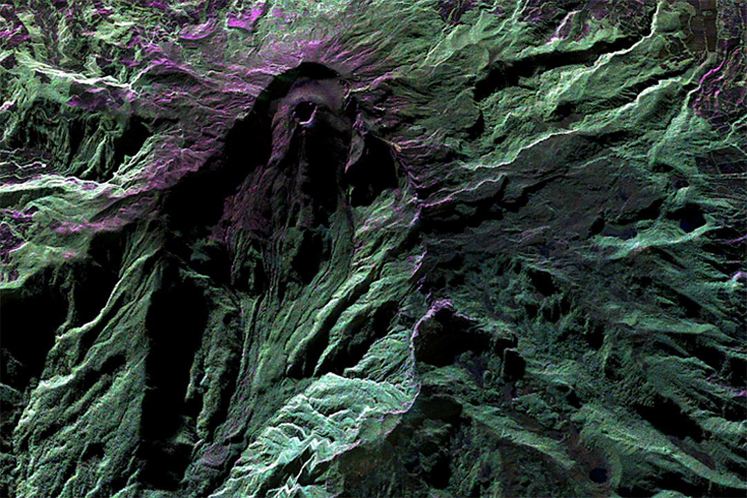
RIT researcher receives Department of Energy grant to develop synthetic aperture radar technology
Sandia National Laboratories awards a grant to James Albano, a researcher/engineer at RIT's Chester F. Carlson Center for Imaging Science, for remote sensing projects.
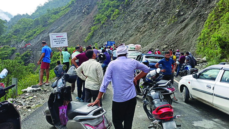
Ph.D. student applies imaging science to preventing disasters
Kamal Rana, an imaging science Ph.D. student from India has helped create algorithms to identify upcoming landslides.
Student Research
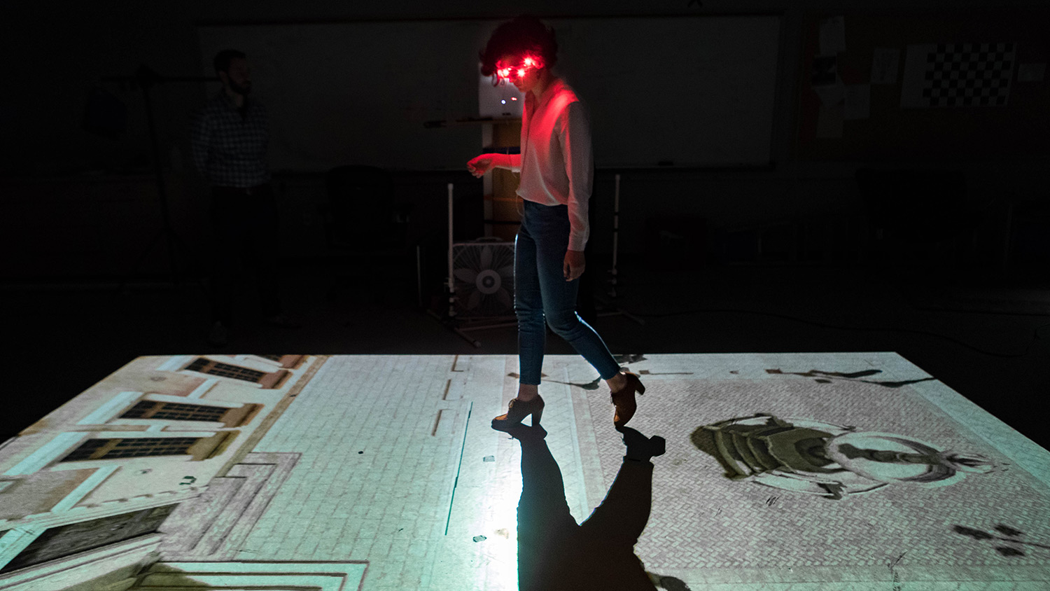
Cayla Fromm
Cayla Fromm, imaging science Ph.D. student, uses this apparatus in the PerForm Lab to study the visually guided strategies for walking and stepping over obstacles—a skill that breaks down with age and...
Latest News
April 10, 2024
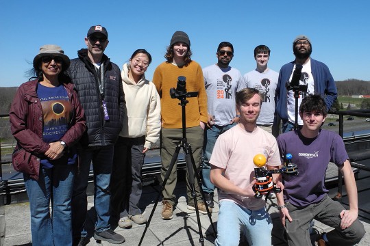
University researchers measure the sun during the eclipse to assess impact on solar arrays
The recent total solar eclipse over Rochester provided a once-in-a-lifetime opportunity on Earth for two faculty-researchers and their students to capture data about the effects of the sun’s energy during a total eclipse.
April 8, 2024

Researchers introduce new way to study, help prevent landslides
Landslides are one of the most destructive natural disasters on the planet, causing billions of dollars of damage and devastating loss of life every year. A global team of researchers has provided help for those who work to predict landslides and risk evaluations.
January 29, 2024
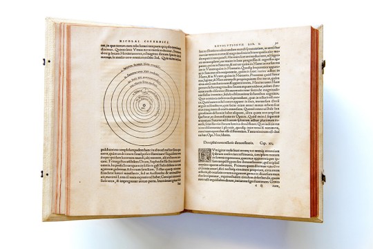
Centuries-old texts penned by early astronomers Copernicus and Sacrobosco find new home at RIT
The ancient astronomer Nicolaus Copernicus was the first scientist to document the theory that the sun is the center of the universe in his book, De Revolutionibus Orbium Coelestium (On the Revolutions of the Heavenly Spheres). That first edition book, along with a delicate manuscript from astronomer Johannes de Sacrobosco, that is contrary to Copernicus’ groundbreaking theory, has now found a permanent home at Rochester Institute of Technology.
Curriculum for 2023-2024 for Imaging Science Ph.D.
Current Students: See Curriculum Requirements
Imaging Science, Ph.D. degree, typical course sequence
* Students opting to take the IMGS elective in the first year would take 2 units of IMGS-PHD in the final year. Students opting not to take the IMGS elective would take 5 units of IMGS-PHD in the final year.
Admissions and Financial Aid
This program is available on-campus only.
Full-time study is 9+ semester credit hours. International students requiring a visa to study at the RIT Rochester campus must study full‑time.
Application Details
To be considered for admission to the Imaging Science Ph.D. program, candidates must fulfill the following requirements:
- Complete an online graduate application .
- Submit copies of official transcript(s) (in English) of all previously completed undergraduate and graduate course work, including any transfer credit earned.
- Hold a baccalaureate degree (or US equivalent) from an accredited university or college in the physical sciences, mathematics, computer science, or engineering.
- A recommended minimum cumulative GPA of 3.0 (or equivalent).
- Submit a current resume or curriculum vitae.
- Submit a statement of purpose for research which will allow the Admissions Committee to learn the most about you as a prospective researcher.
- Submit two letters of recommendation .
- Entrance exam requirements: GRE optional but recommended. No minimum score requirement.
- Writing samples are optional.
- Submit English language test scores (TOEFL, IELTS, PTE Academic), if required. Details are below.
English Language Test Scores
International applicants whose native language is not English must submit one of the following official English language test scores. Some international applicants may be considered for an English test requirement waiver .
International students below the minimum requirement may be considered for conditional admission. Each program requires balanced sub-scores when determining an applicant’s need for additional English language courses.
How to Apply Start or Manage Your Application
Cost and Financial Aid
An RIT graduate degree is an investment with lifelong returns. Ph.D. students typically receive full tuition and an RIT Graduate Assistantship that will consist of a research assistantship (stipend) or a teaching assistantship (salary).
Imaging Science Thesis Defense: Methodology for Volumetric Estimation of Condensed Water Vapor Plumes from Remotely Sensed Imagery
- IEEE Xplore Digital Library
- IEEE Standards
- IEEE Spectrum
- Resource Center
- Create Account
What is Signal Processing?
- Board of Governors
- Executive Committee
- Awards Board
- Conferences Board
- Membership Board
- Publications Board
- Technical Directions Board
- Standing Committees
- Liaisons & Representatives
- Education Board
- Our Members
- Society History
- State of the Society
- SPS Branding Materials
- Publications & Resources
- IEEE Signal Processing Magazine
- IEEE Journal of Selected Topics in Signal Processing
- IEEE Signal Processing Letters
- IEEE/ACM Transactions on Audio Speech and Language Processing
- IEEE Transactions on Computational Imaging
- IEEE Transactions on Image Processing
- IEEE Transactions on Information Forensics and Security
- IEEE Transactions on Multimedia
- IEEE Transactions on Signal and Information Processing over Networks
- IEEE Transactions on Signal Processing
- Data & Challenges
- Submit Manuscript
- Information for Authors
- Special Issue Deadlines
- Overview Articles
- Top Accessed Articles
- SPS Newsletter
- SPS Resource Center
- Publications Feedback
- Publications FAQ
- Dataset Papers
- Conferences & Events
- Conferences
- Attend an Event
- Conference Call for Papers
- Calls for Proposals
- Conference Sponsorship Info
- Conference Resources
- SPS Travel Grants
- Conferences FAQ
- Getting Involved
- Young Professionals
- Our Technical Committees
- Contact Technical Committee
- Technical Committees FAQ
- Data Science Initiative
- Join Technical Committee
- Young Professionals Resources
- Member-driven Initiatives
- Chapter Locator
- Award Recipients
- IEEE Fellows Program
- Call for Nominations
- Professional Development
- Distinguished Lecturers
- Past Lecturers
- Nominations
- DIS Nominations
- Seasonal Schools
- Industry Resources
- Job Submission Form
- IEEE Training Materials
- For Volunteers
- Board Agenda/Minutes
- Chapter Resources
- Governance Documents
- Membership Development Reports
- TC Best Practices
- SPS Directory
- Society FAQ
- Information for Authors-OJSP
ISBI_2025.jpg

(ISBI 2025) 2025 IEEE International Symposium on Biomedical Imaging
Farhan_baqai.jpg.

Distinguished Lecture: Prof. Farhan Baqai (Apple, USA)
- Celebrating 75 Years of IEEE SPS
- Diversity, Equity, and Inclusion
- General SP Multimedia Content
- Submit a Manuscript
- Editorial Board Nominations
- Challenges & Data Collections
- Publication Guidelines
- Unified EDICS
- Signal Processing Magazine The premier publication of the society.
- SPS Newsletter Monthly updates in Signal Processing
- SPS Resource Center Online library of tutorials, lectures, and presentations.
- SigPort Online repository for reports, papers, and more.
- SPS Feed The latest news, events, and more from the world of Signal Processing.
- IEEE SP Magazine
- IEEE SPS Content Gazette
- IEEE SP Letters
- IEEE/ACM TASLP
- All SPS Publications
- SPS Entrepreneurship Forum
- Call for Papers
- Call for Proposals
- Request Sponsorship
- Conference Organizer Resources
- Past Conferences & Events
- Event Calendar
- Conferences Meet colleagues and advance your career.
- Webinars Register for upcoming webinars.
- Distinguished Lectures Learn from experts in signal processing.
- Seasonal Schools For graduate students and early stage researchers.
- All Events Browse all upcoming events.
- Join SPS The IEEE Signal Processing Magazine, Conference, Discounts, Awards, Collaborations, and more!
- Chapter Locator Find your local chapter and connect with fellow industry professionals, academics and students
- Women in Signal Processing Networking and engagement opportunities for women across signal processing disciplines
- Students Scholarships, conference discounts, travel grants, SP Cup, VIP Cup, 5-MICC
- Young Professionals Career development opportunities, networking
- Chapters & Communities
- Member Advocate
- Awards & Submit an Award Nomination
- Volunteer Opportunities
- Organize Local Initiatives
- Autonomous Systems Initiative
- Applied Signal Processing Systems
- Audio and Acoustic Signal Processing
- Bio Imaging and Signal Processing
- Computational Imaging
- Image Video and Multidimensional Signal Processing
- Information Forensics and Security
- Machine Learning for Signal Processing
- Multimedia Signal Processing
- Sensor Array and Multichannel
- Signal Processing for Communication and Networking
- Signal Processing Theory and Methods
- Speech and Language Processing
- Synthetic Aperture Technical Working Group
- Industry Technical Working Group
- Integrated Sensing and Communication Technical Working Group
- TC Affiliate Membership
- Co-Sponsorship of Non-Conference TC Events
- Mentoring Experiences for Underrepresented Young Researchers (ME-UYR)
- Micro Mentoring Experience Program (MiME)
- Distinguished Lecturer Program
- Distinguished Lecturer Nominations
- Distinguished Industry Speaker Program
- Distinguished Industry Speakers
- Distinguished Industry Speaker Nominations
- Jobs in Signal Processing: IEEE Job Site
- SPS Education Program Educational content in signal processing and related fields.
- Distinguished Lecturer Program Chapters have access to educators and authors in the fields of Signal Processing
- PROGRESS Initiative Promoting diversity in the field of signal processing.
- Job Opportunities Signal Processing and Technical Committee specific job opportunities
- Job Submission Form Employers may submit opportunities in the area of Signal Processing.
- Technical Committee Best Practices
- Conflict of Interest
- Policy and Procedures Manual
- Constitution
- Board Agenda/Minutes* Agendas, minutes and supporting documentation for Board and Committee Members
- SPS Directory* Directory of volunteers, society and division directory for Board and Committee Members.
- Membership Development Reports* Insight into the Society’s month-over-month and year-over-year growths and declines for Board and Committee Members
Popular Pages
- (MLSP 2024) 2024 IEEE International Workshop on Machine Learning for Signal Processing
- (SLT 2024) 2024 IEEE Spoken Language Technology Workshop
- (ICASSP 2024) 2024 IEEE International Conference on Acoustics, Speech and Signal Processing
- Information for Authors-SPL
- Signal Processing 101
Last viewed:
- A Free Machine Learning Lecture Series on the SPS Resource Center!
- A Message from the New Society President
- Signal Processing Cup
- ALAN: Self-Attention Is Not All You Need for Image Super-Resolution
- About TASLP
- A Study of Subjective and Objective Quality Assessment of HDR Videos
- SA TWG Activities
- About SP Letters
PhD Position in Medical Image Analysis
Search form, you are here.
- Mentoring Experiences for Underrepresented Young Researchers Program (ME-UYR)
- SPS Education Program
- PROGRESS Initiative
- Young Professionals in Signal Processing
Top Reasons to Join SPS Today!
1. IEEE Signal Processing Magazine 2. Signal Processing Digital Library* 3. Inside Signal Processing Newsletter 4. SPS Resource Center 5. Career advancement & recognition 6. Discounts on conferences and publications 7. Professional networking 8. Communities for students, young professionals, and women 9. Volunteer opportunities 10. Coming soon! PDH/CEU credits Click here to learn more .
PhD Position : Image Analysis of dynamic MRI data to study musculoskeletal disorders
Research at IMT Atlantique involves nearly 800 people, including 290 teachers and researchers and 300 PhD students, and is on digital technology, energy and environment. It covers all disciplines (from the physical sciences to humanities and social sciences through those of information and knowledge) and covers all fields of science and information and communications technology.
The thesis will take place in the laboratory LaTIM (INSERM U1101), at Brest campus under the supervision of François Rousseau and Douraied Ben Salem.
Starting date: October 2019
Funding : IMT Atlantique - Philips
Project description
Musculoskeletal disorders have a debilitating impact on quality of life as well as on healthcare costs. Accurate clinical diagnosis and patient specific treatment are the key areas that play utmost important role in the management of musculoskeletal disorders. Individuals with musculoskeletal disorders often exhibit joint or pain and/or weakness for simple daily tasks or motions. Although using such pain-inducing tasks could be a good strategy to collect dynamic MRI data, a quick and non-repetitive technique to acquire dynamic data is most important. The causative relationship of many disorders spanning almost all human joints have not yet fully understood, and imaging efforts are mostly focused on static diagnosis and treatment follow-ups. Thus, dynamic MRI based evaluation of musculoskeletal disorders could have huge impact in not only understanding the joint pathomechanics but also guiding surgical or rehabilitation therapy [1].
To this extent, we have developed dynamic MRI sequences on the current 3.0T Philips MRI scanner. Both these sequences are real-time sequences with one based on Fast Field Echo (FFE) and another on balanced-FFE. We have also developed a post-processing technique to convert the low-resolution dynamic MRI images to high resolution images using a log-euclidean polyrigid framework.
This PhD thesis is focused on developing a framework to analyze the joint mechanics. It will benefit from the development already done on the ankle joint in children and will focus on resolving challenges faced in image acquisition, and processing. Following specific aims are sought within the scope of this Project:
1) Development of image processing algorithms for specific joint and acquisition (3D+t image reconstruction [2] and segmentation),
2) Application of the developed algorithms on targeted musculoskeletal disorders [3] (ankle joint development in cerebral palsy (see figure below)),
3) Study the complementary between MRI sequences and the potential of diffusion MRI for musculoskeletal system study.
- Master degree in image processing or applied mathematics
- Required skills: machine learning, image processing, programming (C++ & Python).
Net income/month : ~1500€
François Rousseau
email : [email protected]
www : http://perso.telecom-bretagne.eu/francoisrousseau
How to apply
Candidates are invited to email (to François Rousseau) a motivation letter and CV detailing in full your academic background, including all modules taken and grades assigned.
Bibliography
- B. Borotikar, M. Lempereur, M. Lelievre, V. Burdin, D. Ben Salem, S. Brochard. Dynamic MRI to quantify musculoskeletal motion: A systematic review of concurrent validity and reliability, and perspectives for evaluation of musculoskeletal disorders. Plos One 12(12), 2017.
- K. Makki, B. Borotikar, M. Garetier, S. Brochard, D. Ben Salem, F. Rousseau. In vivo ankle joint kinematics from dynamic magnetic resonance imaging using a registration-based framework. Journal of Biomechanics, 86, 193-203, 2019.
- K. Makki, B. Borotikar, M. Garetier, O. Acosta, S. Brochard, D. Ben Salem, F. Rousseau. 4D in vivo quantification of ankle joint space width using dynamic MRI. IEEE EMBC, 2019.
SPS on Twitter
- DEADLINE EXTENDED: The 2023 IEEE International Workshop on Machine Learning for Signal Processing is now accepting… https://t.co/NLH2u19a3y
- ONE MONTH OUT! We are celebrating the inaugural SPS Day on 2 June, honoring the date the Society was established in… https://t.co/V6Z3wKGK1O
- The new SPS Scholarship Program welcomes applications from students interested in pursuing signal processing educat… https://t.co/0aYPMDSWDj
- CALL FOR PAPERS: The IEEE Journal of Selected Topics in Signal Processing is now seeking submissions for a Special… https://t.co/NPCGrSjQbh
- Test your knowledge of signal processing history with our April trivia! Our 75th anniversary celebration continues:… https://t.co/4xal7voFER
IEEE SPS Educational Resources

Home | Sitemap | Contact | Accessibility | Nondiscrimination Policy | IEEE Ethics Reporting | IEEE Privacy Policy | Terms | Feedback
© Copyright 2024 IEEE – All rights reserved. Use of this website signifies your agreement to the IEEE Terms and Conditions . A not-for-profit organization, IEEE is the world's largest technical professional organization dedicated to advancing technology for the benefit of humanity.

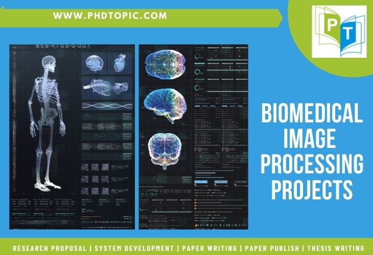
- Biomedical Image Processing Projects
Biomedical image processing projects deals with analyzing of captured internal human body images for clinical treatment and diagnosis. The information of physiological and physiology processes are collected through advanced sensors and processed by suitable computing technology . There are several methodologies to study the present state and disorder of specific human organs/tissue.
For this, it utilizes the following different types of biomedical images. And, they are:
- Ultrasound (sound)
- MRI (magnetism)
- CT scans (x-rays)
- OCT and Endoscopy (light)
- SPECT and PET (nuclear medicine: radioactive pharmaceuticals)
The handling of medical imaging is performed by the computerized system through efficient techniques and algorithms . As well, these techniques are incorporated with many advantages such as scalability, reliability, adaptability, privacy and etc. In general, imaging processes consist of image acquisition, computing, communication, storage, and visualization .
For your information, we shared the summary of the fundamentals of biomedical image processing. All these data make you understand the important terminologies, recent imaging modalities, image processing procedure, formation of medical image, and other futuristic image processing methodologies . Here, we have itemized the expected future developments by our expert’s suggestion.
Let’s have a quick glance over the recent research developments in medical image processing . These areas gain the attention of current active scholars who are pursuing their research careers in the bio-medical image processing research field.
Top 20 Major Research Topics in Biomedical Image Processing
Below we have mentioned most interesting biomedical image processing projects , we can guide you to formulate best research topics based on biomedical; reach us to know more information.
- Advances in Medical Image Computation
- Registration and Fusion of 3D Multimodal Medical Image
- Virtual and Augmented Reality in Medical Applications
- X-Ray Phase Contrast Technology and Tomography
- Automatic 3D Lungs Segmentation in Chest Scan
- 3D Superpixels Computation on Volumetric Intensity Image
- Deep Conventional Networks for Single Image Super-Resolution
- Partitioning 3D surface in Multi-modality Medical Imaging
- Medical Image Registration, Processing and Analysis
- Real-time Ultrasound, MRI and CT Imaging and Investigation
- Improved Non-Radioisotope Imaging Analysis
- Impact of Digital Image Communication in Medicine (DICOM)
- Segmentation of MR image Intensity Inhomogeneity
- Classify Huge-scale Multi-resolution Images using Deep Learning
- Deep Learning Model based 3D based Brain Tumor Segmentation
- Fast Wavelet based Normalized MRI Reconstruction
- Accuracy in Teeth Structures Segmentation on MRI and CT Images
- Techniques and Applications of Magnetic Resonance Imaging (MRI)
- Implementation of DL Algorithms over Multimodal Images for Semantic Segmentation
- Security Challenges in Storing and Exchanging Medical Information
Future Trends in Medical Imaging
- Real Implication of Retinal Diseases
- Wireless Tiny Single-Chip Ultrasound Sensing
- Rise of Artificial Intelligence (AI) in Radiology
- Heart or Cardiovascular Imaging Technology
- Future of Ultrasound Device and System Portability
- Large Superconducting Magnet System Applications
- 4D Printing Bio-medical Models and Applications
- Development of MRIs between Slow and Fast Fuzzy
- 3D Mathematical and Anatomical Models
- Migration and Advancement of X-Rays Films into Digital Files
- Skull Stripping Brain Tumor Segmentation
Based on our recent research on image processing over current journals, we found that most of the scholars are interested in using image datasets like spatial scales, which range from cellular/molecular imaging to organ/tissue imaging for their proposed topics. Other than this, scholars are familiar with the dataset from the followings,
- Colon Cancer
- Nuclear Medicine
- Computerized Tomography
- Magnetic Resonance
- Confocal and Optical Microscopy
- Diabetic Retinopathy
- Range Image and Video Data sets
- Computed Tomography (CT)
- Nuclear Magnetic Resonance (NMR)
For your knowledge, we have enumerated some datasets that are popularly used in developing Biomedical Image Processing Projects . Further, we have grouped the following based on category, modalities, network type with their corresponding datasets for your ease.
Datasets for Medical Image Modalities
- Modalities: MRI
- Network Type: LU-Net
- Data Set: ACDC Stacom 2017
- Modalities : CT, CXR and MRI
- Network Type : FCN, SCAN, dense-FCN and U-Net
- Data set : JSRT, Montgomery, JSRT, 172 sparsely annotated CT scan data set, TCIA, NSCLC
- Modalities: Microscopic
- Network Type: SegNet
- Data Set: ALL-IDB1 database
- Modalities : MRI
- Network Type : 3D U-Net, FCN, cGAN, V-Net, 3d FCN, SegAN-CAT and GAN
- Data Set : MRBrainS13, 62 Healthy Brain Images, BRATS2015, BRATS2017, BRATS2018, BRATS2019, Infant Brain Images, ANDI and NITRC data set
- Modalities : CT
- Network Type : 3D U-Net and CFUN
- Data Set : MICCAI 2017 whole heart and MM-WHS2017
- Modalities : Funduscopy
- Network Type : GAN, PixelBNN, FCN, Res-UNet and U-Net
- Data Set : STARE, DRIVE, RIM-ONE, CHASEDB1 and Drishti-GS data set
- Modalities: CT
- Network Type : Kid-Net
- Data Set – 236 Subjects
- Network Type : SSNet
- Data Set : 60 abdominal MRI Scans
- Modalities: 3D echocardiography
- Network Type: VoxelAtlasGAN
- Data Set: 60 Subjects over 3D echocardiography
- Network Type : Attention U-Net
- Data Set : TCIA
- Network Type : FCN
- Data Set : LVSC, RVSC and SCD
- Modalities : Dermoscopy
- Network Type : FCN and GAN
- Data Set : ISBI 2017 and DermoFit
- Network Type: 3D U-Net
- Data Set: LASC2018
- Modalities : MRI and CT
- Network Type : DI2IN-AN, FCN and DCNN
- Data Set : 1000 CT volumes, LiTS, 3DIRCADb and others
- Data Set: MICCAI Challenge Data Set
- Modalities : Histopathology
- Network Type : GAN
- Data Set : IPMCH
- Network Type : FCN, V-Net, USE-Net,
- Data Set : 152 MRI images, 3 T2-weighted MRI data sets and PROMISE2012
- Modalities: MRI and CT
- Network Type: Spine-GAN and Btrfly Net
- Data Set: 253 multi-center medical patients and 302 CT Scan
For instance: In order to identify the tumor patterns/features, several improved ML algorithms are used for spatial and temporal analysis. Also, it enables to find of the organ/tumor (vasculature, volume, and diameter), fluid/blood flow parameters, and microscopic changes based on the selected appropriate imaging approaches used in wireless body area network projects .

This article is specifically discussed research trends in Biomedical Image Processing Projects along with their technological advancements!!!
Biomedical Image Processing Toolboxes
- Image Acquisition Toolbox
- Mapping Toolbox
- Image Processing Toolbox
- Medical Image Processing Toolbox
- Deep Learning Toolbox
- Computer / Machine Vision Toolbox
- Vision HDL Toolbox
Next, we can see the significant development tools and software for implementing any kind of Biomedical Image Processing projects . More than this, there exist many technologies. So, one should choose the optimal tool based on the requirements of the handpicked project.
Development Tools and Software’s for Biomedical Image Processing
In recent research, our experts have recognized image informatics and cloud computing as the top-demanding research fields by scholars. Also, we just want you to know the other important research areas from the followings,
Cloud based Topics in Medical Image Processing
- Cloud assisted Medical Imaging Services for Social Medias
- Virtual Setting in Mammogram Testing and Interpretation
- MIIP: Web Solution for Medical Image Analysis and Understanding
- Crowdsourcing based Virtual Colonoscopy Video Analysis (For instances: polyp identification and annotation)
In addition, we have also given you the latest image informatics application in the healthcare sector for illustration purposes. Beyond this, it spreads its fame in all many research fields.
Image Informatics Healthcare Applications
- Applying Local Global Classifier for Screening Chest X-Rays
- Digital Pathology Image Analysis using Advanced DNN classification
- 3D-CNN based Malignant Breast Cancer Identification in MR images
- Frequency assisted Human Brain Network Similarity Detection
- DAX Next Generation is near to 1 million processes over commodity hardware
- Automated Thyroid Segmentation in 3D Ultrasound Images
- CBIR based Automatic Multi-Label Annotation in Abdominal CT images
- Blood Vessels Inpainting based on Optic Disc / Cup Segmentation Methodologies
On the whole, we are glad to give our end-to-end research guidance on Biomedical Image Processing Projects topic under the supervision of our technical experts. So, make use of this opportunity and hold your hand with us to create a masterpiece of research.
Related Pages
Services we offer.
Mathematical proof
Pseudo code
Conference Paper
Research Proposal
System Design
Literature Survey
Data Collection
Thesis Writing
Data Analysis
Rough Draft
Paper Collection
Code and Programs
Paper Writing
Course Work
An official website of the United States government
The .gov means it’s official. Federal government websites often end in .gov or .mil. Before sharing sensitive information, make sure you’re on a federal government site.
The site is secure. The https:// ensures that you are connecting to the official website and that any information you provide is encrypted and transmitted securely.
- Publications
- Account settings
Preview improvements coming to the PMC website in October 2024. Learn More or Try it out now .
- Advanced Search
- Journal List

The Constantly Evolving Role of Medical Image Processing in Oncology: From Traditional Medical Image Processing to Imaging Biomarkers and Radiomics
Kostas marias.
1 Department of Electrical and Computer Engineering, Hellenic Mediterranean University, 71410 Heraklion, Greece; rg.umh@sairamk
2 Computational Biomedicine Laboratory (CBML), Foundation for Research and Technology—Hellas (FORTH), 70013 Heraklion, Greece
The role of medical image computing in oncology is growing stronger, not least due to the unprecedented advancement of computational AI techniques, providing a technological bridge between radiology and oncology, which could significantly accelerate the advancement of precision medicine throughout the cancer care continuum. Medical image processing has been an active field of research for more than three decades, focusing initially on traditional image analysis tasks such as registration segmentation, fusion, and contrast optimization. However, with the advancement of model-based medical image processing, the field of imaging biomarker discovery has focused on transforming functional imaging data into meaningful biomarkers that are able to provide insight into a tumor’s pathophysiology. More recently, the advancement of high-performance computing, in conjunction with the availability of large medical imaging datasets, has enabled the deployment of sophisticated machine learning techniques in the context of radiomics and deep learning modeling. This paper reviews and discusses the evolving role of image analysis and processing through the lens of the abovementioned developments, which hold promise for accelerating precision oncology, in the sense of improved diagnosis, prognosis, and treatment planning of cancer.
1. Introduction
To better understand the evolution of medical image processing in oncology, it is necessary to explain the importance of measuring tumor appearance from medical images. Medical image processing approaches contain useful diagnostic and prognostic information that can add precision in cancer care. In addition, because biology is a system of systems, it is reasonable to assume that image-based information may convey multi-level pathophysiology information. This has led to the establishment of many sophisticated predictive and diagnostic image-based biomarker extraction approaches in cancer. In more detail, medical image processing efforts are focused on extracting imaging biomarkers able to decipher the variation within individuals in terms of imaging phenotype, enabling the identification of patient subgroups for precision medicine strategies [ 1 ]. From the very beginning, the main prerequisite for clinical use was that quantitative biomarkers must be precise and reproducible. If these conditions are met, imaging biomarkers have the potential to aid clinicians in assessing the pathophysiologic changes in patients and better planning personalized therapy. This is important, as in clinical practice subjective characterizations might be used (e.g., average heterogeneity, speculated mass, necrotic core) which can decrease the precision of diagnostic processes.
Based on the above considerations, the extraction of quantitative parameters characterizing size, shape, texture, and activity can enhance the role of medical imaging in assisting in diagnosis or therapy response assessment. However, in clinical practice, only simpler image metrics (e.g., linear) are often used in oncology, especially in the evaluation of solid tumor response to therapy (e.g., a longer lesion diameter in RECIST). Both RECIST and WHO evaluation criteria rely on anatomical image measurements, mainly in CT or MRI data, and were originally developed mainly for cytotoxic therapies. Such linear measures suffer from high intra/inter-observed variability, which in some cases can compromise the accurate assessment of tumor response, since some studies report inter-observer RECIST variability of up to 30% [ 2 ]. Several studies have shown that 3D quantitative response assessments are better correlated with disease progression than those based on 1D linear measurements [ 3 ]. Nevertheless, traditional tumor quantification approaches based on linear or 3D tumor measures have experienced substantial difficulties in assessing response to newer oncology therapies, such as targeted, anti-angiogenic treatments and immunotherapies [ 2 ]. Size-based tumor assessments do not always represent tumor response to therapy, since, for example, tumors may display internal necrosis formation, with or without reduction in lesion size (as in traditional cytotoxic treatments). Even if RECIST criteria are constantly updated to address these issues, as in the case of Immune RECIST [ 4 ], such approaches still do not take into consideration a tumor’s image structure and texture over time. In addition the size and the location of metastases have been reported to play a significant role in assessing early tumor shrinkage and depth of response [ 5 ]. To address these limitations, medical image processing has provided over the last few decades the means to extract tumor texture and size descriptors for obtaining more detailed (e.g., pixel-based) descriptors of tissue structure and for discovering feature patterns connected to disease or response. In this paper, it is argued that the evolution of medical image processing has been a gradual process, and the diverse factors that contributed to unprecedented progress in the field with the use of AI are explained. Initially, simplistic approaches to classify benign and malignant masses, e.g., in mammograms, were based on traditional feature extraction and pattern recognition methods. Functional tomographic imaging such as PET gave rise to more sophisticated, model-based approaches from which quantitative markers from tissue properties could be extracted in an effort to optimize diagnosis, treatment stratification, and personalize response criteria. Lastly, the advancement of artificial intelligence enabled the more exhaustive search of imaging phenotype descriptors and led to the increased performance of modern diagnostic and predictive models.
2. Traditional Image Analysis: The First Efforts towards CAD Systems
In the 1990s, one of the first challenges in medical image analysis was to facilitate the interpretation of mammograms in the context of national screening programs for breast cancer. In the United Kingdom, the design of the first screening program was undertaken by a working group under Sir Patrick Forrest, whose report was accepted by the government in 1986. As a consequence, the UK screening program was established for women between 50 and 64 in 1990 [ 6 ]. The implementation of such screening programs throughout Europe led to the establishment of specialist breast screening centers and the formal training of both radiographers and radiologists. X-ray mammography proved to be a cost-effective imaging modality for national screening, and population screening led to smaller and usually non-palpable masses being increasingly detected.
As a result, the radiologist’s task became more complex, since the interpretation of a mammogram is challenging, due to the projective nature of mammography, while at the same time the need for early and accurate detection of cancer became pressing. To address these needs, medical image analysis became an active field of research in the early nineties, giving rise to numerous research efforts into cancer and microcalcification detection, as well as mammogram registration for improving the comparison of temporal mammograms. Figure 1 depicts the temporal mammogram registration concept towards CAD systems that would facilitate comparison and aid clinicians in early diagnose of cancer in screening mammography [ 7 ]. When the ImageChecker system was certified by the FDA for screening mammography in 1998, R2 Technology became the first company to employ computer-assisted diagnosis (CAD) for mammography, and later for digital mammography as well.

Traditional medical image processing on temporal mammograms. From left to right: the most recent mammogram ( a ) is registered to the previous mammogram ( b ), which is shown in ( c ). After registration there is one predominant region of significant difference in the subtraction image ( d ), which corresponds to a mass developed in the breast.
However, early diagnostic decision support systems suffered from low precision, which in turn could potentially lead to a negative impact in the number of unnecessary biopsies. In a relevant study [ 8 ], the positive predictive values of the interpretations worsened from 100%, 92.7%, and 95.5%, to 86.4%, 97.3%, and 91.1%, when mammograms were analyzed by three independent observers, with and without the CAD. This limitation was representative of the low generalizability of such cancer detection tools in these early days. At the same time the lack of more sophisticated imaging modalities hampered the research efforts towards predicting therapy response and optimizing therapy based on imaging data.

3. Quantitative Imaging Based on Models
With the advent of more sophisticated imaging modalities enabling functional imaging, medical image analysis efforts shifted towards the quantification of tissue properties. This opened new horizons in CAD systems towards translating image signals to cancer tissue properties such as perfusion and cellularity and developing more intuitive imaging biomarkers for several cancer imaging applications. For example, in the case of MRI, complex phenomena that occur after excitation are amenable to mathematical modeling, taking into consideration tissue interactions within the tumor microenvironment. In the context of evaluating a model-based approach, the model can be regarded reliable when the predicted data converges on the observed signal intensities and at the same time provides useful insights to radiologists and oncologists. MRI perfusion and diffusion imaging has been the main focus of such modeling efforts, not least due to fact that MRI is ionizing radiation-free.
Diffusion weighted MRI (DWI-MRI) is based on sequences sensitized to microscopic water mobility by means of strong gradient pulses and can provide quantitative information on tumor environment and architecture. Diffusivity can be assessed in the intracellular, extracellular, and intravascular spaces. Apparent diffusion coefficient (ADC) per pixel values derived from DWI-MRI theoretically have an inverse relationship to tumor cell density. In addition, with the introduction of the intravoxel incoherent motion (IVIM) model, both cellularity and microvascular perfusion information could be assessed after parametric modeling [ 9 ]. Figure 2 presents a parametric map of the stretching parameter α from the DWI-MRI stretched-exponential model (SEM), revealing highly heterogeneous parts of a dedifferentiated liposarcoma (DDLS) of Grade 3 [ 9 ].

DWI-MRI stretched-exponential (SEM) DWI-MRI parametric map, revealing highly heterogeneous parts of a dedifferentiated liposarcoma (with permission from the department of Medical Imaging, Heraklion University Hospital). Heterogeneity index α ranges from 0 to 1, with lower values of α indicating microstructural heterogeneity.
DWI-MRI has been tested in most solid tumors for discriminating malignant from benign lesions, to automatize tumor grading, and to predict treatment response and post-treatment monitoring [ 10 ].
However, there is still a lack of standardization and generalization of these results, as well as validation against histopathology. While in clinical routine, in-depth DWI-MRI biomarker validation is difficult, recent pre-clinical studies have found that derived parametric maps can serve as a non-invasive marker of cell death and apoptosis in response to treatment [ 11 ]. To this end, they also confirmed significant correlations of ADC with immunohistochemistry measurements of cell density, cell death, and apoptosis.
In a similar fashion, in dynamic contrast-enhanced MRI (DCE-MRI), T1-weighted sequences are acquired before, during, and after the administration of a paramagnetic contrast agent (CA). Tissue-specific information about pathophysiology can be inferred from the dynamics of signal intensity in every pixel of the studied area. Usually this is performed by visual or semi-quantitative analysis from the signal time curves in selected regions of interest. However, with the use of pharmacokinetic modeling, e.g., between the intravascular and the extravascular extracellular space, it became possible to map signal intensities per pixel to CA concentration and then fit model parameters describing, e.g., interstitial space and transfer constant (ktrans). This enabled the generation of parametric maps, e.g., for ktrans providing more quantitative representation of tumor perfusion and heterogeneity within the tumor image region of interest. Although promising, e.g., for assessing treatment efficacy, such approaches have found limited use in clinical practice, not least due to the low reported reproducibility of model parameter estimation. One aspect of this problems is presented in the example shown in Figure 3 , where the use of image-driven methods based on multiple-flip angles produces a parametric map of a tumor with different contrast compared to the one produced with the Fritz–Hansen population based AIF [ 12 ]. This issue has several implications, including for the accuracy of assessing breast cancer response to neoadjuvant chemotherapy [ 13 ].

( a ) ktrans map of a tumor from PK analysis using AIF measured directly from the MR image, while for the conversion from signal to CA concentration the multiple flip angles method (mFAs) was used, ( b ) ktrans map of the same tumor using a population based AIF from Fritz and Hansen.
In conclusion, the clinical translation of DWI and DCE MRI is hampered by low repeatability and reproducibility across several studies in oncology. To address this problem initiatives such as the Quantitative Imaging Biomarkers Alliance (QIBA) propose clinical and technological requirements for quantitative DWI and DCE-derived imaging biomarkers, as well as image acquisition, processing, and quality control recommendations aimed at improving reproducibility error, precision, and accuracy [ 14 ]. It is argued that this active area of medical image processing has not yet reached its full potential and still represents a complementary approach to AI driven methods, towards CAD systems for promoting precision oncology. In addition, the exploitation of multimodality imaging strategies (e.g., PET/MRI) can provide added value through the combination of anatomical and functional information.
4. Radiomics and Deep Learning Approaches in Oncology through the Cancer Continuum
Traditional cancer medical image analysis was for decades based on human-defined features, usually inspired by low-level image properties, such as intensity, contrast, and a limited number of texture measures. Such methods were successfully used. e.g., in cancer subclassification, but it was hard to capture the high-level, complex patterns that an expert radiologist uses to define the presence or absence of cancer [ 1 ].
However, with the advancement of machine learning and the availability of more powerful, high-performance computational infrastructures, it became possible to exhaustively analyze the texture and shape content of medical images in an effort to decipher high-level pathophysiology patterns. At the same time the evolution of texture representation and feature extraction, through a growing number of techniques during the last decades, played a catalytic role in better capturing tumor appearance through medical image analysis [ 15 ]. Last but not least, the need to decipher the imaging phenotype in cancer became even more pressing, due to the fact that the vast majority of visible phenotypic variation is now considered attributable to non-genetic determinants in chronic and age-associated disorders [ 1 ].
All these factors played a central role in the advancement of radiomics, where in analogy to genomics high-throughput feature extraction followed by ML enabled the development of significant discriminatory and predictive signatures, based on imaging phenotype. Radiomics have been enhanced with deep learning techniques, offering an alternative approach to medical image feature extraction by the learning of complex, high-level features in an automated fashion from a large number of medical images that contain variable instances of a particular tumor. Figure 4 illustrates the main AI/radiomics applications that can assist clinicians in adding precision in the management of cancer patients.

The main medical image processing applications enhanced with AI/radiomics towards precision oncology.
4.1. Cancer Screening
Recent advancements in AI driven medical image processing can have a positive impact in national cancer screening programs, alleviating the heavy workload of radiologists and aiding clinicians to reduce the number of missed cancers and to detect them at an earlier stage. Compared to the initial efforts mentioned in previous sections, recent AI-driven image processing can exceed the limits of human vision and potentially reduce the number of cancers missed in screening, as well as cope with inter-observer variability.
Regarding lung cancer screening, early nodule detection and classification is of paramount importance for improving patient outcomes and quality of life. Despite the existence of such screening programs the majority of lung cancers are detected in the later stages, leading to increased mortality and low 5-year survival rate [ 16 ]. To this end, radiomics and deep-learning-based methods have shown encouraging results towards precision pulmonary nodule evaluation [ 17 ]. A very interesting recent example is reported by Ardill et al., who developed a deep learning algorithm that uses a patient’s current and prior computed tomography volumes to predict the risk of lung cancer. Their model achieved a state-of-the-art performance (94.4% area under the curve) on 6716 cases and performed similarly on an independent clinical validation set of 1139 cases. When prior computed tomography imaging was not available, their model outperformed all six radiologists, with absolute reductions of 11% in false positives and 5% in false negatives [ 18 ].
Regarding breast cancer screening technologies, it is argued that AI may provide the means to limit the inherent drawbacks of mammography and enhance diagnostic performance and robustness. In a prospective clinical study, a commercially available AI algorithm was evaluated as an independent reader of screening mammograms, and adequate diagnostic performance was reported [ 19 ].
4.2. Precision Cancer Diagnosis
During the last decades CAD-driven precision diagnosis has been the holy grail of medical image processing research efforts. However, the clinical interest in such applications has significantly grown only recently with the advancement of AI-driven efforts to generalize performance across diverse datasets. AI systems have reported unprecedented performance regarding the segmentation and classification of cancer. A recent study reported increased performance in segmenting and classifying brain tumors into meningioma, glioma, and pituitary tumors [ 20 ].
In addition, a growing number of studies are concerned with automated tumor grading, which is a prerequisite for optimal therapy planning. Yang et al. presented a retrospective glioma grading study (grade II and grade III concerning low grade glioma and high grade glioma) on one hundred and thirteen glioma patients and used transfer learning with AlexNet and GoogLeNet architectures, achieving up to 0.939 AUC [ 21 ].
At the same time, the quest to decode imaging phenotype has given rise to efforts to correlate imaging features with molecular and genetic markers in the context of radio-genomics [ 22 ]. This promising field of research can provide surrogate molecular information directly from medical images and is not prone to biopsy sampling errors, as the whole tumor can be analyzed. In a recent study, MRI radiomics were able to predict IDH1 mutation with an AUC of up to 90% [ 23 ].
4.3. Treatment Optimization
There are many challenging problems in optimizing treatment for cancer patients, such as accurate segmentation of organs at risk (OAR) in radiotherapy and prediction of neoadjuvant chemotherapy response. Intelligent processing of medical images has opened new horizons to address these clinical needs. In the case of nasopharyngeal carcinoma radiotherapy planning, a deep learning organs-at-risk (OAR) detection and segmentation network provides useful insights for clinicians for the accurate delineation of OARs [ 24 ]. Regarding prediction of neoadjuvant chemotherapy, the use of image-based algorithms to predict outcome has the potential to add precision, not least due to the fact that depending on tumor subtype the outcome can differ significantly. To this end, recent studies report promising preliminary results in applying AI to predict breast cancer neoadjuvant therapy response. Vulchi et al. reported improved prediction of response to HER2-targeted neoadjuvant therapy based on deep learning of DCE-MRI data [ 25 ]. Notably, the AUC dropped from 0.93 to 0.85 in the external validation cohort.
5. Radiomics Limitations Regarding Clinical Translation
While promising, radiomics methodologies are still in a translational phase and thorough clinical validation is needed towards clinical translation. To this end, when these technologies are tested and reviewed, a number of important limitations becomes apparent. In a recent review on MRI based radiomics in nasopharyngeal cancer [ 26 ], the authors reviewed the state of the art and used a radiomic quality score assessment (RQS). Several limitations were highlighted in the reviewed studies, including the absence of a validation cohort in 21% of them, as well as the lack of external validation in 92% of them. In another RQS based evaluation study on radiomics and radio-genomics papers, the RQS was low regarding clinical utility, test-retest analysis, prospective study, and open science [ 27 ]. It was also very interesting that no single study used phantoms to assess the robustness of radiomics features or performed a cost-effectiveness analysis. In a similar fashion, lack of feature robustness assessment and external validation was reported in studies regarding prostate cancer [ 28 ], while the main reported shortcomings in the quality of the MRI lymphoma radiomics studies regarded inconsistencies in the segmentation process and the lack of temporal data to increase model robustness [ 29 ]. All these recent studies clearly indicate that, although medical image processing in oncology has evolved significantly, the clinical translation of radiomics is still hampered by the lack of extensive, high quality validation studies. In addition, the lack of standardization in radiomics extraction remains a problem, which is currently being investigated by several studies, with respect to different software packages [ 30 ] and the reproducibility of standardized radiomics features using multi-modality patient data [ 31 ].
6. Discussion
Contrary to common belief, medical image processing has been evolving for the last few decades and its main application is cancer image analysis. Traditional medical image processing was founded on classical image processing and computer vision principles, focusing on low-level feature extraction and simple classification tasks, e.g., benign vs. malignant, or in the geometrical alignment of temporal images and the segmentation of tumors for volumetric analyses. This early stage in the 1990s was an important milestone for further development, since several radiologists and oncologists understood the future potential and helped in the creation of a multidisciplinary community on medical image analysis and processing. More importantly, it laid the foundations of radiomics by proposing the shape and textural analysis of tumors as useful patterns for detection, segmentation, and classification. However, the main limitation was the high degree of fragmentation in such efforts, the limited computational resources, and the very low availability of cancer image data; usually being mammograms or MRIs.
Functional imaging was another important milestone for medical image computing, since the idea of transforming dynamic image signals to tissue properties paved the way for the discovery of reliable and reproducible image biomarkers for oncology. To achieve this goal, non-conventional medical image processing was deployed based on compartmental models to link the imaging phenotype with microscopic tumor environment properties, based on diffusion and perfusion. Such model-based approaches include compartment pharmacokinetic models for DCE-MRI and the IVIM model for DWI-MRI, often requiring laborious pre-processing to transform the original signal to quantitative parametric maps able to convey perfusion and cellularity information to the clinician. It is argued that this is still an evolving research field and that the potential for clinical translation is significant, especially since techniques such as DWI-MRI do not involve ionizing radiation or the administration of contrast agent. That said, significant standardization efforts are still required in order to converge on stable imaging protocols and model implementations that will guarantee reproducible parametric maps and robust cancer biomarkers. Another limitation when comparing to modern radiomics/deep learning efforts is that the processing of such functional data with compartmental models is a very demanding task, requiring a deeper understanding of imaging protocols, as well as of numerical analysis methods for model fitting.
The gradual advancements of high-performance computing and machine learning and neural networks have revolutionized research in the field, especially during the last decade. The field of radiomics has extended the cancer medical image processing concepts regarding texture and shape descriptors to massive feature extraction and modeling. Such radiomics approaches have also been enhanced by convolutional neural networks, which outperformed the traditional image analysis methods in tasks such as lesion segmentation, while introducing more sophisticated predictive, diagnostic, and correlative pipelines towards precision diagnostics, therapy optimization, and synergistic radio-genomic biomarker discovery. The availability of open access computational tools for machine and deep learning, in combination with public cancer image resources such as the Cancer Imaging Archive (TCIA), has led to an unprecedented number of publications, AI start-ups, and accelerated discussions for the establishment of AI regulatory processes and clinical translation of such technologies. At the same time, the main limitation of these impressive technologies has been their low explainability, which came as a tradeoff for the impressive performances in oncological applications throughout the cancer continuum. Low explainability also contributed to reduced trust in these models, while the vast number of features explored made generalization difficult, especially due to the large variability of image quality and imaging protocols across vendors and clinical sites.
Medical image processing is still evolving and will continue to provide useful tools and methodological concepts for improving cancer image analysis and interpretation. Data science approaches focusing on radiomics have paved the way for accelerating precision oncology [ 32 ]. However, most of the efforts to date only use imaging data, which limits the performance of diagnostic and prognostic tools. To this end, novel data integration paradigms, exploiting both imaging and multi-omics data, is a very promising field for future research [ 33 ]. Recent studies have started exploring the synergy of deep learning with quantitative parametric maps. In [ 34 ], the authors present a deep learning method to predict good responders of locally advanced rectal cancer trained on apparent diffusion coefficient (ADC) parametric scans from different vendors. The fusion of standard imaging representations with parametric maps, as well as integrative diagnostic approaches [ 35 ] involving medical image and other cancer related data, hold promise for increasing accuracy and trustworthiness.
This research received no external funding.
Institutional Review Board Statement
Not applicable.
Informed Consent Statement
Conflicts of interest.
The author declares no conflict of interest.
Publisher’s Note: MDPI stays neutral with regard to jurisdictional claims in published maps and institutional affiliations.
- Our Promise
- Our Achievements
- Our Mission
- Proposal Writing
- System Development
- Paper Writing
- Paper Publish
- Synopsis Writing
- Thesis Writing
- Assignments
- Survey Paper
- Conference Paper
- Journal Paper
- Empirical Paper
- Journal Support
PhD Projects in Medical Image Processing
“Medical Image Processing will get the facts from bio-medical images that are in any form. Generally, it will give a visual view of healthy organs.” PhD Projects in Medical Image Processing give the ever widely held services for the PhD/MS entrants.
To emphasize the scope of this field, our pros have done so many medical image processing thesis . PhD Projects in Medical Image Processing has the latent to give the best works to the students. Evidently, we have given some of the study issues in this area.
EMBRYONIC RESEARCH TOPICS
- Multi-Modal Image Reconstruction
- Image Processing for Glaucoma Detection
- Denoising of 3D Medical Images
- Deformable Image Registration for Contour Propagation
- Swarm Intelligence for Medical Image Recognition
- Automated Image Segmentation
- Medical Image Classification
- Medical Image Analysis with Neural Networks
- Computerized Decision Making
- Cryptography Encryption of Medical Images
As shown above, there are many topics in trend. Only if you want to do a task, then you can call us. We will give worthy aid for your project in the effect of work. In reality, there exists a wide range of medical catalogs. In a word, we have done projects with all those files.
MEDICAL DATABASES
PhD Projects in Medical Image Processing has optimal solutions for all the projects. As can be seen, imaging is a vast area with so many research works still going on. Contrarily, it also has a huge plea in the middle of the PhD students.
In the long run, our doyens know the ways to store the images in a lossless format. At the same time, your success lies at the end of our work.
An innovative function for Predicting CT Image From MRI Data Through Feature Matching With Learned Nonlinear Local Descriptors system
An ingenious method for Simple Measure for Acuity in Medical Images
A new design function for Application of 4-D XCAT Phantoms in Biomedical Imaging and Beyond method
An effectual process of ODHigh-capacity Reversible Watermarking Algorithm Based on the Region of Interest of Medical Images
The novel scheme for Performance and power consumption evaluation in smartphone based image processing for medical applications
An imaginative method for Synergistic PET and SENSE MR Image Reconstruction Using Joint Sparsity Regularization
A new function for Diagnogsis of Diabetic Retinopathy Using Image Processing and Convolutional Neural Network
An effective source for Calcium removal from cardiac ct images using deep convolutional neural network
A novel function for Lung Field Segmentation in Chest Radiographs From Boundary Maps by a Structured Edge Detector
The new mechanism for Groupwise Multichannel Image Registration scheme
A creative source for 3-D Reconstruction in Canonical Co-Ordinate Space From Arbitrarily Oriented 2-D Images
An inventive function for Improved DenseNet Method Based on Transfer Learning for Fundus Medical Images
A new method for Review function based on Multi-Model Medical Image Fusion system
An effective function for Shape-aware Medical Image Enhancement by Weighted Total Variation
The new process for Medical image segmentation based on improved watershed algorithm
An innovative mechanism for Fuzzy Based on Grey Wolf Optimization for Effective Medical Image Retrieval System
A new function for Motion Quantification and Automated Correction in Clinical RSOM
The novel things for Self-learning to detect and segment cysts in lung CT images without manual annotation
On the use for Compression of Medical Images by their Symmetry
An ingenious mechanism for Empirical Aspect of Big Data to Enhance Medical Images Using HIPI
MILESTONE 1: Research Proposal
Finalize journal (indexing).
Before sit down to research proposal writing, we need to decide exact journals. For e.g. SCI, SCI-E, ISI, SCOPUS.
Research Subject Selection
As a doctoral student, subject selection is a big problem. Phdservices.org has the team of world class experts who experience in assisting all subjects. When you decide to work in networking, we assign our experts in your specific area for assistance.
Research Topic Selection
We helping you with right and perfect topic selection, which sound interesting to the other fellows of your committee. For e.g. if your interest in networking, the research topic is VANET / MANET / any other
Literature Survey Writing
To ensure the novelty of research, we find research gaps in 50+ latest benchmark papers (IEEE, Springer, Elsevier, MDPI, Hindawi, etc.)
Case Study Writing
After literature survey, we get the main issue/problem that your research topic will aim to resolve and elegant writing support to identify relevance of the issue.
Problem Statement
Based on the research gaps finding and importance of your research, we conclude the appropriate and specific problem statement.
Writing Research Proposal
Writing a good research proposal has need of lot of time. We only span a few to cover all major aspects (reference papers collection, deficiency finding, drawing system architecture, highlights novelty)
MILESTONE 2: System Development
Fix implementation plan.
We prepare a clear project implementation plan that narrates your proposal in step-by step and it contains Software and OS specification. We recommend you very suitable tools/software that fit for your concept.
Tools/Plan Approval
We get the approval for implementation tool, software, programing language and finally implementation plan to start development process.
Pseudocode Description
Our source code is original since we write the code after pseudocodes, algorithm writing and mathematical equation derivations.
Develop Proposal Idea
We implement our novel idea in step-by-step process that given in implementation plan. We can help scholars in implementation.
Comparison/Experiments
We perform the comparison between proposed and existing schemes in both quantitative and qualitative manner since it is most crucial part of any journal paper.
Graphs, Results, Analysis Table
We evaluate and analyze the project results by plotting graphs, numerical results computation, and broader discussion of quantitative results in table.
Project Deliverables
For every project order, we deliver the following: reference papers, source codes screenshots, project video, installation and running procedures.
MILESTONE 3: Paper Writing
Choosing right format.
We intend to write a paper in customized layout. If you are interesting in any specific journal, we ready to support you. Otherwise we prepare in IEEE transaction level.
Collecting Reliable Resources
Before paper writing, we collect reliable resources such as 50+ journal papers, magazines, news, encyclopedia (books), benchmark datasets, and online resources.
Writing Rough Draft
We create an outline of a paper at first and then writing under each heading and sub-headings. It consists of novel idea and resources
Proofreading & Formatting
We must proofread and formatting a paper to fix typesetting errors, and avoiding misspelled words, misplaced punctuation marks, and so on
Native English Writing
We check the communication of a paper by rewriting with native English writers who accomplish their English literature in University of Oxford.
Scrutinizing Paper Quality
We examine the paper quality by top-experts who can easily fix the issues in journal paper writing and also confirm the level of journal paper (SCI, Scopus or Normal).
Plagiarism Checking
We at phdservices.org is 100% guarantee for original journal paper writing. We never use previously published works.
MILESTONE 4: Paper Publication
Finding apt journal.
We play crucial role in this step since this is very important for scholar’s future. Our experts will help you in choosing high Impact Factor (SJR) journals for publishing.
Lay Paper to Submit
We organize your paper for journal submission, which covers the preparation of Authors Biography, Cover Letter, Highlights of Novelty, and Suggested Reviewers.
Paper Submission
We upload paper with submit all prerequisites that are required in journal. We completely remove frustration in paper publishing.
Paper Status Tracking
We track your paper status and answering the questions raise before review process and also we giving you frequent updates for your paper received from journal.
Revising Paper Precisely
When we receive decision for revising paper, we get ready to prepare the point-point response to address all reviewers query and resubmit it to catch final acceptance.
Get Accept & e-Proofing
We receive final mail for acceptance confirmation letter and editors send e-proofing and licensing to ensure the originality.
Publishing Paper
Paper published in online and we inform you with paper title, authors information, journal name volume, issue number, page number, and DOI link
MILESTONE 5: Thesis Writing
Identifying university format.
We pay special attention for your thesis writing and our 100+ thesis writers are proficient and clear in writing thesis for all university formats.
Gathering Adequate Resources
We collect primary and adequate resources for writing well-structured thesis using published research articles, 150+ reputed reference papers, writing plan, and so on.
Writing Thesis (Preliminary)
We write thesis in chapter-by-chapter without any empirical mistakes and we completely provide plagiarism-free thesis.
Skimming & Reading
Skimming involve reading the thesis and looking abstract, conclusions, sections, & sub-sections, paragraphs, sentences & words and writing thesis chorological order of papers.
Fixing Crosscutting Issues
This step is tricky when write thesis by amateurs. Proofreading and formatting is made by our world class thesis writers who avoid verbose, and brainstorming for significant writing.
Organize Thesis Chapters
We organize thesis chapters by completing the following: elaborate chapter, structuring chapters, flow of writing, citations correction, etc.
Writing Thesis (Final Version)
We attention to details of importance of thesis contribution, well-illustrated literature review, sharp and broad results and discussion and relevant applications study.
How PhDservices.org deal with significant issues ?
1. novel ideas.
Novelty is essential for a PhD degree. Our experts are bringing quality of being novel ideas in the particular research area. It can be only determined by after thorough literature search (state-of-the-art works published in IEEE, Springer, Elsevier, ACM, ScienceDirect, Inderscience, and so on). SCI and SCOPUS journals reviewers and editors will always demand “Novelty” for each publishing work. Our experts have in-depth knowledge in all major and sub-research fields to introduce New Methods and Ideas. MAKING NOVEL IDEAS IS THE ONLY WAY OF WINNING PHD.
2. Plagiarism-Free
To improve the quality and originality of works, we are strictly avoiding plagiarism since plagiarism is not allowed and acceptable for any type journals (SCI, SCI-E, or Scopus) in editorial and reviewer point of view. We have software named as “Anti-Plagiarism Software” that examines the similarity score for documents with good accuracy. We consist of various plagiarism tools like Viper, Turnitin, Students and scholars can get your work in Zero Tolerance to Plagiarism. DONT WORRY ABOUT PHD, WE WILL TAKE CARE OF EVERYTHING.
3. Confidential Info
We intended to keep your personal and technical information in secret and it is a basic worry for all scholars.
- Technical Info: We never share your technical details to any other scholar since we know the importance of time and resources that are giving us by scholars.
- Personal Info: We restricted to access scholars personal details by our experts. Our organization leading team will have your basic and necessary info for scholars.
CONFIDENTIALITY AND PRIVACY OF INFORMATION HELD IS OF VITAL IMPORTANCE AT PHDSERVICES.ORG. WE HONEST FOR ALL CUSTOMERS.
4. Publication
Most of the PhD consultancy services will end their services in Paper Writing, but our PhDservices.org is different from others by giving guarantee for both paper writing and publication in reputed journals. With our 18+ year of experience in delivering PhD services, we meet all requirements of journals (reviewers, editors, and editor-in-chief) for rapid publications. From the beginning of paper writing, we lay our smart works. PUBLICATION IS A ROOT FOR PHD DEGREE. WE LIKE A FRUIT FOR GIVING SWEET FEELING FOR ALL SCHOLARS.
5. No Duplication
After completion of your work, it does not available in our library i.e. we erased after completion of your PhD work so we avoid of giving duplicate contents for scholars. This step makes our experts to bringing new ideas, applications, methodologies and algorithms. Our work is more standard, quality and universal. Everything we make it as a new for all scholars. INNOVATION IS THE ABILITY TO SEE THE ORIGINALITY. EXPLORATION IS OUR ENGINE THAT DRIVES INNOVATION SO LET’S ALL GO EXPLORING.
Client Reviews
I ordered a research proposal in the research area of Wireless Communications and it was as very good as I can catch it.
I had wishes to complete implementation using latest software/tools and I had no idea of where to order it. My friend suggested this place and it delivers what I expect.
It really good platform to get all PhD services and I have used it many times because of reasonable price, best customer services, and high quality.
My colleague recommended this service to me and I’m delighted their services. They guide me a lot and given worthy contents for my research paper.
I’m never disappointed at any kind of service. Till I’m work with professional writers and getting lot of opportunities.
- Christopher
Once I am entered this organization I was just felt relax because lots of my colleagues and family relations were suggested to use this service and I received best thesis writing.
I recommend phdservices.org. They have professional writers for all type of writing (proposal, paper, thesis, assignment) support at affordable price.
You guys did a great job saved more money and time. I will keep working with you and I recommend to others also.
These experts are fast, knowledgeable, and dedicated to work under a short deadline. I had get good conference paper in short span.
Guys! You are the great and real experts for paper writing since it exactly matches with my demand. I will approach again.
I am fully satisfied with thesis writing. Thank you for your faultless service and soon I come back again.
Trusted customer service that you offer for me. I don’t have any cons to say.
I was at the edge of my doctorate graduation since my thesis is totally unconnected chapters. You people did a magic and I get my complete thesis!!!
- Abdul Mohammed
Good family environment with collaboration, and lot of hardworking team who actually share their knowledge by offering PhD Services.
I enjoyed huge when working with PhD services. I was asked several questions about my system development and I had wondered of smooth, dedication and caring.
I had not provided any specific requirements for my proposal work, but you guys are very awesome because I’m received proper proposal. Thank you!
- Bhanuprasad
I was read my entire research proposal and I liked concept suits for my research issues. Thank you so much for your efforts.
- Ghulam Nabi
I am extremely happy with your project development support and source codes are easily understanding and executed.
Hi!!! You guys supported me a lot. Thank you and I am 100% satisfied with publication service.
- Abhimanyu
I had found this as a wonderful platform for scholars so I highly recommend this service to all. I ordered thesis proposal and they covered everything. Thank you so much!!!
Related Pages
Phd Projects In Artificial Neural Network
Phd Projects In Electrical Matlab Simulink
Phd Projects In Neural Networks
Phd Research Topics In Image Processing
Phd Projects In Dip
Phd Projects In Audio Speech And Language Processing
Phd Projects In Augmented Reality
Phd Projects In Biomedical
Phd Projects In Matlab
Phd Projects In Audio Speech Language Processing
Phd Projects In Matlab Simulink
Phd Projects In Biomedical Engineering
Phd Projects In Digital Forensics
Phd Projects In Digital Image Processing
Phd Projects In Ece

IMAGES
VIDEO
COMMENTS
This thesis demonstrates the potential of deep learning for medical image interpretation through three different applications, including AI-assisted bone age assessment to improve human's accuracy and variability, finding previously unrecognized patterns to perform bone sex classification in hand radiographs, and processing raw computed ...
PhD dissertation, Department of Electrical and Computer Engineering, Georgia Institute ... The use of AI methods in image processing, specifically for medical-related images, is a low cost, fast ...
In this type of microscopy, tissue is illuminated using a white light source, commonly a halogen lamp in the microscope stand. Unstained tissue will appear as transparent (background color). For this reason the tissue has to be sliced into thin sections, fixated and then stained to display the desired object.
This thesis develops deep learning models and techniques for medical image analysis, reconstruction and synthesis. In medical image analysis, we concentrate on understanding the content of the medical images and giving guidance to medical practitioners. In particular, we investigate deep learning ways to address classification, detection ...
Medical image processing had grown to include computer vision, pattern recognition, image mining, and also machine learning in several directions [ 3 ]. Deep learning is one methodology that is commonly used to provide the accuracy of the aft state. This opened new doors for medical image analysis [ 4 ].
DLA can help physicians to minimize medical errors and increase medical efficiency in the processing of medical image analysis. DL-based automated diagnostic results using medical images for patient treatment are widely used in the next few decades. Therefore, physicians and scientists should seek the best ways to provide better care to the ...
This technology has found. tremendous success in many fields involving data analysis such as images, shapes, texts, audio. and video signals and so on. In medical applications, images have been regularly used by. physicians for diagnosis of diseases, making treatment plans, and tracking progress of patient.
Summary. There have been rapid advances at the intersection of deep learning and medicine over the last few years, especially for the interpretation of medical images. In this thesis, I describe three key directions that present challenges and opportunities for the development of deep learning technologies for medical image interpretation.
The medical image processing and analysis approaches were identified, examined, and discussed, including preprocessing, segmentation, feature extraction, classification, evaluation metrics, and diagnosis techniques. This article also sheds light on machine learning and deep learning approaches. We also focused on the most important medical ...
Loughborough University Department of Computer Science. The goal of this Ph.D. project is to leverage machine learning and deep learning techniques to enhance medical image analysis, disease diagnostics, and prognostics. Read more. Supervisor: Prof B Li. 1 October 2024 PhD Research Project Self-Funded PhD Students Only.
This dissertation addresses these challenges and presents novel deep convolutional neural network (CNN) techniques for two different medical applications. In addressing the first application of ...
Medical Image Classification using Deep Learning Techniques and Uncertainty Quantification Author: Zakaria SENOUSY Supervisor: Dr. Mohammed ABDELSAMEA Prof. Mohamed GABER A thesis submitted in fulfilment of the requirements for the degree of Doctor of Philosophy in the School of Computing and Digital Technology Birmingham City University ...
PHD. Synopsis. Medical image analysis is a research field where advanced image analysis techniques are developed to solve or analyse medical problems, e.g., designing models to predict, diagnose or monitor diseases. Computer-assisted automatic processing and analysis of medical images is in high demand due to its better precision, repeatability ...
The image processing techniques were founded in the 1960s. Those techniques were used for different fields such as Space, clinical purposes, arts, and TV image improvement. In the 1970s with the ...
This Master Thesis provides a summary overview on the use of current deep learning-based object detection methods for the analysis of medical images, in particular from microscopic tissue sections. An accentuating peculiarity of medical image analysis and likewise a pronounced challenge, arises from the fact, that, datasets from patients are ...
Explanations justify the development and adoption of algorithmic solutions for prediction problems in medical image analysis. This thesis introduces two guiding principles for creating and exploiting explanations of deep networks and medical image data. The first guiding principle is to use explanations to expose inefficiencies in the design of models and image datasets. The second principle ...
In fact, medical image processing has been established as a core field of innovation in modern health care combining medical informatics, ... [PhD Thesis]. University Erlangen-Nuremberg. 2006 [Google Scholar] 60. Wegner I, Vetter M, Schoebinger M, Wolf I, Meinzer HP. Development of a navigation system for endoluminal brachytherapy in human lungs.
The Ph.D. in imaging science signifies high achievement in scholarship and independent investigation in the diverse aspects of imaging science. Students contribute their fundamental body of knowledge in science and engineering that is associated with this field of study. As an imaging Ph.D. candidate, you'll acquire the capabilities, skills ...
IMAGE PROCESSING AS APPLIED TO MEDICAL DIAGNOSTICS by KRISTINE A. THOMAS A THESIS Presented to the Department of Computer and Information Science and the Graduate School of the University of Oregon in partial fulfillment of the requirements for the degree of Master of Science June 2010
PhD Position : Image Analysis of dynamic MRI data to study musculoskeletal disorders Lab Research at IMT Atlantique involves nearly 800 people, including 290 teachers and researchers and 300 PhD students, and is on digital technology, energy and environment. It covers all disciplines (from the physical sciences to humanities and social sciences through those of information and knowledge) and ...
X-Ray Phase Contrast Technology and Tomography. Automatic 3D Lungs Segmentation in Chest Scan. 3D Superpixels Computation on Volumetric Intensity Image. Deep Conventional Networks for Single Image Super-Resolution. Partitioning 3D surface in Multi-modality Medical Imaging. Medical Image Registration, Processing and Analysis.
Traditional medical image processing was founded on classical image processing and computer vision principles, focusing on low-level feature extraction and simple classification tasks, e.g., benign vs. malignant, or in the geometrical alignment of temporal images and the segmentation of tumors for volumetric analyses. This early stage in the ...
PhD Projects in Medical Image Processing give the ever widely held services for the PhD/MS entrants. To emphasize the scope of this field, our pros have done so many medical image processing thesis. PhD Projects in Medical Image Processing has the latent to give the best works to the students. Evidently, we have given some of the study issues ...